Advertisement

Main menu

  • Home
  • Content
    • Current Issue
    • Accepted Manuscripts
    • Article Preview
    • Past Issue Archive
    • AJNR Case Collection
    • Case of the Week Archive
    • Classic Case Archive
    • Case of the Month Archive
  • Special Collections
    • Spinal CSF Leak Articles (Jan 2020-June 2024)
    • 2024 AJNR Journal Awards
    • Most Impactful AJNR Articles
  • Multimedia
    • AJNR Podcast
    • AJNR Scantastics
    • Video Articles
  • For Authors
    • Submit a Manuscript
    • Author Policies
    • Fast publishing of Accepted Manuscripts
    • Graphical Abstract Preparation
    • Manuscript Submission Guidelines
    • Imaging Protocol Submission
    • Submit a Case for the Case Collection
  • About Us
    • About AJNR
    • Editorial Board
  • More
    • Become a Reviewer/Academy of Reviewers
    • Subscribers
    • Permissions
    • Alerts
    • Feedback
    • Advertisers
    • ASNR Home
  • Other Publications
    • ajnr

User menu

  • Alerts
  • Log in

Search

  • Advanced search
American Journal of Neuroradiology

American Journal of Neuroradiology

ASHNR American Society of Functional Neuroradiology ASHNR American Society of Pediatric Neuroradiology ASSR
  • Alerts
  • Log in

Advanced Search

  • Home
  • Content
    • Current Issue
    • Accepted Manuscripts
    • Article Preview
    • Past Issue Archive
    • AJNR Case Collection
    • Case of the Week Archive
    • Classic Case Archive
    • Case of the Month Archive
  • Special Collections
    • Spinal CSF Leak Articles (Jan 2020-June 2024)
    • 2024 AJNR Journal Awards
    • Most Impactful AJNR Articles
  • Multimedia
    • AJNR Podcast
    • AJNR Scantastics
    • Video Articles
  • For Authors
    • Submit a Manuscript
    • Author Policies
    • Fast publishing of Accepted Manuscripts
    • Graphical Abstract Preparation
    • Manuscript Submission Guidelines
    • Imaging Protocol Submission
    • Submit a Case for the Case Collection
  • About Us
    • About AJNR
    • Editorial Board
  • More
    • Become a Reviewer/Academy of Reviewers
    • Subscribers
    • Permissions
    • Alerts
    • Feedback
    • Advertisers
    • ASNR Home
  • Follow AJNR on Twitter
  • Visit AJNR on Facebook
  • Follow AJNR on Instagram
  • Join AJNR on LinkedIn
  • RSS Feeds

Welcome to the new AJNR, Updated Hall of Fame, and more. Read the full announcements.


AJNR is seeking candidates for the position of Associate Section Editor, AJNR Case Collection. Read the full announcement.


Research ArticleHead and Neck
Open Access

Superior Semicircular Canal Dehiscence: Congenital or Acquired Condition?

R.N. Nadgir, A. Ozonoff, A.K. Devaiah, A.A. Halderman and O. Sakai
American Journal of Neuroradiology May 2011, 32 (5) 947-949; DOI: https://doi.org/10.3174/ajnr.A2437
R.N. Nadgir
  • Find this author on Google Scholar
  • Find this author on PubMed
  • Search for this author on this site
A. Ozonoff
  • Find this author on Google Scholar
  • Find this author on PubMed
  • Search for this author on this site
A.K. Devaiah
  • Find this author on Google Scholar
  • Find this author on PubMed
  • Search for this author on this site
A.A. Halderman
  • Find this author on Google Scholar
  • Find this author on PubMed
  • Search for this author on this site
O. Sakai
  • Find this author on Google Scholar
  • Find this author on PubMed
  • Search for this author on this site
  • Article
  • Figures & Data
  • Info & Metrics
  • Responses
  • References
  • PDF
Loading

Abstract

BACKGROUND AND PURPOSE: It remains unclear whether SSCD syndrome, characterized by onset of vestibular symptoms in the setting of loud noises, is a congenital or acquired condition. The purpose of this investigation was to assess the prevalence of SSCD on imaging among multiple age groups to determine whether this condition is more likely to be congenital or acquired.

MATERIALS AND METHODS: Following approval of the institutional review board, 306 consecutive temporal bone CT examinations performed between July 2005 and March 2007 were retrospectively reviewed. Of these, 2 patients were eliminated due to destructive processes in the inner ear. Patients ranged in age from 7 months to 89 years. Images were independently evaluated by 2 neuroradiologists, and the superior semicircular canal was characterized as normal, thin, or frankly dehiscent in each temporal bone. Any discrepancies were resolved by consensus. The patient list was then subcategorized into 5 age groups, and the prevalence of SSCD was calculated for each group.

RESULTS: Twenty-four patients were identified with SSCD, of which 6 demonstrated dehiscence bilaterally. One hundred thirty-seven subjects were identified with thinning, of which 50 demonstrated thinning bilaterally. Each successively older age category experienced a 93% increase (95% CI, 30%–187%) in the prevalence of SSCD (P = .001) and a 9% increase (95% CI, −5%–25%) in the prevalence of thinning (P = .21). Neither crude nor age-adjusted models demonstrated a significant association between thinning and contralateral dehiscence or vice versa.

CONCLUSIONS: The increased radiologic prevalence of SSCD among older age groups suggests that this is more commonly an acquired rather than congenital condition.

Abbreviations

CI
confidence interval
SSCD
superior semicircular canal dehiscence

SSCD syndrome was initially described by Minor et al1 and is classically characterized by vertiginous symptoms, oscillopsia, and rotatory and vertical nystagmus occurring in the setting of loud noises (Tullio phenomenon). The symptoms are thought to result from a bony defect in the roof of the superior semicircular canal, creating a “third window” effect with disruption of endolymphatic homeostasis particularly with loud auditory stimulations. While the classic presentation can be suspected on clinical and audiometric data, imaging plays an important role in the evaluation of these patients. Advances in CT now allow submillimeter high-resolution images to demonstrate the bony defect, while multiplanar reformations can also aid in the radiologic diagnosis.

Despite the increasing volume of literature on this condition, it remains unclear whether the bony defect is congenital or acquired. Certainly, a congenital/developmental basis for the condition has been described.2–6 To our knowledge, however, there are no large-scale investigations specifically addressing age and the prevalence of SSCD on imaging. The purpose of this study was, therefore, to investigate the prevalence of SSCD on imaging among different age groups and to assess whether there may be an acquired component to the condition.

Materials and Methods

Following approval of the institutional review board, consecutive temporal bone CT studies performed between July 2005 and March 2007 were retrospectively reviewed. All examinations were referred by the otolaryngology service at our institution. Temporal bone studies ordered through the emergency department were not included because these were typically performed in conjunction with head CT, and data are acquired from an axial rather than the typical helical technique used for dedicated temporal bone CT imaging at our institution. The examinations were performed for a variety of clinical indications, including conductive, sensorineural, and mixed hearing loss; tinnitus; and vestibular symptoms. All studies were performed on a 64−detector row CT scanner by using 0.625-mm section thickness and 0.3-mm interval reconstructions. All studies were helically acquired without contrast and were reconstructed by using a bone plus algorithm. Coronal and sagittal (1 pixel thickness) interactive reconstructions were performed at the workstation at the time of retrospective analysis (Centricity AW Suite, Version 5.5.3; GE Healthcare, Milwaukee, Wisconsin). In equivocal cases, additional oblique reconstructions, including Poschl and Stenver planes, were evaluated.

All CT examinations were independently reviewed by 2 fellowship-trained neuroradiologists, and the right and left superior semicircular canal was characterized as “normal,” “thin,” or frankly “dehiscent” in each case. The radiologists were blinded to the clinical history and formal radiologic impression at the time of review. Results were recorded in table format, and any discrepancies were resolved by consensus. The age of each patient was also recorded.

Data were analyzed by using the SAS statistical system, Version 9.1 (SAS Institute, Cary, North Carolina). All hypothesis tests were 2-sided by using an .05 significance level. Age was categorized into 5 groups: 0–20 years, 21–40 years, 41–60 years, 61–80 years, and 81–100 years. The radiologic prevalence of SSCD and thinning was calculated for each age group. Log-linear regression models were used to estimate a trend in prevalence ratios by age category. Logistic regression models were used to evaluate the association of thinning with contralateral dehiscence and vice versa. These models were fit with generalized estimating equations to account for multiple observations per subject.

Results

Three hundred six patients (612 temporal bones) were initially identified as meeting eligibility criteria. Of these, 2 patients were excluded due to unilateral destructive skull base masses involving the inner ear. Thus, the final sample was 304 patients (608 temporal bones). Patients ranged in age from 7 months to 89 years. The patient population consisted of 163 females and 141 males. There was little interobserver variability with high (>90%) concordance of classifications.

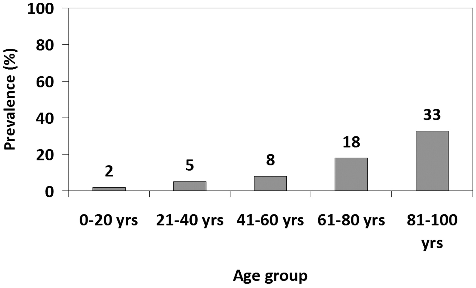
Of the 304 patients evaluated for dehiscence and thinning, 24 patients were identified with SSCD, of which 6 demonstrated dehiscence bilaterally. Categorization of these 24 patients with respect to age group is outlined in Table 1. The prevalence of canal dehiscence was calculated with respect to each age group with results outlined in Fig 1.

View this table:
  • View inline
  • View popup
Table 1:

Age groups of patients with SSCD

Fig 1.
  • Download figure
  • Open in new tab
  • Download powerpoint
Fig 1.

Prevalence of patients with SSCD and age.

Of the 304 patients evaluated, 137 were identified with thinning of the superior semicircular canal, of whom 50 demonstrated the finding bilaterally. Categorization of these 137 patients with respect to age group is outlined in Table 2. Prevalence of canal thinning was calculated with respect to each age group with results as outlined in Fig 2.

View this table:
  • View inline
  • View popup
Table 2:

Age groups of patients with canal thinning

Fig 2.
  • Download figure
  • Open in new tab
  • Download powerpoint
Fig 2.

Prevalence of patients with canal thinning and age.

On average each successively older age category experienced a 93% increase (95% CI, 30%–187%) in the prevalence of the SSCD (P = .001) and a 9% increase (95% CI, −5%–25%) in the prevalence of thinning (P = .21). Neither crude nor age-adjusted models demonstrated a significant association between thinning and contralateral dehiscence (P = .63) nor between dehiscence and contralateral thinning (P = .73).

Discussion

Despite the large volume of literature regarding diagnosis and treatment of the SSCD syndrome, it still remains unclear whether the condition has a predominantly congenital/developmental basis or whether it is acquired in life. There is some literature suggesting a congenital basis for the condition, with 1 case report describing the syndrome in a young child.2 A report from a radiologic series suggested a developmental basis for the condition, demonstrating greater prevalence of unilateral canal thinning when the opposite side is affected by dehiscence.3 In a large postmortem series by Carey et al,4 the superior semicircular canal roof was demonstrated to show progressive thickening during the first 3 years of life, with data suggesting that dehiscence arises from a defect in postnatal development. Certainly, congenital aplasia of inner ear structures, including the superior semicircular canal, has been described in both syndromic and nonsyndromic cases of congenital hearing loss.5,6 However, in practical experience at our institution, the radiologic diagnosis of SSCD is made more commonly among middle and older age groups. To our knowledge, there have been no large-scale investigations to date addressing imaging prevalence of SSCD with respect to age.

Our data demonstrate a statistically significant increase in the prevalence of radiographic dehiscence as age increases and an increasing trend but no significant increase in prevalence of thinning with age. Furthermore, our data show no significant association between thinning and contralateral dehiscence or vice versa, suggesting that thinning occurs independently of dehiscence. While there may be a congenital basis for some cases of SSCD, our data support the impression that SSCD is more commonly an acquired rather than a congenital/developmental condition.

The etiology for the increased prevalence of dehiscence with age remains unclear; while literature on the clinical diagnosis and treatment of canal dehiscence is plentiful, there is a paucity of literature on possible etiologies, to our knowledge. We conjecture that our finding of the increasing prevalence of SSCD with age may be related to systemic bony demineralization generally observed to increase with age. However, while bone resorption in the inner ear in the setting of osteoporosis has been shown in animal models,7 to our knowledge, it has not been confirmed in human studies. Certainly, the utility of CT bone attenuation measurement in the diagnosis of otosclerosis has been investigated, with some but not all groups showing focal diminished bone attenuation in patients with otosclerosis compared with controls.8–11 Change in bone attenuation with age in the inner ear is of great interest to the authors and is currently under investigation by our group. Alternatively, we conjecture that the condition may be the cumulative result of repetitive microtrauma, which would increase in number throughout the course of life, but this is yet to be proved.

While this investigation reviewed the radiologic prevalence of SSCD, we did not specifically address the long-term clinical outcome analysis of our patients in this particular investigation, which could be considered a limitation. The relevance of the imaging finding of SSCD, therefore, remains unclear but is currently under investigation by our group.

Conclusions

While there may be a congenital basis for some cases of SSCD, our data support the impression that SSCD is more commonly an acquired rather than congenital or developmental condition.

Footnotes

  • Paper previously presented at: Annual Meeting of the American Society of Neuroradiology and the Neuroradiology Education and Research Foundation Symposium, May 31–June 5, 2008; New Orleans, Louisiana.

Indicates open access to non-subscribers at www.ajnr.org

References

  1. 1.↵
    1. Minor LB,
    2. Solomon D,
    3. Zinreich JS,
    4. et al
    . Sound- and/or pressure-induced vertigo due to bone dehiscence of the superior semicircular canal. Arch Otolaryngol Head Neck Surg 1998;124:249–58
    CrossRefPubMed
  2. 2.↵
    1. Zhou G,
    2. Ohlms L,
    3. Liberman J,
    4. et al
    . Superior semicircular canal dehiscence in a young child: implication of developmental defect. Int J Pediatr Otorhinolaryngol 2007;71:1925–28
    CrossRefPubMed
  3. 3.↵
    1. Hirvonen TP,
    2. Weg N,
    3. Zinreich SJ,
    4. et al
    . High-resolution CT findings suggest a developmental abnormality underlying superior canal dehiscence syndrome. Acta Otolaryngol 2003;123:447–81
  4. 4.↵
    1. Carey JP,
    2. Minor LB,
    3. Nager GT
    . Dehiscence or thinning of bone overlying the superior semicircular canal in a temporal bone survey. Arch Otolaryngol Head Neck Surg 2000;126:137–47
    CrossRefPubMed
  5. 5.↵
    1. Morimoto AK,
    2. Wiggins RH 3rd.,
    3. Hudgins PA,
    4. et al
    . Absent semicircular canals in CHARGE syndrome: radiologic spectrum of findings. AJNR Am J Neuroradiol 2006;27:1663–71
    Abstract/FREE Full Text
  6. 6.↵
    1. Satar B,
    2. Mukherji SK,
    3. Telian SA
    . Congenital aplasia of the semicircular canals. Otol Neurotol 2003;24:437–46
    CrossRefPubMed
  7. 7.↵
    1. Horner KC
    . The effect of sex hormones on bone metabolism of the otic capsule: an overview. Hear Res 2009;252:56–60. Epub 2008 Dec 24
    CrossRefPubMed
  8. 8.↵
    1. Tringali S,
    2. Pouget JF,
    3. Bertholon P,
    4. et al
    . Value of temporal bone density measurements in otosclerosis patients with normal-appearing computed tomographic scan. Ann Otol Rhinol Laryngol 2007;116:195–98
    Abstract/FREE Full Text
  9. 9.↵
    1. Kawase S,
    2. Naganawa S,
    3. Sone M,
    4. et al
    . Relationship between CT densitometry with a slice thickness of 0.5 mm and audiometry in otosclerosis. Eur Radiol 2006;16:1367–73
    CrossRefPubMed
  10. 10.↵
    1. Grayeli AB,
    2. Yrieix CS,
    3. Imauchi Y,
    4. et al
    . Temporal bone density measurements using CT in otosclerosis. Acta Otolaryngol 2004;124:1136–40
    CrossRefPubMed
  11. 11.↵
    1. Huizing EH,
    2. de Groot JA
    . Densitometry of the cochlear capsule and correlation between bone density loss and bone conduction hearing loss in otosclerosis. Acta Otolaryngol 1987;103:464–68
    PubMed
  • Received May 13, 2010.
  • Accepted after revision September 19, 2010.
  • Copyright © American Society of Neuroradiology
View Abstract
PreviousNext
Back to top

In this issue

American Journal of Neuroradiology: 32 (5)
American Journal of Neuroradiology
Vol. 32, Issue 5
1 May 2011
  • Table of Contents
  • Index by author
Advertisement
Print
Download PDF
Email Article

Thank you for your interest in spreading the word on American Journal of Neuroradiology.

NOTE: We only request your email address so that the person you are recommending the page to knows that you wanted them to see it, and that it is not junk mail. We do not capture any email address.

Enter multiple addresses on separate lines or separate them with commas.
Superior Semicircular Canal Dehiscence: Congenital or Acquired Condition?
(Your Name) has sent you a message from American Journal of Neuroradiology
(Your Name) thought you would like to see the American Journal of Neuroradiology web site.
CAPTCHA
This question is for testing whether or not you are a human visitor and to prevent automated spam submissions.
Cite this article
R.N. Nadgir, A. Ozonoff, A.K. Devaiah, A.A. Halderman, O. Sakai
Superior Semicircular Canal Dehiscence: Congenital or Acquired Condition?
American Journal of Neuroradiology May 2011, 32 (5) 947-949; DOI: 10.3174/ajnr.A2437

Citation Manager Formats

  • BibTeX
  • Bookends
  • EasyBib
  • EndNote (tagged)
  • EndNote 8 (xml)
  • Medlars
  • Mendeley
  • Papers
  • RefWorks Tagged
  • Ref Manager
  • RIS
  • Zotero
0 Responses
Respond to this article
Share
Bookmark this article
Superior Semicircular Canal Dehiscence: Congenital or Acquired Condition?
R.N. Nadgir, A. Ozonoff, A.K. Devaiah, A.A. Halderman, O. Sakai
American Journal of Neuroradiology May 2011, 32 (5) 947-949; DOI: 10.3174/ajnr.A2437
del.icio.us logo Twitter logo Facebook logo Mendeley logo
  • Tweet Widget
  • Facebook Like
  • Google Plus One
Purchase

Jump to section

  • Article
    • Abstract
    • Abbreviations
    • Materials and Methods
    • Results
    • Discussion
    • Conclusions
    • Footnotes
    • References
  • Figures & Data
  • Info & Metrics
  • Responses
  • References
  • PDF

Related Articles

  • No related articles found.
  • PubMed
  • Google Scholar

Cited By...

  • Bilateral superior semicircular canal dehiscence: bilateral conductive hearing loss with subtle vestibular symptoms
  • Prevalence of Superior Semicircular Canal Dehiscence on High-Resolution CT Imaging in Patients without Vestibular or Auditory Abnormalities
  • Crossref (95)
  • Google Scholar

This article has been cited by the following articles in journals that are participating in Crossref Cited-by Linking.

  • Superior semicircular canal dehiscence: Diagnosis and management
    Christine Mau, Naveed Kamal, Saiaditya Badeti, Renuka Reddy, Yu-Lan Mary Ying, Robert W. Jyung, James K. Liu
    Journal of Clinical Neuroscience 2018 48
  • Superior semicircular canal dehiscence syndrome
    Wenya Linda Bi, Ryan Brewster, Dennis Poe, David Vernick, Daniel J. Lee, C. Eduardo Corrales, Ian F. Dunn
    Journal of Neurosurgery 2017 127 6
  • Endoscopic‐assisted repair of superior canal dehiscence syndrome
    Margaret S. Carter, Sarah Lookabaugh, Daniel J. Lee
    The Laryngoscope 2014 124 6
  • Prevalence of radiographic semicircular canal dehiscence in very young children: an evaluation using high-resolution computed tomography of the temporal bones
    Mari Hagiwara, Jamil A. Shaikh, Yixin Fang, Girish Fatterpekar, Pamela C. Roehm
    Pediatric Radiology 2012 42 12
  • Radiological prevalence of superior and posterior semicircular canal dehiscence in children
    Alexander J. Saxby, Claire Gowdy, Marcela Fandiño, Neil K. Chadha, Frederick K. Kozak, Michael A. Sargent, Jane Lea
    International Journal of Pediatric Otorhinolaryngology 2015 79 3
  • A radiologic and anatomic study of the superior semicircular canal
    N. Klopp-Dutote, C. Kolski, A. Biet, V. Strunski, C. Page
    European Annals of Otorhinolaryngology, Head and Neck Diseases 2016 133 2
  • Outcomes of middle fossa craniotomy for the repair of superior semicircular canal dehiscence
    Nolan Ung, Lawrance K. Chung, Carlito Lagman, Nikhilesh S. Bhatt, Natalie E. Barnette, Vera Ong, Quinton Gopen, Isaac Yang
    Journal of Clinical Neuroscience 2017 43
  • Recent advances in superior semicircular canal dehiscence syndrome
    G Chilvers, I McKay-Davies
    The Journal of Laryngology & Otology 2015 129 3
  • Surgical capping of superior semicircular canal dehiscence
    S. A. Mueller, D. Vibert, R. Haeusler, A. Raabe, M. Caversaccio
    European Archives of Oto-Rhino-Laryngology 2014 271 6
  • Incidence of Pediatric Superior Semicircular Canal Dehiscence and Inner Ear Anomalies
    Eric M. Sugihara, Seilesh C. Babu, Dennis J. Kitsko, Michael S. Haupert, Prasad J. Thottam
    Otology & Neurotology 2016 37 9

More in this TOC Section

  • Correlation of Apparent Diffusion Coefficient at 3T with Prognostic Parameters of Retinoblastoma
  • Parathyroid Lesions: Characterization with Dual-Phase Arterial and Venous Enhanced CT of the Neck
  • Efficacy of Diffusion-Weighted Imaging for the Differentiation between Lymphomas and Carcinomas of the Nasopharynx and Oropharynx: Correlations of Apparent Diffusion Coefficients and Histologic Features
Show more Head and Neck

Similar Articles

Advertisement

Indexed Content

  • Current Issue
  • Accepted Manuscripts
  • Article Preview
  • Past Issues
  • Editorials
  • Editors Choice
  • Fellow Journal Club
  • Letters to the Editor

Cases

  • Case Collection
  • Archive - Case of the Week
  • Archive - Case of the Month
  • Archive - Classic Case

Special Collections

  • Special Collections

Resources

  • News and Updates
  • Turn around Times
  • Submit a Manuscript
  • Author Policies
  • Manuscript Submission Guidelines
  • Evidence-Based Medicine Level Guide
  • Publishing Checklists
  • Graphical Abstract Preparation
  • Imaging Protocol Submission
  • Submit a Case
  • Become a Reviewer/Academy of Reviewers
  • Get Peer Review Credit from Publons

Multimedia

  • AJNR Podcast
  • AJNR SCANtastic
  • Video Articles

About Us

  • About AJNR
  • Editorial Board
  • Not an AJNR Subscriber? Join Now
  • Alerts
  • Feedback
  • Advertise with us
  • Librarian Resources
  • Permissions
  • Terms and Conditions

American Society of Neuroradiology

  • Not an ASNR Member? Join Now

© 2025 by the American Society of Neuroradiology All rights, including for text and data mining, AI training, and similar technologies, are reserved.
Print ISSN: 0195-6108 Online ISSN: 1936-959X

Powered by HighWire