Advertisement

Main menu

  • Home
  • Content
    • Current Issue
    • Accepted Manuscripts
    • Article Preview
    • Past Issue Archive
    • Video Articles
    • AJNR Case Collection
    • Case of the Week Archive
    • Case of the Month Archive
    • Classic Case Archive
  • Special Collections
    • AJNR Awards
    • Low-Field MRI
    • Alzheimer Disease
    • ASNR Foundation Special Collection
    • Photon-Counting CT
    • View All
  • Multimedia
    • AJNR Podcasts
    • AJNR SCANtastic
    • Trainee Corner
    • MRI Safety Corner
    • Imaging Protocols
  • For Authors
    • Submit a Manuscript
    • Submit a Video Article
    • Submit an eLetter to the Editor/Response
    • Manuscript Submission Guidelines
    • Statistical Tips
    • Fast Publishing of Accepted Manuscripts
    • Graphical Abstract Preparation
    • Imaging Protocol Submission
    • Author Policies
  • About Us
    • About AJNR
    • Editorial Board
    • Editorial Board Alumni
  • More
    • Become a Reviewer/Academy of Reviewers
    • Subscribers
    • Permissions
    • Alerts
    • Feedback
    • Advertisers
    • ASNR Home

User menu

  • Alerts
  • Log in

Search

  • Advanced search
American Journal of Neuroradiology

American Journal of Neuroradiology

ASHNR American Society of Functional Neuroradiology ASHNR American Society of Pediatric Neuroradiology ASSR
  • Alerts
  • Log in

Advanced Search

  • Home
  • Content
    • Current Issue
    • Accepted Manuscripts
    • Article Preview
    • Past Issue Archive
    • Video Articles
    • AJNR Case Collection
    • Case of the Week Archive
    • Case of the Month Archive
    • Classic Case Archive
  • Special Collections
    • AJNR Awards
    • Low-Field MRI
    • Alzheimer Disease
    • ASNR Foundation Special Collection
    • Photon-Counting CT
    • View All
  • Multimedia
    • AJNR Podcasts
    • AJNR SCANtastic
    • Trainee Corner
    • MRI Safety Corner
    • Imaging Protocols
  • For Authors
    • Submit a Manuscript
    • Submit a Video Article
    • Submit an eLetter to the Editor/Response
    • Manuscript Submission Guidelines
    • Statistical Tips
    • Fast Publishing of Accepted Manuscripts
    • Graphical Abstract Preparation
    • Imaging Protocol Submission
    • Author Policies
  • About Us
    • About AJNR
    • Editorial Board
    • Editorial Board Alumni
  • More
    • Become a Reviewer/Academy of Reviewers
    • Subscribers
    • Permissions
    • Alerts
    • Feedback
    • Advertisers
    • ASNR Home
  • Follow AJNR on Twitter
  • Visit AJNR on Facebook
  • Follow AJNR on Instagram
  • Join AJNR on LinkedIn
  • RSS Feeds

AJNR Awards, New Junior Editors, and more. Read the latest AJNR updates

Research ArticleBrain

Evaluation of Parenchymal Neuro-Behçet Disease by Using Susceptibility-Weighted Imaging

S. Albayram, S. Saip, Z.I. Hasiloglu, M. Teke, E. Ceyhan, M. Tutuncu, H. Selcuk, A. Kina and A. Siva
American Journal of Neuroradiology June 2011, 32 (6) 1050-1055; DOI: https://doi.org/10.3174/ajnr.A2477
S. Albayram
  • Find this author on Google Scholar
  • Find this author on PubMed
  • Search for this author on this site
S. Saip
  • Find this author on Google Scholar
  • Find this author on PubMed
  • Search for this author on this site
Z.I. Hasiloglu
  • Find this author on Google Scholar
  • Find this author on PubMed
  • Search for this author on this site
M. Teke
  • Find this author on Google Scholar
  • Find this author on PubMed
  • Search for this author on this site
E. Ceyhan
  • Find this author on Google Scholar
  • Find this author on PubMed
  • Search for this author on this site
M. Tutuncu
  • Find this author on Google Scholar
  • Find this author on PubMed
  • Search for this author on this site
H. Selcuk
  • Find this author on Google Scholar
  • Find this author on PubMed
  • Search for this author on this site
A. Kina
  • Find this author on Google Scholar
  • Find this author on PubMed
  • Search for this author on this site
A. Siva
  • Find this author on Google Scholar
  • Find this author on PubMed
  • Search for this author on this site
  • Article
  • Figures & Data
  • Info & Metrics
  • Responses
  • References
  • PDF
Loading

Article Figures & Data

Figures

  • Tables
  • Fig 1.
    • Download figure
    • Open in new tab
    • Download powerpoint
    Fig 1.

    Case 16. Lesion in the right capsula interna is observed as hyperintense on T2 FSE (A) and T2*GE (B) images. In this localization, multiple hemorrhagic foci are observed as hypointense on SWI magnitude (C) and SWI mIP (D) images. In addition, prominent venous structures adjacent to the left corona radiata lesion are observed with continuity in consecutive sections on the SWI mIP (E) image.

  • Fig 2.
    • Download figure
    • Open in new tab
    • Download powerpoint
    Fig 2.

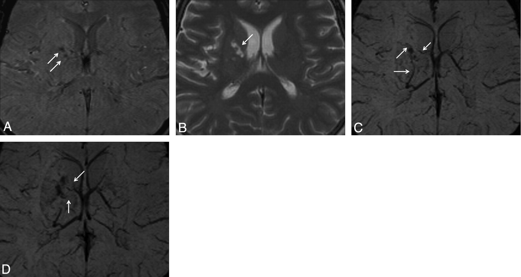
    Case 20. Hemorrhagic lesion in the right putamen is observed as hypointense on the SWI magnitude (A) image. This lesion is observed as hyperintense on the T2 FSE (B) image. In the lesion vicinity, occlusion of thalamostriate veins (C) and marked collateral venous structure (D) are observed on the SWI mIP images.

Tables

  • Figures
    • View popup
    Table 1:

    MR findings and characteristics of the lesions in parenchymal NBD

    Case No.AgeSexLesion No.Locations of LesionsSignal Intensity in LesionsOther Findings
    T2 FSET2*GESWI MagnitudeSWI mIP
    144F1PutamenHyperHyperHyperHyper
    2MesencephalonHypoHypoHypo
    239F3,4,5PonsHyperHyperHypoHypo
    6Medulla oblongataHyperHyper
    331M7PonsHypoHypo
    8PonsHyperHyperHypoHypo
    440M9PutamenHypoHypoHypoHypo
    531M10PonsHyperHyperHypoHypo
    644F11ThalamusHypoHypo
    12ThalamusHypoHypoPVS
    736MBSA
    852F13,14PutamenHyperHypoHypo
    15PutamenHyperHyperHyperHyper
    941M16PonsHypoHypoBSA
    17,18Medulla oblongataHyperHyperHyperHyperBSA
    1046FBSA
    1138M19ThalamusHyperHyperHypoHypoBSA
    20,21PonsHyperHypoHypoBSA
    22Medulla oblongataHyperHyperBSA
    23PonsHyperHyperHypoHypoBSA
    1230M24ThalamusHypoHypo
    25,26PonsHyperHypoHypo
    1337F27PonsHyperHyperHypoHypoBSA
    28Medulla oblongataHypoHypoBSA
    1445M29ThalamusHyperHyperHypoHypoPVS
    1527F30ThalamusHypoHypoPVS
    31MesencephalonHypoHypoHypoHypo
    1644M32Corona radiataHyperHyperHypoHypoPVS
    33Capsula internaHyperHyperHypoHypo
    1720F34Splenium of CCHypoHypo
    1836F35Caudate nucleusHyperHyperHypoHypoPVS
    36PonsHyperHyperHypoHypo
    1934F37Corona radiataHyperHyperHyperHyper
    38ThalamusHyperHyperHypoHypo
    39,40PonsHyperHyperHyperHyper
    41Medulla oblongataHyperHyperHyperHyper
    2041M42Caudate nucleusHyperHyperHypoHypoOcclusion of TSV and CV
    43PutamenHyperHyperHypoHypo
    44MesencephalonHyperHyperHypoHypo
    45PonsHyperHyperHypoHypo
    2140F46Caudate nucleusHyperHyperHypoHypoOcclusion of TSV
    47,48MesencephalonHyperHyperHypoHypo
    49,50PonsHyperHyperHypoHypo
    2239F51PonsHyperHyperHypoHypoPVS, BSA
    52Medulla oblongataHyperHyperHypoHypoBSA
    2335M53Temporal lobeArterial ischemia
PreviousNext
Back to top

In this issue

American Journal of Neuroradiology: 32 (6)
American Journal of Neuroradiology
Vol. 32, Issue 6
1 Jun 2011
  • Table of Contents
  • Index by author
Advertisement
Print
Download PDF
Email Article

Thank you for your interest in spreading the word on American Journal of Neuroradiology.

NOTE: We only request your email address so that the person you are recommending the page to knows that you wanted them to see it, and that it is not junk mail. We do not capture any email address.

Enter multiple addresses on separate lines or separate them with commas.
Evaluation of Parenchymal Neuro-Behçet Disease by Using Susceptibility-Weighted Imaging
(Your Name) has sent you a message from American Journal of Neuroradiology
(Your Name) thought you would like to see the American Journal of Neuroradiology web site.
CAPTCHA
This question is for testing whether or not you are a human visitor and to prevent automated spam submissions.
Cite this article
S. Albayram, S. Saip, Z.I. Hasiloglu, M. Teke, E. Ceyhan, M. Tutuncu, H. Selcuk, A. Kina, A. Siva
Evaluation of Parenchymal Neuro-Behçet Disease by Using Susceptibility-Weighted Imaging
American Journal of Neuroradiology Jun 2011, 32 (6) 1050-1055; DOI: 10.3174/ajnr.A2477

Citation Manager Formats

  • BibTeX
  • Bookends
  • EasyBib
  • EndNote (tagged)
  • EndNote 8 (xml)
  • Medlars
  • Mendeley
  • Papers
  • RefWorks Tagged
  • Ref Manager
  • RIS
  • Zotero
0 Responses
Respond to this article
Share
Bookmark this article
Evaluation of Parenchymal Neuro-Behçet Disease by Using Susceptibility-Weighted Imaging
S. Albayram, S. Saip, Z.I. Hasiloglu, M. Teke, E. Ceyhan, M. Tutuncu, H. Selcuk, A. Kina, A. Siva
American Journal of Neuroradiology Jun 2011, 32 (6) 1050-1055; DOI: 10.3174/ajnr.A2477
del.icio.us logo Twitter logo Facebook logo Mendeley logo
  • Tweet Widget
  • Facebook Like
  • Google Plus One
Purchase

Jump to section

  • Article
    • Abstract
    • Abbreviations
    • Materials and Methods
    • Results
    • Discussion
    • Conclusions
    • Footnotes
    • References
  • Figures & Data
  • Info & Metrics
  • Responses
  • References
  • PDF

Related Articles

  • No related articles found.
  • PubMed
  • Google Scholar

Cited By...

  • Neurological involvement by Behcets syndrome: clinical features, diagnosis, treatment and outcome
  • The corpus callosum in the diagnosis of multiple sclerosis and other CNS demyelinating and inflammatory diseases
  • Crossref
  • Google Scholar

This article has not yet been cited by articles in journals that are participating in Crossref Cited-by Linking.

More in this TOC Section

  • Usefulness of Quantitative Susceptibility Mapping for the Diagnosis of Parkinson Disease
  • White Matter Alterations in the Brains of Patients with Active, Remitted, and Cured Cushing Syndrome: A DTI Study
  • Qualitative and Quantitative Analysis of MR Imaging Findings in Patients with Middle Cerebral Artery Stroke Implanted with Mesenchymal Stem Cells
Show more Brain

Similar Articles

Advertisement

Indexed Content

  • Current Issue
  • Accepted Manuscripts
  • Article Preview
  • Past Issues
  • Editorials
  • Editor's Choice
  • Fellows' Journal Club
  • Letters to the Editor
  • Video Articles

Cases

  • Case Collection
  • Archive - Case of the Week
  • Archive - Case of the Month
  • Archive - Classic Case

More from AJNR

  • Trainee Corner
  • Imaging Protocols
  • MRI Safety Corner
  • Book Reviews

Multimedia

  • AJNR Podcasts
  • AJNR Scantastics

Resources

  • Turnaround Time
  • Submit a Manuscript
  • Submit a Video Article
  • Submit an eLetter to the Editor/Response
  • Manuscript Submission Guidelines
  • Statistical Tips
  • Fast Publishing of Accepted Manuscripts
  • Graphical Abstract Preparation
  • Imaging Protocol Submission
  • Evidence-Based Medicine Level Guide
  • Publishing Checklists
  • Author Policies
  • Become a Reviewer/Academy of Reviewers
  • News and Updates

About Us

  • About AJNR
  • Editorial Board
  • Editorial Board Alumni
  • Alerts
  • Permissions
  • Not an AJNR Subscriber? Join Now
  • Advertise with Us
  • Librarian Resources
  • Feedback
  • Terms and Conditions
  • AJNR Editorial Board Alumni

American Society of Neuroradiology

  • Not an ASNR Member? Join Now

© 2025 by the American Society of Neuroradiology All rights, including for text and data mining, AI training, and similar technologies, are reserved.
Print ISSN: 0195-6108 Online ISSN: 1936-959X

Powered by HighWire