Advertisement

Main menu

  • Home
  • Content
    • Current Issue
    • Accepted Manuscripts
    • Article Preview
    • Past Issue Archive
    • AJNR Case Collection
    • Case of the Week Archive
    • Classic Case Archive
    • Case of the Month Archive
  • Special Collections
    • Spinal CSF Leak Articles (Jan 2020-June 2024)
    • 2024 AJNR Journal Awards
    • Most Impactful AJNR Articles
  • Multimedia
    • AJNR Podcast
    • AJNR Scantastics
    • Video Articles
  • For Authors
    • Submit a Manuscript
    • Author Policies
    • Fast publishing of Accepted Manuscripts
    • Graphical Abstract Preparation
    • Manuscript Submission Guidelines
    • Imaging Protocol Submission
    • Submit a Case for the Case Collection
  • About Us
    • About AJNR
    • Editorial Board
  • More
    • Become a Reviewer/Academy of Reviewers
    • Subscribers
    • Permissions
    • Alerts
    • Feedback
    • Advertisers
    • ASNR Home
  • Other Publications
    • ajnr

User menu

  • Alerts
  • Log in

Search

  • Advanced search
American Journal of Neuroradiology

American Journal of Neuroradiology

ASHNR American Society of Functional Neuroradiology ASHNR American Society of Pediatric Neuroradiology ASSR
  • Alerts
  • Log in

Advanced Search

  • Home
  • Content
    • Current Issue
    • Accepted Manuscripts
    • Article Preview
    • Past Issue Archive
    • AJNR Case Collection
    • Case of the Week Archive
    • Classic Case Archive
    • Case of the Month Archive
  • Special Collections
    • Spinal CSF Leak Articles (Jan 2020-June 2024)
    • 2024 AJNR Journal Awards
    • Most Impactful AJNR Articles
  • Multimedia
    • AJNR Podcast
    • AJNR Scantastics
    • Video Articles
  • For Authors
    • Submit a Manuscript
    • Author Policies
    • Fast publishing of Accepted Manuscripts
    • Graphical Abstract Preparation
    • Manuscript Submission Guidelines
    • Imaging Protocol Submission
    • Submit a Case for the Case Collection
  • About Us
    • About AJNR
    • Editorial Board
  • More
    • Become a Reviewer/Academy of Reviewers
    • Subscribers
    • Permissions
    • Alerts
    • Feedback
    • Advertisers
    • ASNR Home
  • Follow AJNR on Twitter
  • Visit AJNR on Facebook
  • Follow AJNR on Instagram
  • Join AJNR on LinkedIn
  • RSS Feeds

Welcome to the new AJNR, Updated Hall of Fame, and more. Read the full announcements.


AJNR is seeking candidates for the position of Associate Section Editor, AJNR Case Collection. Read the full announcement.


Research Article50th Anniversary Perspectives
Open Access

Neuroradiology Back to the Future: Head and Neck Imaging

E.G. Hoeffner, S.K. Mukherji, A. Srinivasan and D.J. Quint
American Journal of Neuroradiology December 2012, 33 (11) 2026-2032; DOI: https://doi.org/10.3174/ajnr.A3365
E.G. Hoeffner
aFrom the Division of Neuroradiology, Department of Radiology, University of Michigan Health System, Ann Arbor, Michigan.
  • Find this author on Google Scholar
  • Find this author on PubMed
  • Search for this author on this site
S.K. Mukherji
aFrom the Division of Neuroradiology, Department of Radiology, University of Michigan Health System, Ann Arbor, Michigan.
  • Find this author on Google Scholar
  • Find this author on PubMed
  • Search for this author on this site
A. Srinivasan
aFrom the Division of Neuroradiology, Department of Radiology, University of Michigan Health System, Ann Arbor, Michigan.
  • Find this author on Google Scholar
  • Find this author on PubMed
  • Search for this author on this site
D.J. Quint
aFrom the Division of Neuroradiology, Department of Radiology, University of Michigan Health System, Ann Arbor, Michigan.
  • Find this author on Google Scholar
  • Find this author on PubMed
  • Search for this author on this site
  • Article
  • Figures & Data
  • Info & Metrics
  • Responses
  • References
  • PDF
Loading

Article Figures & Data

Figures

  • Fig 1.
    • Download figure
    • Open in new tab
    • Download powerpoint
    Fig 1.

    A, Waters view of the paranasal sinuses with a depressed right orbital floor fracture (white arrow) and an opacified right maxillary sinus (black arrow). B, Coronal tomogram in a different patient shows a left orbital floor fracture (white arrows) with partial left maxillary sinus opacification (black arrows).

  • Fig 2.
    • Download figure
    • Open in new tab
    • Download powerpoint
    Fig 2.

    Schüller view of the temporal bone showing a fracture (short white arrows) extending through the squamosal temporal bone, anterior to the mastoid process (long white arrow) and into the external auditory canal (double white arrows). A second fracture (short black arrows) extends through the parietal bone, into the squamosal temporal bone, and then into the skull base just anterior to the articular eminence (large black arrow) of the temporal bone.

  • Fig 3.
    • Download figure
    • Open in new tab
    • Download powerpoint
    Fig 3.

    A 20-year-old man with a lump near the angle of the mandible. Oblique view from a parotid sialogram shows distortion of secondary ducts and a “ductless” appearance (arrows) near the angle of the mandible, related to a pleomorphic adenoma of the parotid gland.
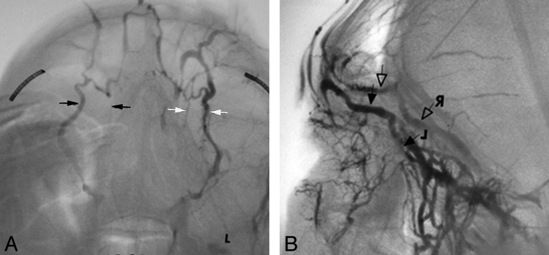
  • Fig 4.
    • Download figure
    • Open in new tab
    • Download powerpoint
    Fig 4.

    A 59-year-old man with left proptosis and negative findings on orbital x-rays and tomograms. Orbital venography was performed following percutaneous puncture of a lateral frontal vein. Frontal view from an orbital venogram (A) shows medial displacement of the anterior and midportions of the left superior ophthalmic vein (SOV) (white arrows) compared with the right (black arrows). Lateral view from an orbital venogram (B) demonstrates inferior displacement of the left SOV (open arrows) compared with the right (solid arrows). Imaging findings are suggestive of a tumor in the anterior and midportion of the orbit laterally. Biopsy of mass revealed lacrimal gland carcinoma.
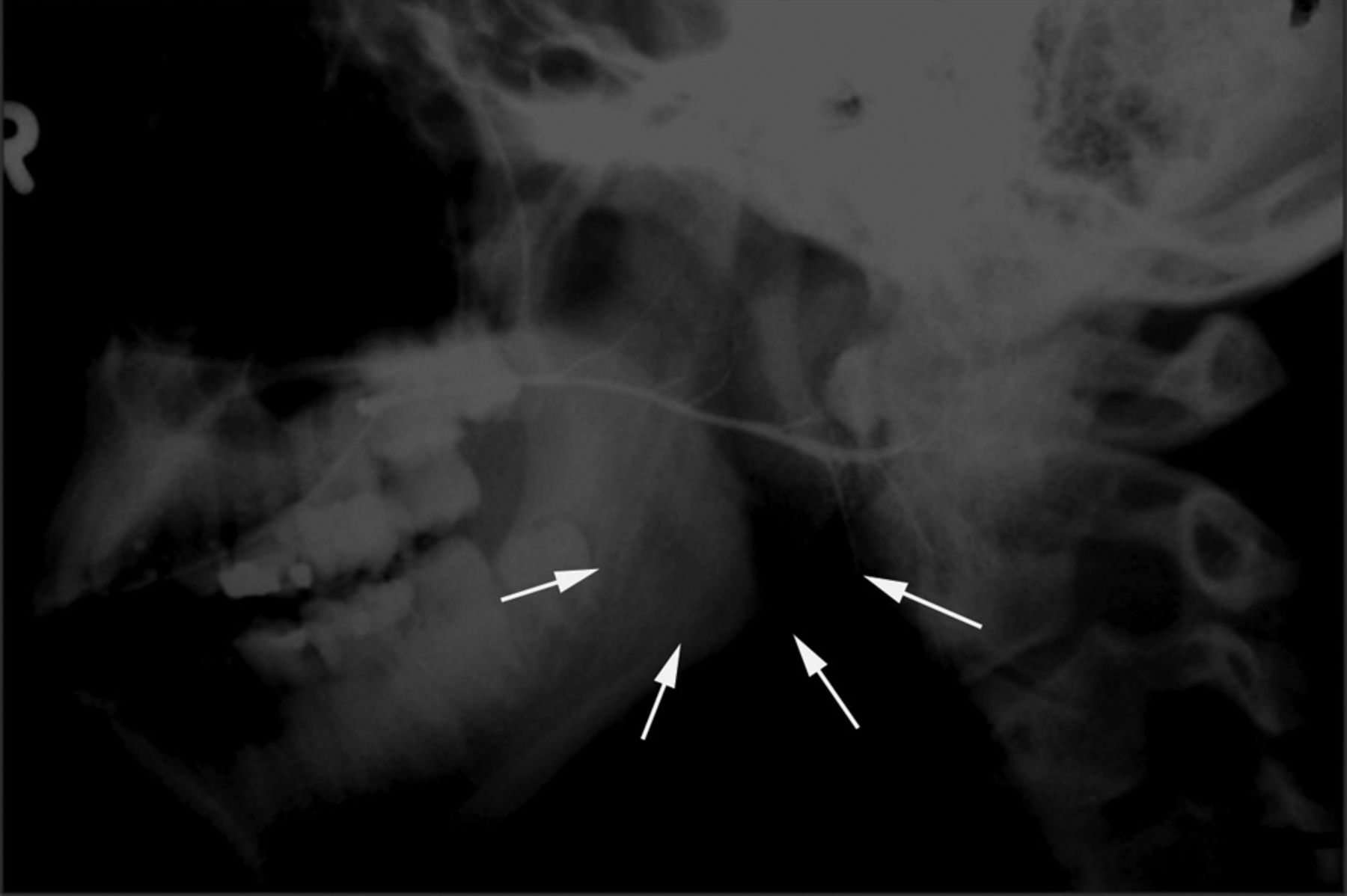
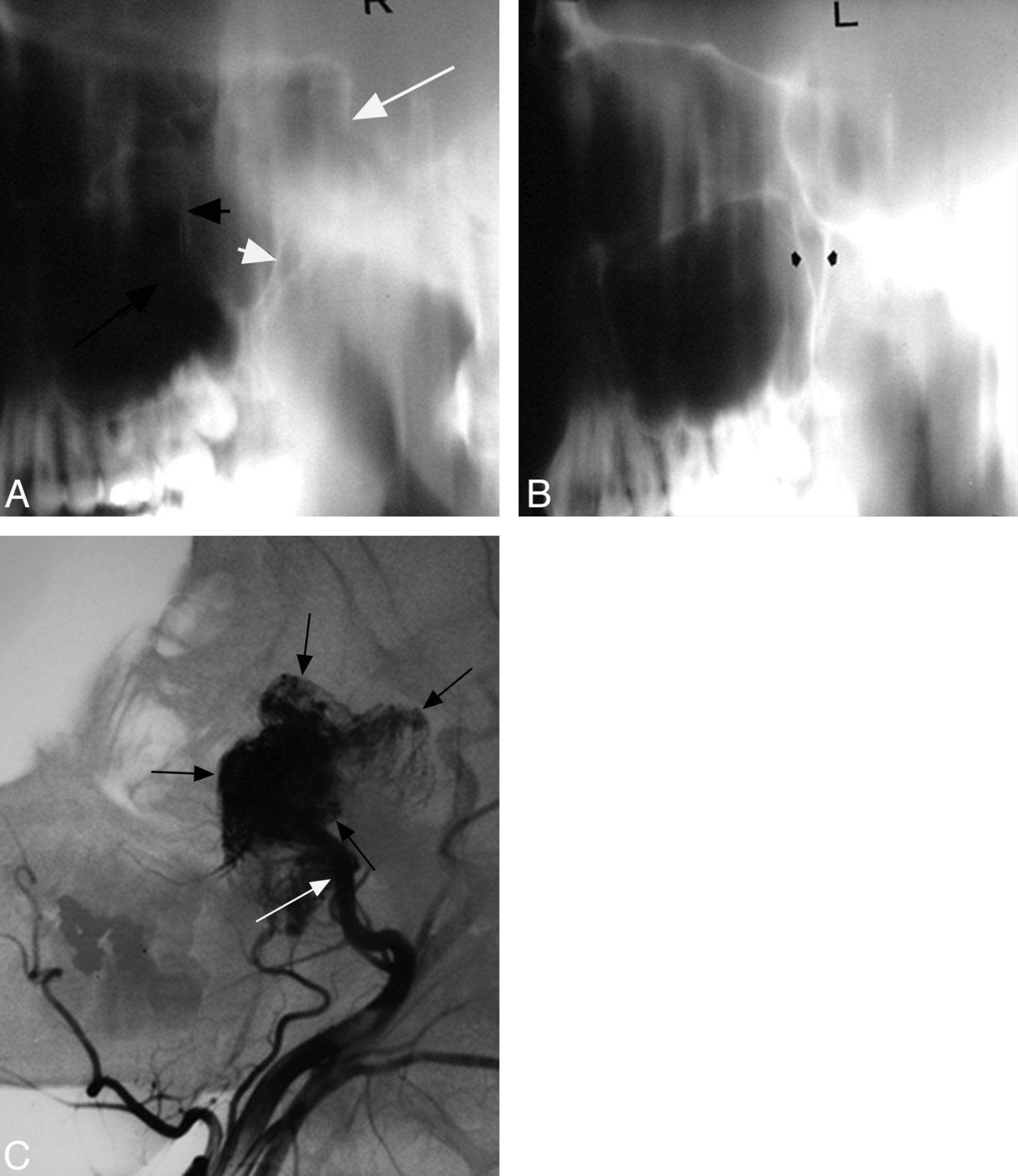
  • Fig 5.
    • Download figure
    • Open in new tab
    • Download powerpoint
    Fig 5.

    Adolescent boy with epistaxis. Lateral tomogram through the right side of the nasopharynx (A) shows a nasopharyngeal mass extending into and enlarging the pterygopalatine fossa with forward bowing of the posterior wall of the maxillary antrum (short black arrow) and posterior displacement of the pterygoid process (short white arrow). The mass also extends into the anterior half of the sphenoid sinus (long white arrow) and right maxillary antrum (long black arrow). B, Lateral tomogram through the left side of the nasopharynx shows a normal pterygopalatine fossa (arrows). C, Lateral view from the right external carotid artery angiogram shows a very vascular mass (black arrows) in the nasopharynx, right pterygopalatine fossa, anterior sphenoid sinus, posterior right maxillary sinus, and posterior inferior right orbit. Supply is from the internal maxillary artery (white arrow).

PreviousNext
Back to top

In this issue

American Journal of Neuroradiology: 33 (11)
American Journal of Neuroradiology
Vol. 33, Issue 11
1 Dec 2012
  • Table of Contents
  • Index by author
Advertisement
Print
Download PDF
Email Article

Thank you for your interest in spreading the word on American Journal of Neuroradiology.

NOTE: We only request your email address so that the person you are recommending the page to knows that you wanted them to see it, and that it is not junk mail. We do not capture any email address.

Enter multiple addresses on separate lines or separate them with commas.
Neuroradiology Back to the Future: Head and Neck Imaging
(Your Name) has sent you a message from American Journal of Neuroradiology
(Your Name) thought you would like to see the American Journal of Neuroradiology web site.
CAPTCHA
This question is for testing whether or not you are a human visitor and to prevent automated spam submissions.
Cite this article
E.G. Hoeffner, S.K. Mukherji, A. Srinivasan, D.J. Quint
Neuroradiology Back to the Future: Head and Neck Imaging
American Journal of Neuroradiology Dec 2012, 33 (11) 2026-2032; DOI: 10.3174/ajnr.A3365

Citation Manager Formats

  • BibTeX
  • Bookends
  • EasyBib
  • EndNote (tagged)
  • EndNote 8 (xml)
  • Medlars
  • Mendeley
  • Papers
  • RefWorks Tagged
  • Ref Manager
  • RIS
  • Zotero
0 Responses
Respond to this article
Share
Bookmark this article
Neuroradiology Back to the Future: Head and Neck Imaging
E.G. Hoeffner, S.K. Mukherji, A. Srinivasan, D.J. Quint
American Journal of Neuroradiology Dec 2012, 33 (11) 2026-2032; DOI: 10.3174/ajnr.A3365
del.icio.us logo Twitter logo Facebook logo Mendeley logo
  • Tweet Widget
  • Facebook Like
  • Google Plus One
Purchase

Jump to section

  • Article
    • Abstract
    • Conclusions
    • Footnotes
    • References
  • Figures & Data
  • Info & Metrics
  • Responses
  • References
  • PDF

Related Articles

  • No related articles found.
  • PubMed
  • Google Scholar

Cited By...

  • Large retropharyngeal abscess: a case of mistaken identity
  • Crossref (11)
  • Google Scholar

This article has been cited by the following articles in journals that are participating in Crossref Cited-by Linking.

  • Spine Anatomy Imaging
    Amirali Modir Shanechi, Matthew Kiczek, Majid Khan, Gaurav Jindal
    Neuroimaging Clinics of North America 2019 29 4
  • Challenges in early operative approaches to intramedullary spinal cord tumors: Harvey Cushing’s perspective
    Courtney Pendleton, Jordina Rincon-Torroella, Ziya L. Gokaslan, George I. Jallo, Alfredo Quinones-Hinojosa
    Journal of Neurosurgery: Spine 2015 23 4
  • Algorithmic interpretation of head and neck computerized tomography (CT) images
    Khalid Al Qahtani, Shereen Yankisari, Mohammed Al Shehabi, Fareed Al Ghamdi, Faiza Al Otaibi, Tahera Islam
    Journal of the Anatomical Society of India 2017 66 2
  • Large retropharyngeal abscess: a case of mistaken identity
    N Makaram, R Gohil, Q Gardiner, J Manickavasagam
    BMJ Case Reports 2016 2016
  • Head and Neck Imaging
    S. Connor
    Clinical Radiology 2018 73 1
  • Image Guided Interventions of the Spine
    Matthew D. Alvin, Amirali Modir Shanechi, Matthew Kiczek, Majid Khan
    2021
  • Medulloblastoma - Therapeutic Outcomes and Future Clinical Trials
    Hebah Abudawaba, Vinaya Rajahraman, Maryann Bishop-Jodoin, John M. Buatti
    2025 1
  • Radiopaedia.org
    Dustin Roberts
    2018
  • Revision Lumbar Spine Surgery
    Eric A. Potts
    2022
  • The Role and Contribution of Lumbar Myelography in the Diagnosis and Treatment of Patients With Lumbar Degenerative Disorders: Clinical and Statistical Evaluation of Post-Myelography Treatment of 63 Patients
    Hasan Burak Gündüz, Aysegul Esen Aydin, Aysegul Ozdemir Ovalioglu, Erhan Emel, Ozden Erhan Sofuoglu, Mustafa Levent Uysal, Muslum Gunes, Murad Asiltürk, Talat Cem Ovalioglu
    Cureus 2021

More in this TOC Section

  • Neuroradiology Back to the Future: Spine Imaging
  • Neuroradiology Back to the Future: Brain Imaging
Show more 50th Anniversary Perspectives

Similar Articles

Advertisement

Indexed Content

  • Current Issue
  • Accepted Manuscripts
  • Article Preview
  • Past Issues
  • Editorials
  • Editors Choice
  • Fellow Journal Club
  • Letters to the Editor

Cases

  • Case Collection
  • Archive - Case of the Week
  • Archive - Case of the Month
  • Archive - Classic Case

Special Collections

  • Special Collections

Resources

  • News and Updates
  • Turn around Times
  • Submit a Manuscript
  • Author Policies
  • Manuscript Submission Guidelines
  • Evidence-Based Medicine Level Guide
  • Publishing Checklists
  • Graphical Abstract Preparation
  • Imaging Protocol Submission
  • Submit a Case
  • Become a Reviewer/Academy of Reviewers
  • Get Peer Review Credit from Publons

Multimedia

  • AJNR Podcast
  • AJNR SCANtastic
  • Video Articles

About Us

  • About AJNR
  • Editorial Board
  • Not an AJNR Subscriber? Join Now
  • Alerts
  • Feedback
  • Advertise with us
  • Librarian Resources
  • Permissions
  • Terms and Conditions

American Society of Neuroradiology

  • Not an ASNR Member? Join Now

© 2025 by the American Society of Neuroradiology All rights, including for text and data mining, AI training, and similar technologies, are reserved.
Print ISSN: 0195-6108 Online ISSN: 1936-959X

Powered by HighWire