
Article Figures & Data
Figures
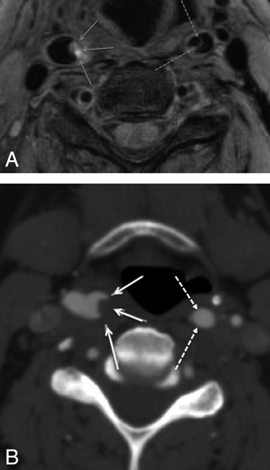
- Fig 1.
BBI (A) performed 1 day subsequent to CTA (B) demonstrates thrombus within the lumen of the right internal carotid artery (continuous arrows) but also a dissection flap in the left internal carotid artery not appreciated on CTA.
- Fig 2.
Double inversion recovery BBI demonstrates crescentic intramural high T1 methemoglobin signal intensity (arrows) in the dissected left VA.
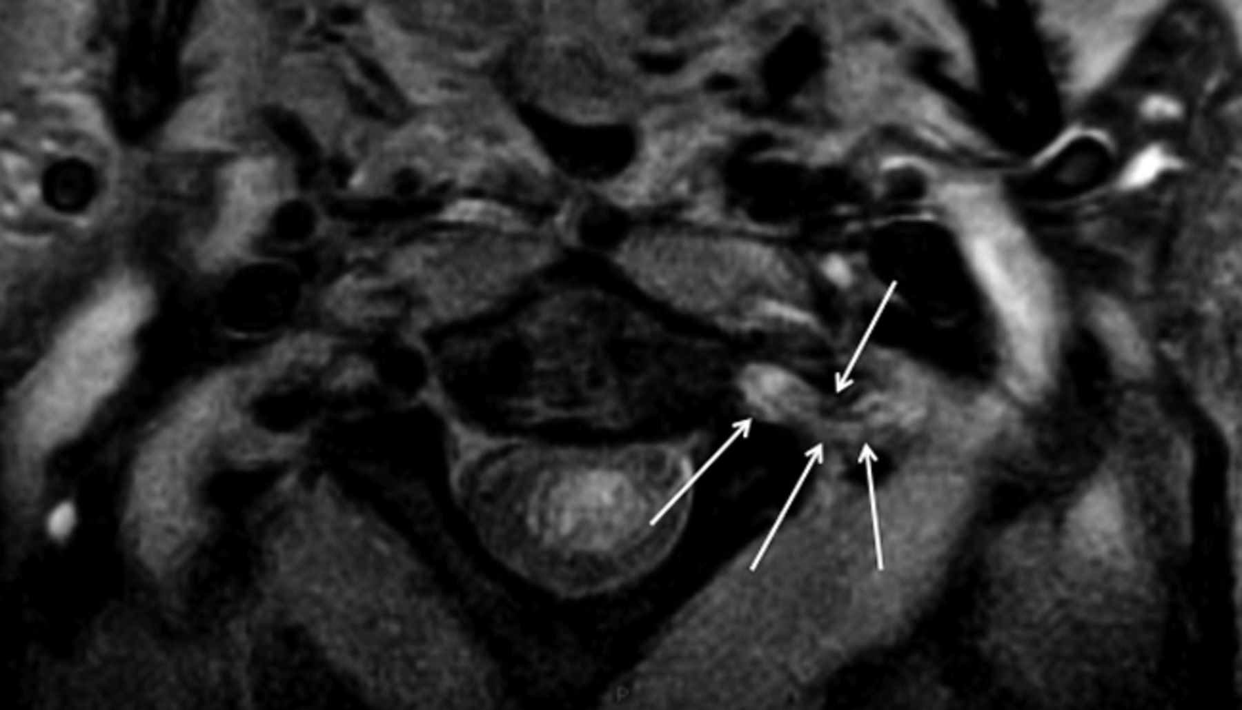
- Fig 3.
T2* sequence demonstrates crescentic intramural deoxyhemoglobin signal intensity (arrows) in the dissected right VA.
- Fig 4.
“Target sign” appearance on BBI caused by hematoma in subintimal dissection. The media (continuous arrow) returns low signal intensity; the hematoma (discontinuous arrow), high signal intensity.
- Fig 5.
Both BBI (A) and CTA (B) demonstrate pseudoaneurysm formation in the skull base of the dissected left internal carotid artery.
- Fig 6.
Periadventitial signal-intensity change (arrows), presumed due to inflammatory response, surrounding the dissected left VA.


Tables
Subject Sex Age (yr) Neurology Antecedents Brain Imaging Findings 1 M 39 Dizziness and diplopia Established right PICA infarct, recent right PCA infarct 2 F 28 Vertigo, diplopia, and left sensory disturbance Right midbrain infarct 3 M 51 Right sensory disturbance with left Horner syndrome Working on overhead light Lateral medullary infarct 4 M 75 Left facial weakness with expressive dysphasia Prior transient ischemic attack, smoker Left occipital infarct 5 M 65 Ataxia, dysphonia, left sensory disturbance Hypertensive, type 2 diabetes, ex-smoker Left medullary infarct 6 F 35 Top of basilar syndrome Right cerebellar and bilateral thalamic infarct 7 M 44 Dysarthria, left homonymous hemianopia Left occipital infarct 8 M 41 Right hemiparesis dysarthria and eye movement disorder Left PCA infarct 9 M 32 Dysarthria, ataxia, and photophobia Playing soccer at time Right occipital infarct 10 F 71 Diplopia and dizziness Hypertensive Bilateral cerebellar and pontine infarcts 11 M 40 Vertigo, right weakness, dysarthria, and nystagmus Right cerebellar infarct 12 F 26 Left facial droop and ptosis Ehlers Danlos syndrome type 1 Left thalamic and bilateral cerebellar infarcts 13 F 19 Collapse, left weakness; dysarthria and homonymous hemianopia Weak history of trauma, on injectable contraceptive Right MCA territory infarct 14 M 60 Left sensorimotor, left homonymous hemianopia Right total anterior circulatory stroke 15 F 64 Right weakness and dizziness Right MCA territory infarct 16 M 45 Left Horner syndrome Heavy laborer with prior chiropractic treatment 17 M 68 Right weakness Left parietal and insular infarct 18 F 55 Right weakness and dysphasia Left basal ganglia infarct 19 M 48 Right weakness and dysphasia Left MCA territory infarct Subject Dissected Vessel Modality Lumena Wallb Vessel Contour Other 1 Rt VA CTA Intimal flap Thickened Stenosis Rt VA BBI Intimal flap Thickened Occlusion Periadvential T2 hyperintensity T1 hyperintensity + 2 Lt VA CTA – Thickened Stenosis Lt VA BBI – Thickened Stenosis T1 hyperintensity ++ T2* hypointensity + Rt VA CTA – Thickened Stenosis Rt VA BBI Intimal flap Thickened Stenosis Periadvential T2 hyperintensity T1 hyperintensity ++ 3 Lt VA CTA – Thickened Stenosis Lt VA BBI – Thickened Stenosis Periadvential T2 hyperintensity T1 hyperintensity ++ 4 Lt VA CTA – Normal Stenosis (Abnormal but equivocal cause) Lt VA BBI – Thickened Occlusion Periadvential T2 hyperintensity T1 hyperintensity ++ 5 Lt VA CTA – Thickened Occlusion Lt VA BBI – Thickened Occlusion Periadvential T2 hyperintensity T1 hyperintensity + 6 Lt VA CTA – Thickened Stenosis Lt VA BBI – Thickened T1 hyperintensity ++ Stenosis Rt VA CTA – Thickened Stenosis Rt VA BBI – Thickened T1 hyperintensity ++ Stenosis T2* hypointensity + 7 Rt VA CTA – Thickened Occlusion Rt VA BBI – Thickened Occlusion Periadvential T2 hyperintensity T1 hyperintensity + 8 Lt VA CTA – Thickened Stenosis Lt VA BBI Intimal flap Thickened Stenosis Periadvential T2 hyperintensity T1 hyperintensity ++ 9 Lt VA CTA Intimal flap Thickened Occlusion Lt VA BBI Intimal flap Thickened Stenosis Periadvential T2 hyperintensity T1 hyperintensity + T2* hypointensity + 10 Lt VA CTA – Thickened Occlusion Lt VA BBI Intimal flap Thickened Stenosis Periadvential T2 hyperintensity T1 hyperintensity +/++ Rt VA CTA – Thickened Stenosis Rt VA BBI – Thickened Stenosis Periadvential T2 hyperintensity T1 hyperintensity + 11 Rt VA CTA Intimal flap Thickened Stenosis (Abnormal but equivocal cause) Rt VA BBI Intimal flap Thickened Stenosis Periadvential T2 hyperintensity T1 hyperintensity +/++ 12 Lt VA CTA – Thickened Occlusion Lt VA BBI Intimal flap Thickened Stenosis Periadvential T2 hyperintensity T2* hypointensity + 13 Rt CA CTA – Thickened Occlusion Rt CA BBI Intimal flap Thickened Occlusion Periadvential T2 hyperintensity T1 hyperintensity + 14 Rt CA CTA – Thickened Occlusion Rt CA BBI Intimal flap Thickened Occlusion Periadvential T2 hyperintensity T1 hyperintensity ++ 15 Lt CA BBI Intimal flap Thickened – Periadvential T2 hyperintensity T1 hyperintensity + Rt CA CTA Intimal flap Thickened Stenosis Rt CA BBI Intimal flap Thickened Stenosis Periadvential T2 hyperintensity T1 hyperintensity + 16 Rt CA CTA Intimal flap Thickened Stenosis Pseudoaneurysm Rt CA BBI – Thickened Stenosis Pseudoaneurysm T1 hyperintensity ++ Periadvential T2 hyperintensity 17 Lt CA CTA – Thickened Occlusion Lt CA BBI Intimal flap Thickened Stenosis Periadvential T2 hyperintensity T1 hyperintensity ++ 18 Lt CA CTA Intimal flap Thickened Occlusion Lt CA BBI Intimal flap Thickened Stenosis Periadvential T2 hyperintensity T1 hyperintensity ++ Rt CA BBI Intimal flap Thickened Stenosis Periadvential T2 hyperintensity T1 hyperintensity ++ 19 Lt CA CTA Intimal flap Thickened Stenosis Lt CA BBI Intimal flap Thickened Stenosis Periadvential T2 hyperintensity T1 hyperintensity ++


