Advertisement

Main menu

  • Home
  • Content
    • Current Issue
    • Accepted Manuscripts
    • Article Preview
    • Past Issue Archive
    • Video Articles
    • AJNR Case Collection
    • Case of the Week Archive
    • Case of the Month Archive
    • Classic Case Archive
  • Special Collections
    • AJNR Awards
    • Low-Field MRI
    • Alzheimer Disease
    • ASNR Foundation Special Collection
    • Photon-Counting CT
    • View All
  • Multimedia
    • AJNR Podcasts
    • AJNR SCANtastic
    • Trainee Corner
    • MRI Safety Corner
    • Imaging Protocols
  • For Authors
    • Submit a Manuscript
    • Submit a Video Article
    • Submit an eLetter to the Editor/Response
    • Manuscript Submission Guidelines
    • Statistical Tips
    • Fast Publishing of Accepted Manuscripts
    • Graphical Abstract Preparation
    • Imaging Protocol Submission
    • Author Policies
  • About Us
    • About AJNR
    • Editorial Board
    • Editorial Board Alumni
  • More
    • Become a Reviewer/Academy of Reviewers
    • Subscribers
    • Permissions
    • Alerts
    • Feedback
    • Advertisers
    • ASNR Home

User menu

  • Alerts
  • Log in

Search

  • Advanced search
American Journal of Neuroradiology

American Journal of Neuroradiology

ASHNR American Society of Functional Neuroradiology ASHNR American Society of Pediatric Neuroradiology ASSR
  • Alerts
  • Log in

Advanced Search

  • Home
  • Content
    • Current Issue
    • Accepted Manuscripts
    • Article Preview
    • Past Issue Archive
    • Video Articles
    • AJNR Case Collection
    • Case of the Week Archive
    • Case of the Month Archive
    • Classic Case Archive
  • Special Collections
    • AJNR Awards
    • Low-Field MRI
    • Alzheimer Disease
    • ASNR Foundation Special Collection
    • Photon-Counting CT
    • View All
  • Multimedia
    • AJNR Podcasts
    • AJNR SCANtastic
    • Trainee Corner
    • MRI Safety Corner
    • Imaging Protocols
  • For Authors
    • Submit a Manuscript
    • Submit a Video Article
    • Submit an eLetter to the Editor/Response
    • Manuscript Submission Guidelines
    • Statistical Tips
    • Fast Publishing of Accepted Manuscripts
    • Graphical Abstract Preparation
    • Imaging Protocol Submission
    • Author Policies
  • About Us
    • About AJNR
    • Editorial Board
    • Editorial Board Alumni
  • More
    • Become a Reviewer/Academy of Reviewers
    • Subscribers
    • Permissions
    • Alerts
    • Feedback
    • Advertisers
    • ASNR Home
  • Follow AJNR on Twitter
  • Visit AJNR on Facebook
  • Follow AJNR on Instagram
  • Join AJNR on LinkedIn
  • RSS Feeds

AJNR Awards, New Junior Editors, and more. Read the latest AJNR updates

Research ArticleSpine Imaging and Spine Image-Guided Interventions

Evaluation of Cervical Myelopathy Using Apparent Diffusion Coefficient Measured by Diffusion-Weighted Imaging

T. Sato, T. Horikoshi, A. Watanabe, M. Uchida, K. Ishigame, T. Araki and H. Kinouchi
American Journal of Neuroradiology February 2012, 33 (2) 388-392; DOI: https://doi.org/10.3174/ajnr.A2756
T. Sato
  • Find this author on Google Scholar
  • Find this author on PubMed
  • Search for this author on this site
T. Horikoshi
  • Find this author on Google Scholar
  • Find this author on PubMed
  • Search for this author on this site
A. Watanabe
  • Find this author on Google Scholar
  • Find this author on PubMed
  • Search for this author on this site
M. Uchida
  • Find this author on Google Scholar
  • Find this author on PubMed
  • Search for this author on this site
K. Ishigame
  • Find this author on Google Scholar
  • Find this author on PubMed
  • Search for this author on this site
T. Araki
  • Find this author on Google Scholar
  • Find this author on PubMed
  • Search for this author on this site
H. Kinouchi
  • Find this author on Google Scholar
  • Find this author on PubMed
  • Search for this author on this site
  • Article
  • Figures & Data
  • Supplemental
  • Info & Metrics
  • Responses
  • References
  • PDF
Loading

Article Figures & Data

Figures

  • Tables
  • Fig 1.
    • Download figure
    • Open in new tab
    • Download powerpoint
    Fig 1.

    Axial view of DWI. The mean ADC was calculated in the region of interest. The region of interest covered most of the cross-section of spinal cord, excluding the CSF space.

  • Fig 2.
    • Download figure
    • Open in new tab
    • Download powerpoint
    Fig 2.

    Grades of spinal cord compression on MR imaging. Type 0 indicates no compression; type 1, disappearance of CSF space; type 2, mild compressive deformity of spinal cord; and type 3, marked deformity (flattening) of the spinal cord.

  • Fig 3.
    • Download figure
    • Open in new tab
    • Download powerpoint
    Fig 3.

    The relationship between preoperative JOA scores and ADC values in all patients. Patients with ADC values of ≥0.9 × 10⁻³ mm2/s had lower JOA scores (P = .0085).

  • Fig 4.
    • Download figure
    • Open in new tab
    • Download powerpoint
    Fig 4.

    Recovery rates of JOA scores and preoperative ADC values in 18 patients followed for >6 months. Patients with an ADC of <0.7 × 10⁻³ mm2/s tended to show better recovery rates than those with an ADC of 0.9 × 10⁻³ mm2/s (P = .0636).

Tables

  • Figures
    • View popup
    Table 1:

    Chronologic changes of mean ADC values in each compression type

    TypePreoperative StagePostoperative Stage (1 wk)Follow-Up Stage (6 mo)
    00.704 ± 0.142 (n = 140)0.692 ± 0.134 (n = 114)0.648 ± 0.143 (n = 84)a
    I0.833 ± 0.228 (n = 75)b0.807 ± 0.171 (n = 63)c0.676 ± 0.093 (n = 49)d
    II0.939 ± 0.330 (n = 74)b0.880 ± 0.238 (n = 67)e0.763 ± 0.150 (n = 57)f,g
    III0.948 ± 0.289 (n = 33)b0.908 ± 0.197 (n = 28)h0.791 ± 0.153 (n = 23)
    Total0.813 ± 0.254 (n = 322)0.723 ± 0.191 (n = 272)0.691 ± 0.145 (n = 213)i
    • Note:—Values are expressed as mean (×10 − 3 mm2/s).

    • ↵a P = .01 versus type 0 in the preoperative stage.

    • ↵b P <.0001 versus type 0 in the preoperative stage.

    • ↵c P = .0008.

    • ↵d P = .0015 versus type I in the preoperative stage.

    • ↵e P < .0001.

    • ↵f P = .004 versus type 0 in the follow-up stage.

    • ↵g P = .009 versus type II in the preoperative stage.

    • ↵h P = .005 versus type 0 in the postoperative stage.

    • ↵i P < .0001 versus total value in the preoperative stage.

    • View popup
    Table 2:

    Type of spinal cord compression and occurrence of high T2 signal intensity

    TypeISI (+)
    0 (n = 140)8 (5.7%)
    I (n = 75)8 (10.7%)
    II (n = 74)14 (18.9%)a
    III (n = 33)18 (54.5%)b
    Total (n = 322)48/322 (14.9%)
    • Note:—ISI indicates increased signal intensity.

    • ↵a P = .004.

    • ↵b P < .0001 versus type 0.

    • View popup
    Table 3:

    Chronologic changes of mean ADC values in high T2 signal intensity

    Preoperative StagePostoperative Stage (1 wk)Follow-Up Stage (6 mo)
    ISI (+)1.002 ± 0.305 (n = 48)0.905 ± 0.255 (n = 41)0.782 ± 0.147 (n = 32)a
    ISI (−)0.775 ± 0.126 (n = 274)b0.760 ± 0.178 (n = 231)c0.674 ± 0.141 (n = 181)d
    Total0.813 ± 0.254 (n = 322)0.723 ± 0.191 (n = 272)0.691 ± 0.145 (n = 213)
    • Note:—ISI indicates increased signal intensity. Values are expressed as mean (×10−3 mm2/s).

    • ↵a P = .02 versus ISI(+) group in the preoperative stage.

    • ↵b P < .0001 versus ISI(+) group in the preoperative stage.

    • ↵c P = .001 versus ISI(+) group in the postoperative stage.

    • ↵d P <.0001 versus ISI(−) group in the preoperative stage.

    • View popup
    Table 4:

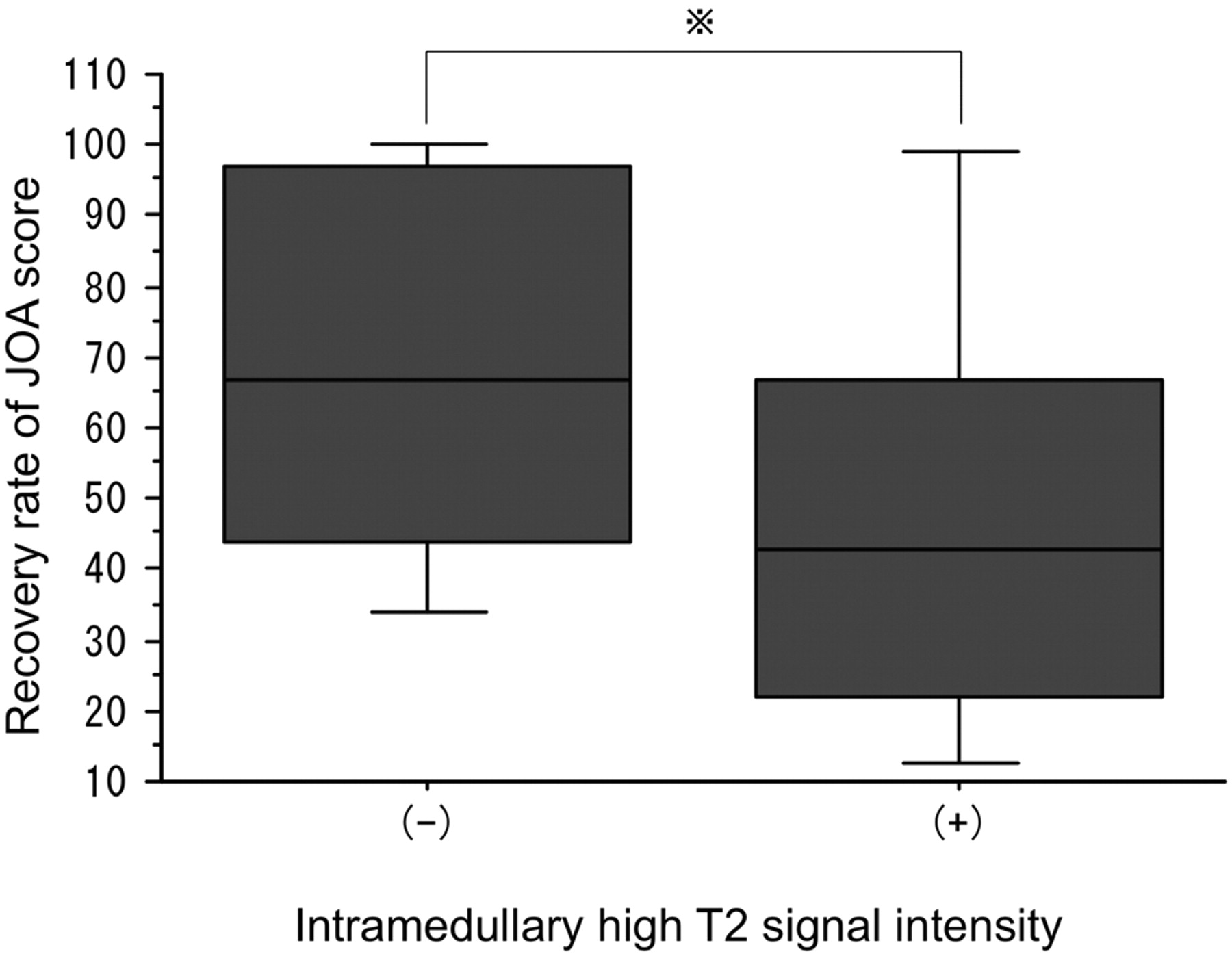
    Chronologic changes of mean JOA score and compression group and high T2 signal intensity

    PreoperativePostoperative (1 wk)Recovery RateFollow-Up (6 mo)Recovery Rate
    Type 013.7 ± 2.75 (n = 4)16.8 ± 0.35 (n = 3)83.4 ± 23.517 (n = 2)100
    Type I14.1 ± 3.50 (n = 8)15.9 ± 2.17 (n = 7)a75.5 ± 28.417 (n = 4)b100
    Type II11.1 ± 3.44 (n = 27)14.1 ± 2.58 (n = 25)c57.5 ± 29.814.9 ± 2.16 (n = 21)d72.7 ± 31.4
    Type III9.9 ± 3.47 (n = 27)e13.2 ± 3.36 (n = 24)f50.3 ± 28.616.1 ± 1.50 (n = 21)g84.3 ± 19.1
    Total11.0 ± 3.6 (n = 66)14.0 ± 2.9 (n = 59)15.7 ± 1.9 (n = 48)
    ISI(−)11.9 ± 3.54 (n = 34)15.2 ± 1.95 (n = 30)h66.2 ± 27.616.4 ± 0.97 (n = 20)i86.6 ± 21.6
    ISI(+)10.2 ± 3.65 (n = 32)13.1 ± 3.28 (n = 29)j,k49.2 ± 29.9l15.5 ± 1.85 (n = 28)m77.6 ± 28.2
    Total11.0 ± 3.6 (n = 66)14.0 ± 2.9 (n = 59)15.7 ± 1.9 (n = 48)
    • Note:—ISI indicates increased signal intensity. Values are expressed as means.

    • ↵a P = .02.

    • ↵b P = < .0001 versus type 1 in the preoperative state.

    • ↵c P = .003.

    • ↵d P = .02 versus type II in the preoperative stage.

    • ↵e P = .007.

    • ↵f P = .0002.

    • ↵g P = .0001 versus type III in the preoperative stage.

    • ↵h P = .0001.

    • ↵i P = .0002 versus ISI(−) group in the preoperative state

    • ↵j P = .009 versus the ISI(−) group in the postoperative stage.

    • ↵k P = .004.

    • ↵l P = .04 versus the ISI(−) group.

    • ↵m P = .0001 versus ISI(+) in the preoperative stage.

PreviousNext
Back to top

In this issue

American Journal of Neuroradiology: 33 (2)
American Journal of Neuroradiology
Vol. 33, Issue 2
1 Feb 2012
  • Table of Contents
  • Index by author
Advertisement
Print
Download PDF
Email Article

Thank you for your interest in spreading the word on American Journal of Neuroradiology.

NOTE: We only request your email address so that the person you are recommending the page to knows that you wanted them to see it, and that it is not junk mail. We do not capture any email address.

Enter multiple addresses on separate lines or separate them with commas.
Evaluation of Cervical Myelopathy Using Apparent Diffusion Coefficient Measured by Diffusion-Weighted Imaging
(Your Name) has sent you a message from American Journal of Neuroradiology
(Your Name) thought you would like to see the American Journal of Neuroradiology web site.
CAPTCHA
This question is for testing whether or not you are a human visitor and to prevent automated spam submissions.
Cite this article
T. Sato, T. Horikoshi, A. Watanabe, M. Uchida, K. Ishigame, T. Araki, H. Kinouchi
Evaluation of Cervical Myelopathy Using Apparent Diffusion Coefficient Measured by Diffusion-Weighted Imaging
American Journal of Neuroradiology Feb 2012, 33 (2) 388-392; DOI: 10.3174/ajnr.A2756

Citation Manager Formats

  • BibTeX
  • Bookends
  • EasyBib
  • EndNote (tagged)
  • EndNote 8 (xml)
  • Medlars
  • Mendeley
  • Papers
  • RefWorks Tagged
  • Ref Manager
  • RIS
  • Zotero
0 Responses
Respond to this article
Share
Bookmark this article
Evaluation of Cervical Myelopathy Using Apparent Diffusion Coefficient Measured by Diffusion-Weighted Imaging
T. Sato, T. Horikoshi, A. Watanabe, M. Uchida, K. Ishigame, T. Araki, H. Kinouchi
American Journal of Neuroradiology Feb 2012, 33 (2) 388-392; DOI: 10.3174/ajnr.A2756
del.icio.us logo Twitter logo Facebook logo Mendeley logo
  • Tweet Widget
  • Facebook Like
  • Google Plus One
Purchase

Jump to section

  • Article
    • Abstract
    • ABBREVIATIONS:
    • Materials and Methods
    • Results
    • Discussion
    • Conclusions
    • References
  • Figures & Data
  • Supplemental
  • Info & Metrics
  • Responses
  • References
  • PDF

Related Articles

  • PubMed
  • Google Scholar

Cited By...

  • Hot Topics in Functional Neuroradiology
  • Crossref
  • Google Scholar

This article has not yet been cited by articles in journals that are participating in Crossref Cited-by Linking.

More in this TOC Section

  • Management Outcomes For VO Spine Biopsy
  • Characteristics of SIH Type I Culprit Lesions
  • Advanced Imaging of Type 2 Spinal CSF Leaks
Show more Spine Imaging and Spine Image-Guided Interventions

Similar Articles

Advertisement

Indexed Content

  • Current Issue
  • Accepted Manuscripts
  • Article Preview
  • Past Issues
  • Editorials
  • Editor's Choice
  • Fellows' Journal Club
  • Letters to the Editor
  • Video Articles

Cases

  • Case Collection
  • Archive - Case of the Week
  • Archive - Case of the Month
  • Archive - Classic Case

More from AJNR

  • Trainee Corner
  • Imaging Protocols
  • MRI Safety Corner
  • Book Reviews

Multimedia

  • AJNR Podcasts
  • AJNR Scantastics

Resources

  • Turnaround Time
  • Submit a Manuscript
  • Submit a Video Article
  • Submit an eLetter to the Editor/Response
  • Manuscript Submission Guidelines
  • Statistical Tips
  • Fast Publishing of Accepted Manuscripts
  • Graphical Abstract Preparation
  • Imaging Protocol Submission
  • Evidence-Based Medicine Level Guide
  • Publishing Checklists
  • Author Policies
  • Become a Reviewer/Academy of Reviewers
  • News and Updates

About Us

  • About AJNR
  • Editorial Board
  • Editorial Board Alumni
  • Alerts
  • Permissions
  • Not an AJNR Subscriber? Join Now
  • Advertise with Us
  • Librarian Resources
  • Feedback
  • Terms and Conditions
  • AJNR Editorial Board Alumni

American Society of Neuroradiology

  • Not an ASNR Member? Join Now

© 2025 by the American Society of Neuroradiology All rights, including for text and data mining, AI training, and similar technologies, are reserved.
Print ISSN: 0195-6108 Online ISSN: 1936-959X

Powered by HighWire