Advertisement

Main menu

  • Home
  • Content
    • Current Issue
    • Accepted Manuscripts
    • Article Preview
    • Past Issue Archive
    • Video Articles
    • AJNR Case Collection
    • Case of the Week Archive
    • Case of the Month Archive
    • Classic Case Archive
  • Special Collections
    • AJNR Awards
    • Low-Field MRI
    • Alzheimer Disease
    • ASNR Foundation Special Collection
    • Photon-Counting CT
    • View All
  • Multimedia
    • AJNR Podcasts
    • AJNR SCANtastic
    • Trainee Corner
    • MRI Safety Corner
    • Imaging Protocols
  • For Authors
    • Submit a Manuscript
    • Submit a Video Article
    • Submit an eLetter to the Editor/Response
    • Manuscript Submission Guidelines
    • Statistical Tips
    • Fast Publishing of Accepted Manuscripts
    • Graphical Abstract Preparation
    • Imaging Protocol Submission
    • Author Policies
  • About Us
    • About AJNR
    • Editorial Board
    • Editorial Board Alumni
  • More
    • Become a Reviewer/Academy of Reviewers
    • Subscribers
    • Permissions
    • Alerts
    • Feedback
    • Advertisers
    • ASNR Home

User menu

  • Alerts
  • Log in

Search

  • Advanced search
American Journal of Neuroradiology

American Journal of Neuroradiology

ASHNR American Society of Functional Neuroradiology ASHNR American Society of Pediatric Neuroradiology ASSR
  • Alerts
  • Log in

Advanced Search

  • Home
  • Content
    • Current Issue
    • Accepted Manuscripts
    • Article Preview
    • Past Issue Archive
    • Video Articles
    • AJNR Case Collection
    • Case of the Week Archive
    • Case of the Month Archive
    • Classic Case Archive
  • Special Collections
    • AJNR Awards
    • Low-Field MRI
    • Alzheimer Disease
    • ASNR Foundation Special Collection
    • Photon-Counting CT
    • View All
  • Multimedia
    • AJNR Podcasts
    • AJNR SCANtastic
    • Trainee Corner
    • MRI Safety Corner
    • Imaging Protocols
  • For Authors
    • Submit a Manuscript
    • Submit a Video Article
    • Submit an eLetter to the Editor/Response
    • Manuscript Submission Guidelines
    • Statistical Tips
    • Fast Publishing of Accepted Manuscripts
    • Graphical Abstract Preparation
    • Imaging Protocol Submission
    • Author Policies
  • About Us
    • About AJNR
    • Editorial Board
    • Editorial Board Alumni
  • More
    • Become a Reviewer/Academy of Reviewers
    • Subscribers
    • Permissions
    • Alerts
    • Feedback
    • Advertisers
    • ASNR Home
  • Follow AJNR on Twitter
  • Visit AJNR on Facebook
  • Follow AJNR on Instagram
  • Join AJNR on LinkedIn
  • RSS Feeds

AJNR Awards, New Junior Editors, and more. Read the latest AJNR updates

Research ArticleSpineE

Clinical Correlation of a New MR Imaging Method for Assessing Lumbar Foraminal Stenosis

H.-J. Park, S.S. Kim, S.-Y. Lee, N.-H. Park, M.-H. Rho, H.-P. Hong, H.-J. Kwag, S.-H. Kook and S.-H. Choi
American Journal of Neuroradiology May 2012, 33 (5) 818-822; DOI: https://doi.org/10.3174/ajnr.A2870
H.-J. Park
  • Find this author on Google Scholar
  • Find this author on PubMed
  • Search for this author on this site
S.S. Kim
  • Find this author on Google Scholar
  • Find this author on PubMed
  • Search for this author on this site
S.-Y. Lee
  • Find this author on Google Scholar
  • Find this author on PubMed
  • Search for this author on this site
N.-H. Park
  • Find this author on Google Scholar
  • Find this author on PubMed
  • Search for this author on this site
M.-H. Rho
  • Find this author on Google Scholar
  • Find this author on PubMed
  • Search for this author on this site
H.-P. Hong
  • Find this author on Google Scholar
  • Find this author on PubMed
  • Search for this author on this site
H.-J. Kwag
  • Find this author on Google Scholar
  • Find this author on PubMed
  • Search for this author on this site
S.-H. Kook
  • Find this author on Google Scholar
  • Find this author on PubMed
  • Search for this author on this site
S.-H. Choi
  • Find this author on Google Scholar
  • Find this author on PubMed
  • Search for this author on this site
  • Article
  • Figures & Data
  • Info & Metrics
  • Responses
  • References
  • PDF
Loading

Article Figures & Data

Figures

  • Tables
  • Fig 1.
    • Download figure
    • Open in new tab
    • Download powerpoint
    Fig 1.

    A and B, The Lee system grade 2 and the Wildermuth system grade 2. T2-weighted images of a 39-year-old man and a 64-year-old man show narrowing of the vertical and transverse width of neural foramina in the left L5–S1 and the right L5–S1. Decreased intervertebral disk space, thickened ligamentum flavum, and disk protrusions are seen (arrows). Perineural fat obliteration is also seen, but nerve root deformity is not noted.
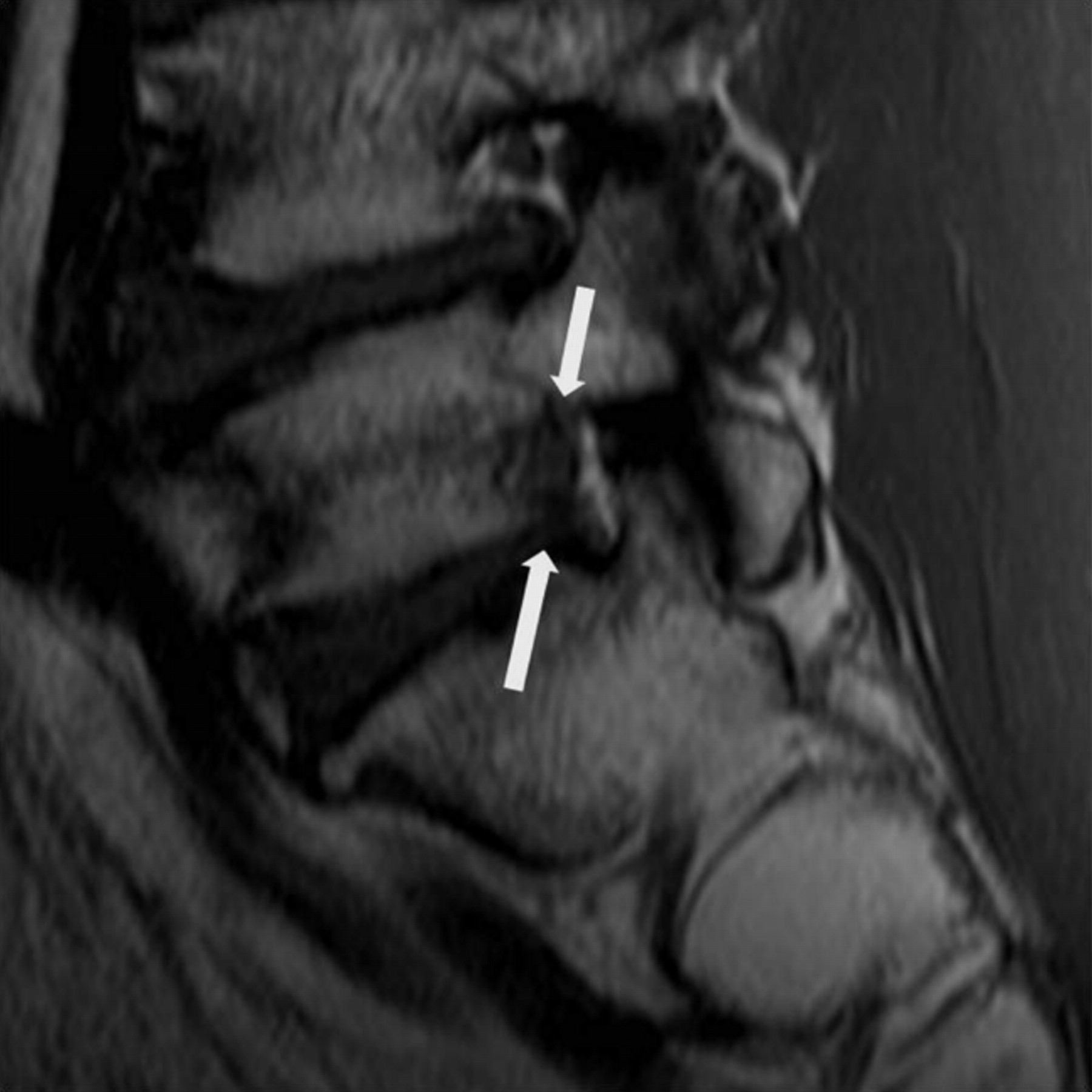
  • Fig 2.
    • Download figure
    • Open in new tab
    • Download powerpoint
    Fig 2.

    The Lee system grade 3 and the Wildermuth system grade 3. T1-weighted image of an 82-year-old woman revealing marked narrowing of the vertical and transverse width of neural foramina at L5–S1. Decreased intervertebral disk space, thickened ligamentum flavum, and disk protrusions are seen (arrows). The nerve root is collapsed and deformed compared with another nerve root at a different level.

  • Fig 3.
    • Download figure
    • Open in new tab
    • Download powerpoint
    Fig 3.

    The Lee system grade 0 and the Wildermuth system grade 1 foraminal stenosis. T2-weighted sagittal image of a 78-year-old woman shows a normal nerve root without compression but mild narrowing of the foramen in the right L4–5. The arrow indicates the protruded disk.

  • Fig 4.
    • Download figure
    • Open in new tab
    • Download powerpoint
    Fig 4.

    The Lee system grade 1 and the Wildermuth system grade 2. T1-weighted image of a 54-year-old man shows narrowing of the vertical width of the neural foramen and decreased intervertebral disk space in the left L5–S1 (arrows). Partial perineural fat obliteration is noted, but deformity of the nerve root is not seen.

Tables

  • Figures
    • View popup
    Table 1:

    Incidence of grades for the Lee system

    Grade 0Grade 1Grade 2Grade 3Total
    Reader 156198891
    Reader 265128691
    • View popup
    Table 2:

    Incidence of grades for the Wildermuth system

    Grade 0Grade 1Grade 2Grade 3Total
    Reader 1501618791
    Reader 260818391
    • View popup
    Table 3:

    Rs for lumbar foraminal stenosisa

    ObserverL System vs W SystemL System vs W SystemW System vs PNM
    Reader 10.8880.7510.800
    Reader 20.8850.7510.680
    • Note:—L system indicates Lee system; W system, Wildermuth system.

    • ↵a The level of correlation significance was .01.

    • View popup
    Table 4:

    Rs for lumbar foraminal stenosis for patients younger than 50 years of agea

    ObserverL System vs W SystemL System vs PNMW System vs PNM
    Reader 10.9730.7110.695
    Reader 20.9850.5110.503
    • Note:—L system indicates Lee system; W system, Wildermuth system.

    • ↵a The level of correlation significance was .01.

    • View popup
    Table 5:

    Rs for lumbar foraminal stenosis for patients older than 50 years of agea

    ObserverL System vs W SystemL System vs PNMW System vs PNM
    Reader 10.8320.6120.742
    Reader 20.8410.5300.637
    • Note:—L system indicates Lee system; W system, Wildermuth system.

    • ↵a The level of correlation significance was .01.

PreviousNext
Back to top

In this issue

American Journal of Neuroradiology: 33 (5)
American Journal of Neuroradiology
Vol. 33, Issue 5
1 May 2012
  • Table of Contents
  • Index by author
Advertisement
Print
Download PDF
Email Article

Thank you for your interest in spreading the word on American Journal of Neuroradiology.

NOTE: We only request your email address so that the person you are recommending the page to knows that you wanted them to see it, and that it is not junk mail. We do not capture any email address.

Enter multiple addresses on separate lines or separate them with commas.
Clinical Correlation of a New MR Imaging Method for Assessing Lumbar Foraminal Stenosis
(Your Name) has sent you a message from American Journal of Neuroradiology
(Your Name) thought you would like to see the American Journal of Neuroradiology web site.
CAPTCHA
This question is for testing whether or not you are a human visitor and to prevent automated spam submissions.
Cite this article
H.-J. Park, S.S. Kim, S.-Y. Lee, N.-H. Park, M.-H. Rho, H.-P. Hong, H.-J. Kwag, S.-H. Kook, S.-H. Choi
Clinical Correlation of a New MR Imaging Method for Assessing Lumbar Foraminal Stenosis
American Journal of Neuroradiology May 2012, 33 (5) 818-822; DOI: 10.3174/ajnr.A2870

Citation Manager Formats

  • BibTeX
  • Bookends
  • EasyBib
  • EndNote (tagged)
  • EndNote 8 (xml)
  • Medlars
  • Mendeley
  • Papers
  • RefWorks Tagged
  • Ref Manager
  • RIS
  • Zotero
0 Responses
Respond to this article
Share
Bookmark this article
Clinical Correlation of a New MR Imaging Method for Assessing Lumbar Foraminal Stenosis
H.-J. Park, S.S. Kim, S.-Y. Lee, N.-H. Park, M.-H. Rho, H.-P. Hong, H.-J. Kwag, S.-H. Kook, S.-H. Choi
American Journal of Neuroradiology May 2012, 33 (5) 818-822; DOI: 10.3174/ajnr.A2870
del.icio.us logo Twitter logo Facebook logo Mendeley logo
  • Tweet Widget
  • Facebook Like
  • Google Plus One
Purchase

Jump to section

  • Article
    • Abstract
    • ABBREVIATIONS:
    • Materials and Methods
    • Results
    • Discussion
    • Conclusions
    • References
  • Figures & Data
  • Info & Metrics
  • Responses
  • References
  • PDF

Related Articles

  • Erratum
  • PubMed
  • Google Scholar

Cited By...

  • Long-term Clinical Outcomes Following Endoscopic Foraminoplasty for Patients With Single-Level Foraminal Stenosis of the Lumbar Spine
  • A practical MRI grading system for cervical foraminal stenosis based on oblique sagittal images
  • Crossref
  • Google Scholar

This article has not yet been cited by articles in journals that are participating in Crossref Cited-by Linking.

More in this TOC Section

  • CT-Guided C2 Dorsal Root Ganglion Radiofrequency Ablation for the Treatment of Cervicogenic Headache: Case Series and Clinical Outcomes
  • The Evaluation and Prediction of Laminoplasty Surgery Outcome in Patients with Degenerative Cervical Myelopathy Using Diffusion Tensor MRI
  • Subject-Specific Studies of CSF Bulk Flow Patterns in the Spinal Canal: Implications for the Dispersion of Solute Particles in Intrathecal Drug Delivery
Show more Spine

Similar Articles

Advertisement

Indexed Content

  • Current Issue
  • Accepted Manuscripts
  • Article Preview
  • Past Issues
  • Editorials
  • Editor's Choice
  • Fellows' Journal Club
  • Letters to the Editor
  • Video Articles

Cases

  • Case Collection
  • Archive - Case of the Week
  • Archive - Case of the Month
  • Archive - Classic Case

More from AJNR

  • Trainee Corner
  • Imaging Protocols
  • MRI Safety Corner
  • Book Reviews

Multimedia

  • AJNR Podcasts
  • AJNR Scantastics

Resources

  • Turnaround Time
  • Submit a Manuscript
  • Submit a Video Article
  • Submit an eLetter to the Editor/Response
  • Manuscript Submission Guidelines
  • Statistical Tips
  • Fast Publishing of Accepted Manuscripts
  • Graphical Abstract Preparation
  • Imaging Protocol Submission
  • Evidence-Based Medicine Level Guide
  • Publishing Checklists
  • Author Policies
  • Become a Reviewer/Academy of Reviewers
  • News and Updates

About Us

  • About AJNR
  • Editorial Board
  • Editorial Board Alumni
  • Alerts
  • Permissions
  • Not an AJNR Subscriber? Join Now
  • Advertise with Us
  • Librarian Resources
  • Feedback
  • Terms and Conditions
  • AJNR Editorial Board Alumni

American Society of Neuroradiology

  • Not an ASNR Member? Join Now

© 2025 by the American Society of Neuroradiology All rights, including for text and data mining, AI training, and similar technologies, are reserved.
Print ISSN: 0195-6108 Online ISSN: 1936-959X

Powered by HighWire