
Article Figures & Data
Figures
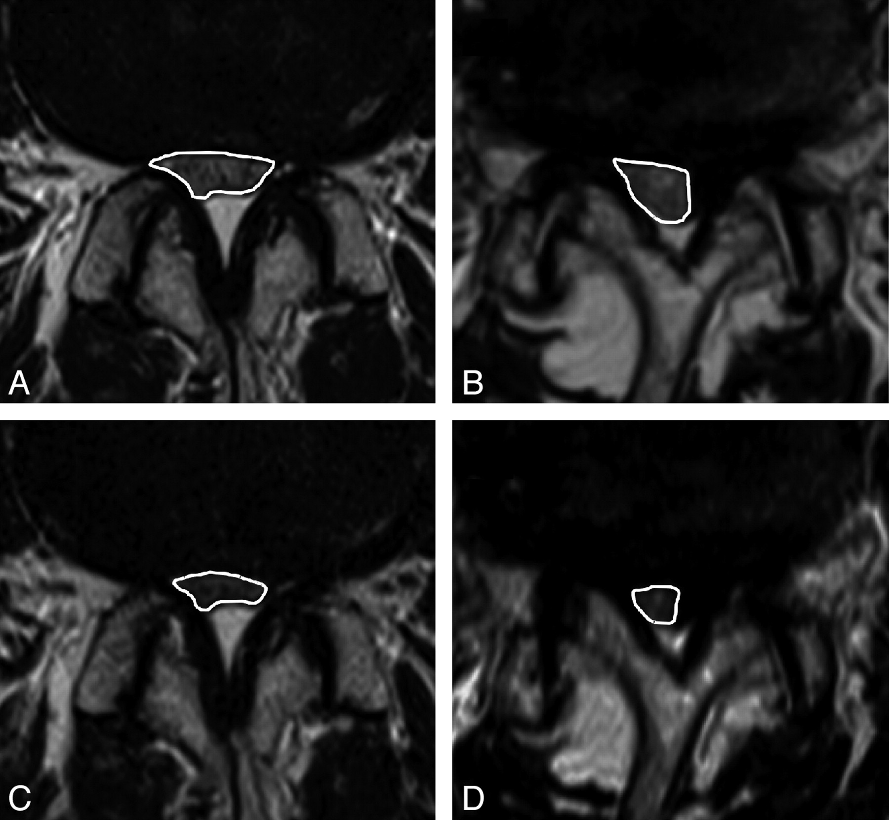
- Fig 1.
The measurement of DCSA on conventional MR imaging and axial loaded MR imaging in representative patients with SpS (A and C) and DS (B and D). The white lines indicate the outlines of the DCSA. In the first patient with SpS, the DCSA changed from 67 (A) to 41 mm2 (C) due to the axial loading. In the second patient with DS, the DCSA changed from 52 (B) to 25 mm2 (D).
- Fig 2.
The values of DCSA for conventional MR imaging and axial loaded MR imaging at the most constricted intervertebral levels for each patient in the SpS (A) and the DS (B) groups. The Pearson correlation coefficients between conventional MR imaging and axial loaded MR imaging were 0.97 in the SpS group and 0.88 in the DS group (P < .001).
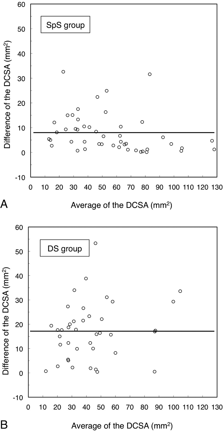
- Fig 3.
Bland-Altman plots for each patient in the SpS (A) and the DS (B) groups. The y-axis shows the difference in the DCSA between conventional MR imaging and axial loaded MR imaging (DCSA in conventional MR imaging − DCSA in axial loaded MR imaging). The x-axis shows the average of the DCSA of the conventional and axial loaded MR imaging [(DCSA in conventional MR imaging + DCSA in axial loaded MR imaging) / 2]. The solid lines indicate the mean differences in the DCSA.
- Fig 4.
Comparison of the DCSA obtained by conventional MR imaging and axial loaded MR imaging between the SpS and the DS groups. The asterisk indicates P < .05; error bars, SD.
- Fig 5.
Comparison of the change in the DCSA induced by axial loading between the SpS and the DS groups. The asterisk indicates P < .05; error bars, SD.
- Fig 6.
Comparison of the DCSA obtained by conventional MR imaging and axial loaded MR imaging between patients with >15- and ≤15-mm2 changes in the DCSA in the SpS (A) and DS (B) groups. The asterisk indicates P < .05; error bars, SD.


Tables
Baseline Characteristics SpS Group (n = 48) DS Group (n = 40) P Valuea Age (yr)b 68 ± 9 68 ± 11 .730 Sex (male) 69% 53% .090 Height (cm)b 161 ± 8 159 ± 10 .336 Body weight (kg)b 64 ± 10 65 ± 13 .702 Body mass index (kg/m2)b 25 ± 3 26 ± 4 .251 Duration of symptoms (mo)b 28 ± 34 39 ± 31 .121 Presence of low back pain 48% 50% .508 The most constricted level .182 L2/3 \n2% \n0% L3/4 21% 23% L4/5 63% 75% L5/S1 15% 3% DCSA on Conventional MR Imaging DCSA on Axial Loaded MR Imaging 1st Measurement 56.9 ± 27.8 43.2 ± 29.3 2nd Measurement 54.7 ± 27.3 42.6 ± 28.0 3rd Measurement 54.2 ± 26.8 43.2 ± 28.3 Averageb 55.3 ± 26.8 43.2 ± 28.2 SpS Group (n = 48) DS Group (n = 40) ≤15 mm2 change in the DCSA 40 (83.3%) 15 (37.5%) >15 mm2 change in the DCSA 8 (16.7%) 25 (62.5%) -
↵a There is a significant difference (χ2 test, P < .001; odds ratio, 8.33).
-


