
Article Figures & Data
Figures
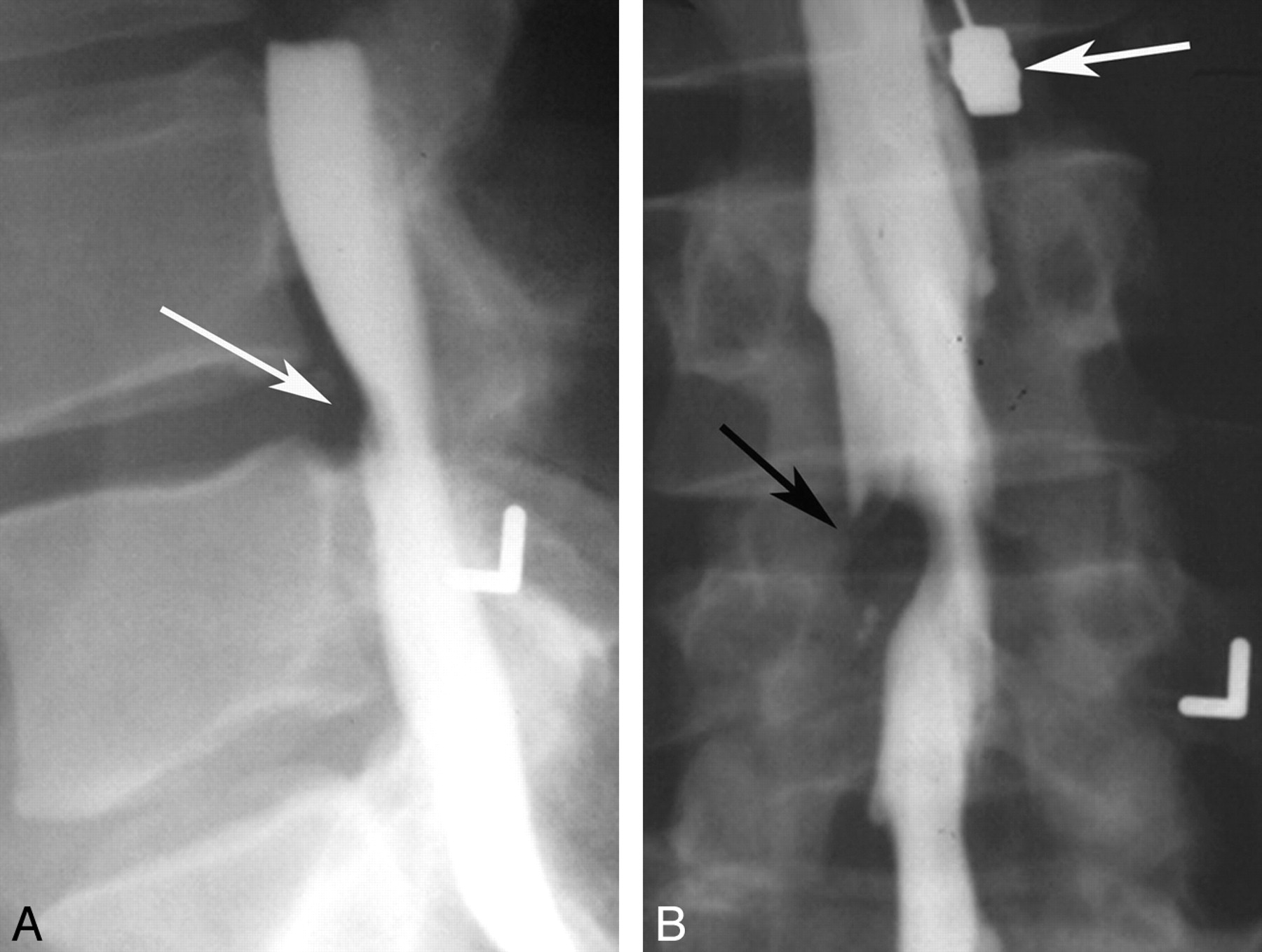
- Fig 1.
Myelogram in a 30-year-old man with radicular pain. A, Lateral lumbar myelographic image shows a typical extradural defect indenting the ventral dural sac at L4-L5 (arrow). B, Frontal lumbar myelographic image shows the defect, which is also resulting in poor filling of the right L5 nerve root sleeve (black arrow). Note that the needle remains in place (white arrow), presumably for removing the contrast.
- Fig 2.
Frontal image from a cervical myelogram showing a typical intradural extramedullary mass. The mass is widening the adjacent subarachnoid space with a meniscus of contrast surrounding the mass (black arrows). The cervical cord is compressed and displaced away from the mass (white arrows).
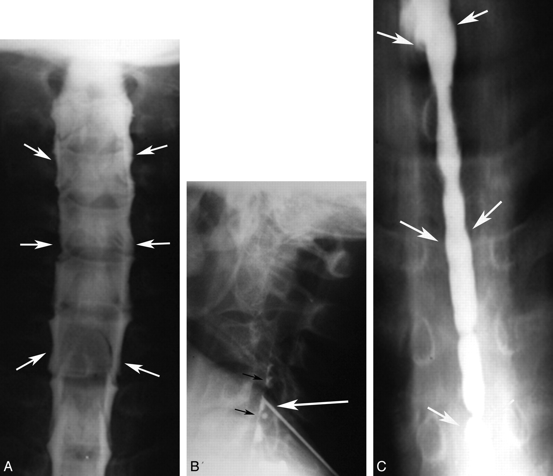
- Fig 3.
Cervical syrinx cavity demonstrated by myelography and myelocystography. A, Frontal cervical myelographic image shows a typical intramedullary mass with enlargement of the cord and thinning of the surrounding subarachnoid space (arrows). B, Lateral image of the cervical spine obtained during cyst puncture at the C4 level shows a needle tip projecting over the middle of spinal canal (white arrow) with a small amount of contrast in the cyst (black arrows). C4 was chosen as a site of puncture because this was where the cord was most expanded. C, Frontal image of the lower cervical and upper thoracic spine obtained following needle removal, showing contrast with a cystic cavity, compatible with a syrinx (arrows) extending inferiorly to approximately T7. It was not uncommon for the syrinx to be more extensive than suggested by myelography.
- Fig 4.
Spinal cord AVM diagnosed with myelography and angiography. A, Frontal myelographic image of thoracic spine shows multiple serpiginous filling defects (arrows) compatible with enlarged vessels from a vascular malformation or vascular neoplasm. B, Frontal image of the thoracic spine from a spinal angiogram shows an abnormal tangle of vessels (arrows) compatible with an AVM.
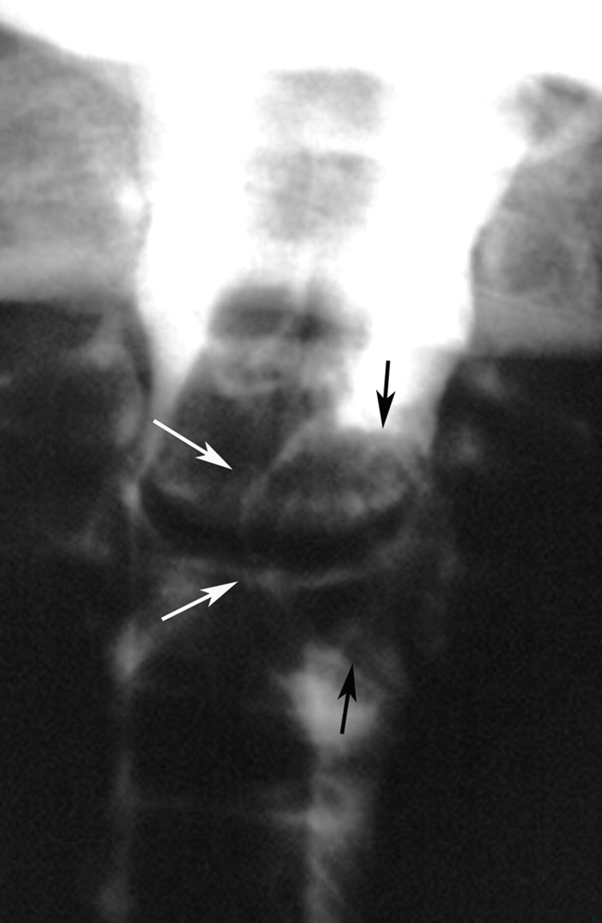
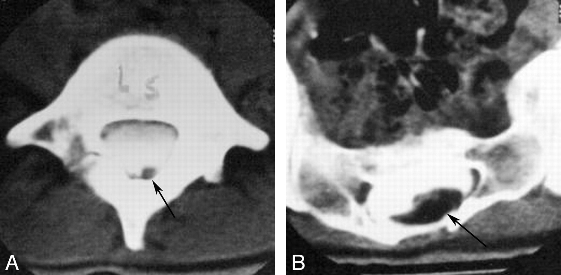
- Fig 5.
Metrizamide CT myelogram. A, Axial image at L5 shows a low-lying tethered cord (arrow). B, Axial image at the level of the sacrum shows an intraspinal lipoma (arrow).
- Fig 6.
Lateral skull x-ray obtained in a 15-day-old boy born with a lumbar myelomeningocele, which ruptured and was infected. The only treatment available for this infant at this time was antibiotics. Note typical findings of lacunar skull or Lückenschädel, with areas of lucency (short black arrows) surrounded by areas of normal attenuation (white arrows), giving a soap bubble appearance to the skull. There is widening of the coronal suture (large black arrows), concerning for increased intracranial pressure.


In this issue


Jump to section
- Article
- Abstract
- ABBREVIATION:
- The Beginning: Spine X-Rays
- Contrast Studies of the Spinal Canal
- Introduction of Additional Contrast Agents
- Introduction of Water-Soluble Contrast Agents
- Endomyelography
- Spinal Angiography
- Cross-Sectional Imaging of the Spine
- Advances in CT and MR Imaging of the Spine
- Conclusions
- Footnotes
- References
- Figures & Data
- Info & Metrics
- Responses
- References