Advertisement

Main menu

  • Home
  • Content
    • Current Issue
    • Accepted Manuscripts
    • Article Preview
    • Past Issue Archive
    • Video Articles
    • AJNR Case Collection
    • Case of the Week Archive
    • Case of the Month Archive
    • Classic Case Archive
  • Special Collections
    • AJNR Awards
    • Low-Field MRI
    • Alzheimer Disease
    • ASNR Foundation Special Collection
    • Photon-Counting CT
    • View All
  • Multimedia
    • AJNR Podcasts
    • AJNR SCANtastic
    • Trainee Corner
    • MRI Safety Corner
    • Imaging Protocols
  • For Authors
    • Submit a Manuscript
    • Submit a Video Article
    • Submit an eLetter to the Editor/Response
    • Manuscript Submission Guidelines
    • Statistical Tips
    • Fast Publishing of Accepted Manuscripts
    • Graphical Abstract Preparation
    • Imaging Protocol Submission
    • Author Policies
  • About Us
    • About AJNR
    • Editorial Board
    • Editorial Board Alumni
  • More
    • Become a Reviewer/Academy of Reviewers
    • Subscribers
    • Permissions
    • Alerts
    • Feedback
    • Advertisers
    • ASNR Home

User menu

  • Alerts
  • Log in

Search

  • Advanced search
American Journal of Neuroradiology

American Journal of Neuroradiology

ASHNR American Society of Functional Neuroradiology ASHNR American Society of Pediatric Neuroradiology ASSR
  • Alerts
  • Log in

Advanced Search

  • Home
  • Content
    • Current Issue
    • Accepted Manuscripts
    • Article Preview
    • Past Issue Archive
    • Video Articles
    • AJNR Case Collection
    • Case of the Week Archive
    • Case of the Month Archive
    • Classic Case Archive
  • Special Collections
    • AJNR Awards
    • Low-Field MRI
    • Alzheimer Disease
    • ASNR Foundation Special Collection
    • Photon-Counting CT
    • View All
  • Multimedia
    • AJNR Podcasts
    • AJNR SCANtastic
    • Trainee Corner
    • MRI Safety Corner
    • Imaging Protocols
  • For Authors
    • Submit a Manuscript
    • Submit a Video Article
    • Submit an eLetter to the Editor/Response
    • Manuscript Submission Guidelines
    • Statistical Tips
    • Fast Publishing of Accepted Manuscripts
    • Graphical Abstract Preparation
    • Imaging Protocol Submission
    • Author Policies
  • About Us
    • About AJNR
    • Editorial Board
    • Editorial Board Alumni
  • More
    • Become a Reviewer/Academy of Reviewers
    • Subscribers
    • Permissions
    • Alerts
    • Feedback
    • Advertisers
    • ASNR Home
  • Follow AJNR on Twitter
  • Visit AJNR on Facebook
  • Follow AJNR on Instagram
  • Join AJNR on LinkedIn
  • RSS Feeds

AJNR Awards, New Junior Editors, and more. Read the latest AJNR updates

Research ArticleBrainF

Active Bleeding in Acute Subarachnoid Hemorrhage Observed by Multiphase Dynamic-Enhanced CT

K. Suzuki, N. Tanaka, S. Morita, H. Machida, E. Ueno and H. Kasuya
American Journal of Neuroradiology August 2012, 33 (7) 1374-1379; DOI: https://doi.org/10.3174/ajnr.A2981
K. Suzuki
aFrom the Departments of Radiology (K.S., H.M., E.U.) and
  • Find this author on Google Scholar
  • Find this author on PubMed
  • Search for this author on this site
N. Tanaka
bNeurosurgery (N.T., H.K.), Tokyo Women's Medical University Medical Center East, Tokyo, Japan; and
  • Find this author on Google Scholar
  • Find this author on PubMed
  • Search for this author on this site
S. Morita
cDepartment of Diagnostic Imaging and Nuclear Medicine (S.M.), Tokyo Women's Medical University, Tokyo, Japan.
  • Find this author on Google Scholar
  • Find this author on PubMed
  • Search for this author on this site
H. Machida
aFrom the Departments of Radiology (K.S., H.M., E.U.) and
  • Find this author on Google Scholar
  • Find this author on PubMed
  • Search for this author on this site
E. Ueno
aFrom the Departments of Radiology (K.S., H.M., E.U.) and
  • Find this author on Google Scholar
  • Find this author on PubMed
  • Search for this author on this site
H. Kasuya
bNeurosurgery (N.T., H.K.), Tokyo Women's Medical University Medical Center East, Tokyo, Japan; and
  • Find this author on Google Scholar
  • Find this author on PubMed
  • Search for this author on this site
  • Article
  • Figures & Data
  • Info & Metrics
  • Responses
  • References
  • PDF
Loading

Article Figures & Data

Figures

  • Tables
  • Fig 1.
    • Download figure
    • Open in new tab
    • Download powerpoint
    Fig 1.

    A 64-year-old man with Claassen grade 3 and WFNS grade 5. Extravasation from the right VA was observed on a multiphase enhanced source image. We selected 4 typical phases obtained at 5, 30.2, 38.6, and 52.6 seconds from among 18-phase scans taken at 2.8-second intervals on multiphase dynamic-enhanced CT. A portion of the right VA near the union was enhanced during the early phase (A, blank arrow), and lobulated extravasation was depicted as an irregularly shaped enhancement (A, white arrow). The premedullary cistern was enhanced in the late phase. Extravasation was well depicted as a wedge-shaped enhancement extending into the subarachnoid space on an additional 4D CT image (B, dark arrow). VA dissection with active bleeding was diagnosed.

  • Fig 2.
    • Download figure
    • Open in new tab
    • Download powerpoint
    Fig 2.

    A 56-year-old woman with Claassen grade 3 and WFNS grade 4. There was a large aneurysm in the MCA. We selected scans obtained at 5, 24.6, 33, and 52.6 seconds, and this case showed typical findings of nebulous enhancement around an aneurysm that was associated with extravasation (A, blank arrow). Hematomas larger than those seen on plain CT were observed during the surgical clipping procedure, and premature rupture occurred intraoperatively. A follow-up plain CT obtained the following day showed increased hematoma (B).
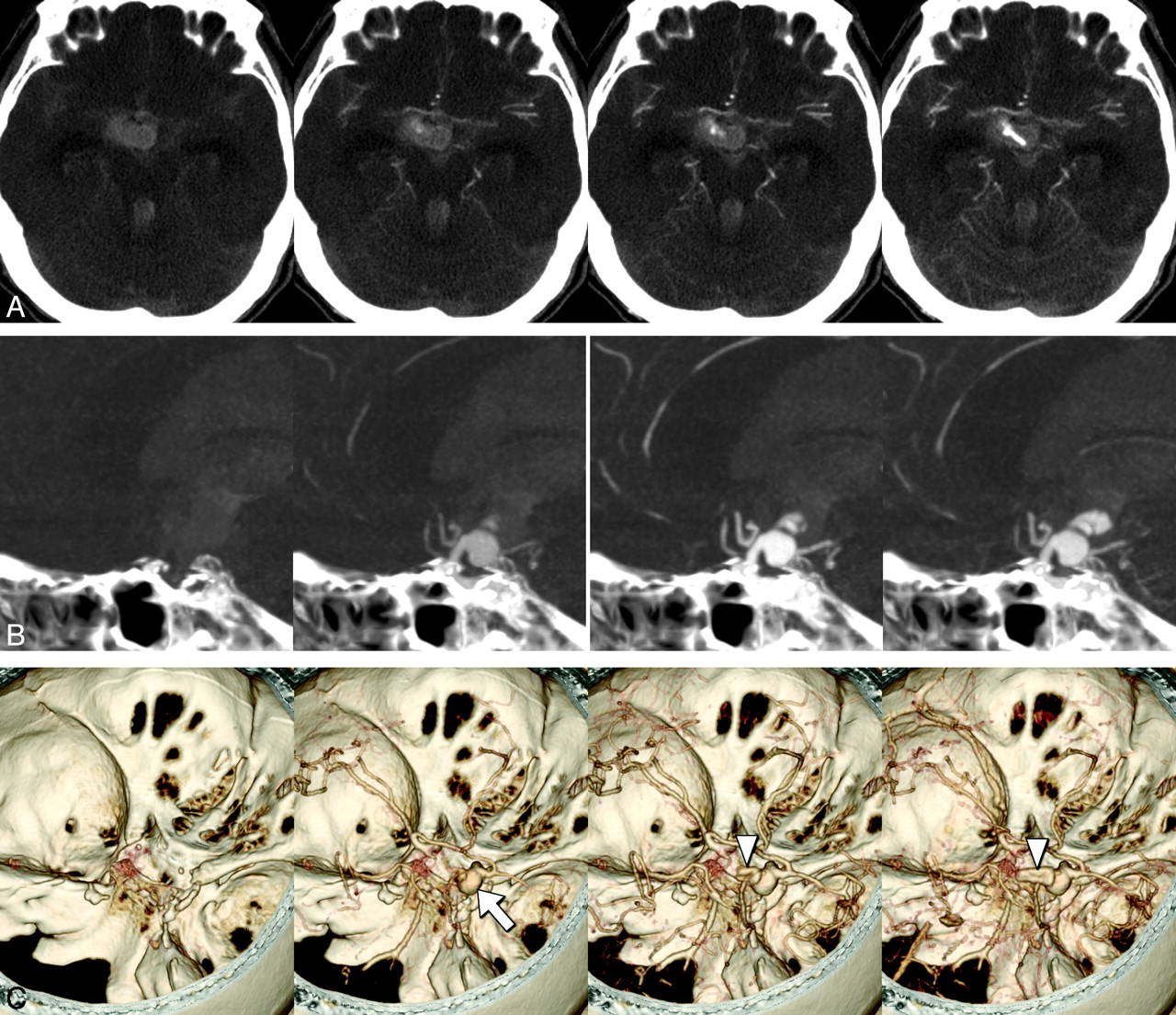
  • Fig 3.
    • Download figure
    • Open in new tab
    • Download powerpoint
    Fig 3.

    A 68-year-old woman with Claassen grade 4 and WFNS grade 5. A 9-mm aneurysm was identified in the right internal carotid posterior communicating portion by CTA. Contrast extravasation at the third ventricle was suspected on the basis of a CTP source image (A). Active bleeding from an aneurysm penetrating the third ventricle was confirmed with sagittal 20-mm-thickness-slab maximum-intensity-projection images (B), and a volume-rendered image was also useful for assessing the shape of the aneurysm (C, white arrow) and the extent of bleeding (C, arrowheads). Contrast medium in the aneurysm was washed out in the delayed phase, but extravasation was not.

  • Fig 4.
    • Download figure
    • Open in new tab
    • Download powerpoint
    Fig 4.

    A 52-year-old man with Claassen grade 4 and WFNS grade 5. An irregularly shaped aneurysm 2 cm in diameter was identified at the anterior communicating artery on CTA. Extravasation had a vessel-like shape at the interhemispheric cistern (A, arrow), and spreading contrast extravasation was observed in the left ventricle (B, arrow). On the reconstructed 4D CT images, the aneurysm showed early enhancement and delayed washout (C, arrow), and string-shaped extravasation from the aneurysm penetrated from the interhemispheric cistern into left lateral ventricle without washout (C, arrowheads). Extravasation of contrast medium was also observed on angiography performed in preparation for a coiling procedure. Follow-up CT after 6 hours indicated enlargement of the hematoma.

Tables

  • Figures
  • Characteristics of patients with and without active bleeding

    FactorActive Bleeding at Initial CT PerfusionP
    With BleedingWithout Bleeding
    Sex.323
        Male316
        Female1022
    Average age (yr)65.862.9.754
    Aneurysm location.154
        ICA211
        ACA317
        MCA45
        VA/BA45
    Claassen gradea.023
        1, 2012
        3, 41326
    WFNS gradea.023
        1, 2012
        3, 4, 51326
    Hours from onseta.007
        ∼21115
        >2013
        Unknown210
    Surgical repair of aneurysm (clip/coil)725.515
    Follow-up CT performed1033.404
    Increased amount of hemorrhagea75.002
    Death within 14 days after admission815.207
    Total1338
    • ↵a Items differ significantly between the 2 patient groups.

PreviousNext
Back to top

In this issue

American Journal of Neuroradiology: 33 (7)
American Journal of Neuroradiology
Vol. 33, Issue 7
1 Aug 2012
  • Table of Contents
  • Index by author
Advertisement
Print
Download PDF
Email Article

Thank you for your interest in spreading the word on American Journal of Neuroradiology.

NOTE: We only request your email address so that the person you are recommending the page to knows that you wanted them to see it, and that it is not junk mail. We do not capture any email address.

Enter multiple addresses on separate lines or separate them with commas.
Active Bleeding in Acute Subarachnoid Hemorrhage Observed by Multiphase Dynamic-Enhanced CT
(Your Name) has sent you a message from American Journal of Neuroradiology
(Your Name) thought you would like to see the American Journal of Neuroradiology web site.
CAPTCHA
This question is for testing whether or not you are a human visitor and to prevent automated spam submissions.
Cite this article
K. Suzuki, N. Tanaka, S. Morita, H. Machida, E. Ueno, H. Kasuya
Active Bleeding in Acute Subarachnoid Hemorrhage Observed by Multiphase Dynamic-Enhanced CT
American Journal of Neuroradiology Aug 2012, 33 (7) 1374-1379; DOI: 10.3174/ajnr.A2981

Citation Manager Formats

  • BibTeX
  • Bookends
  • EasyBib
  • EndNote (tagged)
  • EndNote 8 (xml)
  • Medlars
  • Mendeley
  • Papers
  • RefWorks Tagged
  • Ref Manager
  • RIS
  • Zotero
0 Responses
Respond to this article
Share
Bookmark this article
Active Bleeding in Acute Subarachnoid Hemorrhage Observed by Multiphase Dynamic-Enhanced CT
K. Suzuki, N. Tanaka, S. Morita, H. Machida, E. Ueno, H. Kasuya
American Journal of Neuroradiology Aug 2012, 33 (7) 1374-1379; DOI: 10.3174/ajnr.A2981
del.icio.us logo Twitter logo Facebook logo Mendeley logo
  • Tweet Widget
  • Facebook Like
  • Google Plus One
Purchase

Jump to section

  • Article
    • Abstract
    • ABBREVIATIONS:
    • Materials and Methods
    • Results
    • Discussion
    • Conclusion
    • Acknowledgments
    • Footnotes
    • References
  • Figures & Data
  • Info & Metrics
  • Responses
  • References
  • PDF

Related Articles

  • No related articles found.
  • PubMed
  • Google Scholar

Cited By...

  • No citing articles found.
  • Crossref
  • Google Scholar

This article has not yet been cited by articles in journals that are participating in Crossref Cited-by Linking.

More in this TOC Section

  • Progression of Microstructural Damage in Spinocerebellar Ataxia Type 2: A Longitudinal DTI Study
  • SWI or T2*: Which MRI Sequence to Use in the Detection of Cerebral Microbleeds? The Karolinska Imaging Dementia Study
  • Statin Therapy Does Not Affect the Radiographic and Clinical Profile of Patients with TIA and Minor Stroke
Show more Brain

Similar Articles

Advertisement

Indexed Content

  • Current Issue
  • Accepted Manuscripts
  • Article Preview
  • Past Issues
  • Editorials
  • Editor's Choice
  • Fellows' Journal Club
  • Letters to the Editor
  • Video Articles

Cases

  • Case Collection
  • Archive - Case of the Week
  • Archive - Case of the Month
  • Archive - Classic Case

More from AJNR

  • Trainee Corner
  • Imaging Protocols
  • MRI Safety Corner
  • Book Reviews

Multimedia

  • AJNR Podcasts
  • AJNR Scantastics

Resources

  • Turnaround Time
  • Submit a Manuscript
  • Submit a Video Article
  • Submit an eLetter to the Editor/Response
  • Manuscript Submission Guidelines
  • Statistical Tips
  • Fast Publishing of Accepted Manuscripts
  • Graphical Abstract Preparation
  • Imaging Protocol Submission
  • Evidence-Based Medicine Level Guide
  • Publishing Checklists
  • Author Policies
  • Become a Reviewer/Academy of Reviewers
  • News and Updates

About Us

  • About AJNR
  • Editorial Board
  • Editorial Board Alumni
  • Alerts
  • Permissions
  • Not an AJNR Subscriber? Join Now
  • Advertise with Us
  • Librarian Resources
  • Feedback
  • Terms and Conditions
  • AJNR Editorial Board Alumni

American Society of Neuroradiology

  • Not an ASNR Member? Join Now

© 2025 by the American Society of Neuroradiology All rights, including for text and data mining, AI training, and similar technologies, are reserved.
Print ISSN: 0195-6108 Online ISSN: 1936-959X

Powered by HighWire