Advertisement

Main menu

  • Home
  • Content
    • Current Issue
    • Accepted Manuscripts
    • Article Preview
    • Past Issue Archive
    • AJNR Case Collection
    • Case of the Week Archive
    • Classic Case Archive
    • Case of the Month Archive
  • Special Collections
    • Spinal CSF Leak Articles (Jan 2020-June 2024)
    • 2024 AJNR Journal Awards
    • Most Impactful AJNR Articles
  • Multimedia
    • AJNR Podcast
    • AJNR Scantastics
    • Video Articles
  • For Authors
    • Submit a Manuscript
    • Author Policies
    • Fast publishing of Accepted Manuscripts
    • Graphical Abstract Preparation
    • Manuscript Submission Guidelines
    • Imaging Protocol Submission
    • Submit a Case for the Case Collection
  • About Us
    • About AJNR
    • Editorial Board
  • More
    • Become a Reviewer/Academy of Reviewers
    • Subscribers
    • Permissions
    • Alerts
    • Feedback
    • Advertisers
    • ASNR Home
  • Other Publications
    • ajnr

User menu

  • Alerts
  • Log in

Search

  • Advanced search
American Journal of Neuroradiology

American Journal of Neuroradiology

ASHNR American Society of Functional Neuroradiology ASHNR American Society of Pediatric Neuroradiology ASSR
  • Alerts
  • Log in

Advanced Search

  • Home
  • Content
    • Current Issue
    • Accepted Manuscripts
    • Article Preview
    • Past Issue Archive
    • AJNR Case Collection
    • Case of the Week Archive
    • Classic Case Archive
    • Case of the Month Archive
  • Special Collections
    • Spinal CSF Leak Articles (Jan 2020-June 2024)
    • 2024 AJNR Journal Awards
    • Most Impactful AJNR Articles
  • Multimedia
    • AJNR Podcast
    • AJNR Scantastics
    • Video Articles
  • For Authors
    • Submit a Manuscript
    • Author Policies
    • Fast publishing of Accepted Manuscripts
    • Graphical Abstract Preparation
    • Manuscript Submission Guidelines
    • Imaging Protocol Submission
    • Submit a Case for the Case Collection
  • About Us
    • About AJNR
    • Editorial Board
  • More
    • Become a Reviewer/Academy of Reviewers
    • Subscribers
    • Permissions
    • Alerts
    • Feedback
    • Advertisers
    • ASNR Home
  • Follow AJNR on Twitter
  • Visit AJNR on Facebook
  • Follow AJNR on Instagram
  • Join AJNR on LinkedIn
  • RSS Feeds

Welcome to the new AJNR, Updated Hall of Fame, and more. Read the full announcements.


AJNR is seeking candidates for the position of Associate Section Editor, AJNR Case Collection. Read the full announcement.


Research ArticleSpine

MR Imaging Findings of the Intraspinal Meningeal Melanocytoma: Correlation with Histopathologic Findings

G.Q. Hou, J.C. Sun, X.J. Zhang, B.X. Shen, X.J. Zhu, L. Liang and X.L. Zhang
American Journal of Neuroradiology September 2012, 33 (8) 1525-1529; DOI: https://doi.org/10.3174/ajnr.A2987
G.Q. Hou
aFrom the Department of Medical Image Center (G.Q.H., B.X.S.), Nanshan Hospital, Guangdong Medical College, Guangdong Province, People's Republic of China
  • Find this author on Google Scholar
  • Find this author on PubMed
  • Search for this author on this site
J.C. Sun
bDepartment of Pathology (J.C.S., X.J. Zhang, L.L.), School of Basic Medical Sciences, Southern Medical University, Guangzhou, Guangdong Province, People's Republic of China
cGuangdong Province Key Laboratory of Molecular Tumor Pathology (J.C.S., X.J. Zhang, L.L.), Guangzhou, Guangdong Province, People's Republic of China
  • Find this author on Google Scholar
  • Find this author on PubMed
  • Search for this author on this site
X.J. Zhang
bDepartment of Pathology (J.C.S., X.J. Zhang, L.L.), School of Basic Medical Sciences, Southern Medical University, Guangzhou, Guangdong Province, People's Republic of China
cGuangdong Province Key Laboratory of Molecular Tumor Pathology (J.C.S., X.J. Zhang, L.L.), Guangzhou, Guangdong Province, People's Republic of China
  • Find this author on Google Scholar
  • Find this author on PubMed
  • Search for this author on this site
B.X. Shen
aFrom the Department of Medical Image Center (G.Q.H., B.X.S.), Nanshan Hospital, Guangdong Medical College, Guangdong Province, People's Republic of China
  • Find this author on Google Scholar
  • Find this author on PubMed
  • Search for this author on this site
X.J. Zhu
dDepartment of Medical Image Center (X.L.Z., X.J. Zhu), Nanfang Hospital, Southern Medical University, Guangzhou, Guangdong Province, People's Republic of China
  • Find this author on Google Scholar
  • Find this author on PubMed
  • Search for this author on this site
L. Liang
bDepartment of Pathology (J.C.S., X.J. Zhang, L.L.), School of Basic Medical Sciences, Southern Medical University, Guangzhou, Guangdong Province, People's Republic of China
cGuangdong Province Key Laboratory of Molecular Tumor Pathology (J.C.S., X.J. Zhang, L.L.), Guangzhou, Guangdong Province, People's Republic of China
  • Find this author on Google Scholar
  • Find this author on PubMed
  • Search for this author on this site
X.L. Zhang
dDepartment of Medical Image Center (X.L.Z., X.J. Zhu), Nanfang Hospital, Southern Medical University, Guangzhou, Guangdong Province, People's Republic of China
  • Find this author on Google Scholar
  • Find this author on PubMed
  • Search for this author on this site
  • Article
  • Figures & Data
  • Info & Metrics
  • Responses
  • References
  • PDF
Loading

Abstract

SUMMARY: Our aim was to better understand and improve the accuracy of the preoperative diagnosis of intraspinal MM by a combined analysis of MR imaging and pathologic findings. All 5 patients had undergone unenhanced and contrast-enhanced MR imaging examinations. All tumor samples had immunohistochemical reactions to HMB-45, vimentin, S-100, EMA, and Leu-7 antibodies. All 5 cases were located in the intradural extramedullary compartment. Two cases had multifocal lesions, and 3 cases were solitary. Two cases showed homogeneously strong enhancement, and 3 cases showed moderate enhancement on contrast-enhanced T1WI. The tumor cells had positive reactions to HMB-45, vimentin, and S-100 antibodies. MR imaging plays an important role in the detection and diagnosis of intraspinal MM. Final diagnosis should be based on histopathology and IHC examinations.

ABBREVIATIONS:

EMA
epithelial membrane antigen
HMB-45
human melanoma black-45
IHC
immunohistochemistry
MM
meningeal melanocytoma

Primary melanocytic neoplasms of the CNS are lesions arising from melanocytes of the leptomeninges. Melanocytes are melanin-producing cells that derive from the neural crest during embryogenesis and then migrate to the skin, mucous membranes, and CNS.1 Melanocytes are often concentrated at the base of the brain and on the ventral surface of the cervical spinal cord. Melanocytic neoplasms of the CNS are rare and most frequently are metastatic in origin. Primary melanocytic lesions of the CNS include neurocutaneous melanosis, MM, primary leptomeningeal melanomatosis, and melanoma.

MM is a rare primary benign melanotic tumor of the leptomeninges, which occurs anywhere in the cranial or spinal region but most commonly in the supratentorial and thoracic spine regions.2 It was first described by Limas and Tio3 in 1972 with a prolonged clinical course and benign histologic features.4 From 1972 to 2009, <60 cases of intraspinal MM were reported.5 Here, we report 5 cases of intraspinal MM. To our knowledge, this study is the largest collection of intraspinal MM cases with MR imaging to date.

Materials and Methods

We searched the data base of our hospital for all intraspinal tumors from 2003 to 2010. The clinicopathologic data and MR imaging findings of 5 cases of MM were analyzed retrospectively. The patients included 1 woman and 4 men with the average age of 49 years. The main clinical symptoms were gradually worsening debilitation of the lower limb (n = 5), leg numbness (n = 3), lumbodynia (n = 1), dorsodynia (n = 1), and dizziness (n = 1). The On-line Table shows the clinical data and MR imaging findings of all patients.

The patients were examined with MR imaging at 1.5T (n = 3) or 3T (n = 2). Spin-echo or fast spin-echo T1WI and T2WI in the axial, coronal, or sagittal planes were acquired. The corresponding contrast-enhanced T1WI was obtained after intravenous injection of 0.1 mmol/kg of gadopentetate dimeglumine (Magnevist; Schering, Berlin, Germany).

Two neuroradiologists reviewed the MR images separately. All lesions were removed by open surgery. HMB-45, vimentin, S-100, EMA, and Leu-7 antibodies were purchased from Dako North America (Carpinteria, California). The final diagnoses were established by 2 pathologists after reviewing the clinical process, the macroscopic protocols, and the histologic slides.

Results

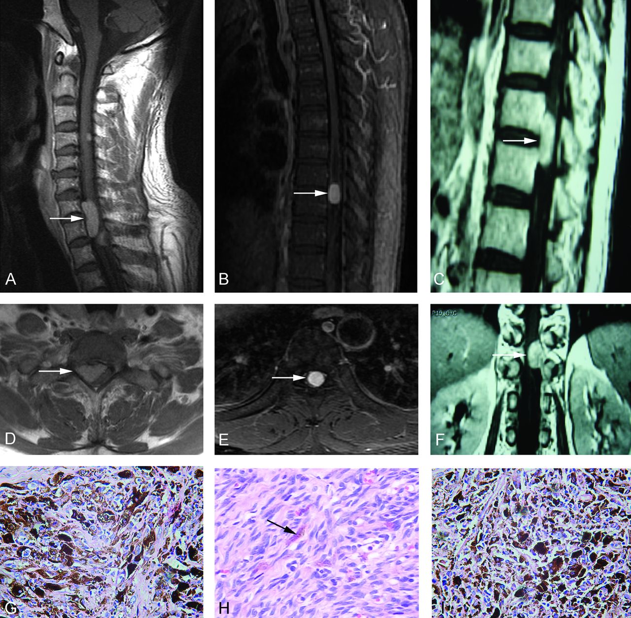
All 5 cases were located in the intradural extramedullary compartment, with the results revealing that the spine was obviously compressed (Figs 1 and 2). All lesions were tightly attached to the dura mater. Two cases were multifocal and others had solitary lesions. The single lateral intervertebral foramen was widened (n = 1). T1WI showed homogeneous hyperintensity (n = 2), moderate hyperintensity (n = 2), and isointensity (n = 1); T2WI showed homogeneous hypointensity (n = 3), moderate hyperintensity (n = 1), and isointensity (n = 1). Two cases showed homogeneously strong enhancement, and others showed moderate enhancement on contrast-enhanced T1WI (Fig 1 and On-line Table).

Fig 1.
  • Download figure
  • Open in new tab
  • Download powerpoint
Fig 1.

In case 1 (A and B), 3 lesions are shown on sagittal nonenhanced MR images of the cervical vertebrae at the C4-T2 level. In case 2 (C and D), the lesion is shown on sagittal nonenhanced MR images of the thoracic vertebrae at the T7-T8 level. In case 3, the lesion is shown on sagittal (E) and coronal (F) nonenhanced MR images of the thoracic vertebrae at the T11-T12 level.

All the tumors showed well-defined, round, or ovoid masses. Three cases were solitary, and others were multifocal. The cut surfaces and peripheral meninges of the tumors were all in black. Tumor cells were spindle, epithelioid, or polygonal with round vesicular nuclei, prominent nucleoli, and plenty of melanin granules in the cytoplasm. They had a uniform cytologic appearance and lacked evidence of atypia or significant mitotic activity. Tumor cells demonstrated storiform vasocentric arrangements. In case 2, the amount of melanin-containing cells was <10%, while in the other 4 cases, >10% of tumor cells had melanin granules in the cytoplasm, even masking the morphology of the nucleus (Fig 2). Tumor cells had a positive reaction to anti-HMB-45, vimentin, and S-100 antibodies but a negative reaction to anti-EMA and Leu-7 (Fig 3). All 5 cases were finally diagnosed as intraspinal MM. All patients underwent surgical resection and radiation therapy. To date, 4 patients are alive (22–37 months after surgery; mean, 28 months), and follow-up MR imaging showed no signs of tumor recurrence. One patient with multifocal lesions developed malignant melanoma and died of metastasis 13 months after surgery.

Fig 2.
  • Download figure
  • Open in new tab
  • Download powerpoint
Fig 2.

In case 1, the sagittal (A) and axial (D) contrast-enhanced T1WI and morphologic changes (G, HE original magnification ×400) of the lesion are seen. In case 2, the sagittal (B) and axial (E) contrast-enhanced T1WI sequences and morphologic changes (H, HE original magnification ×200) of the lesion are seen. In case 3, the sagittal (C) and axial (F) contrast-enhanced T1WI sequence and morphologic changes (I, HE original magnification ×400) of the lesion are shown.

Fig 3.
  • Download figure
  • Open in new tab
  • Download powerpoint
Fig 3.

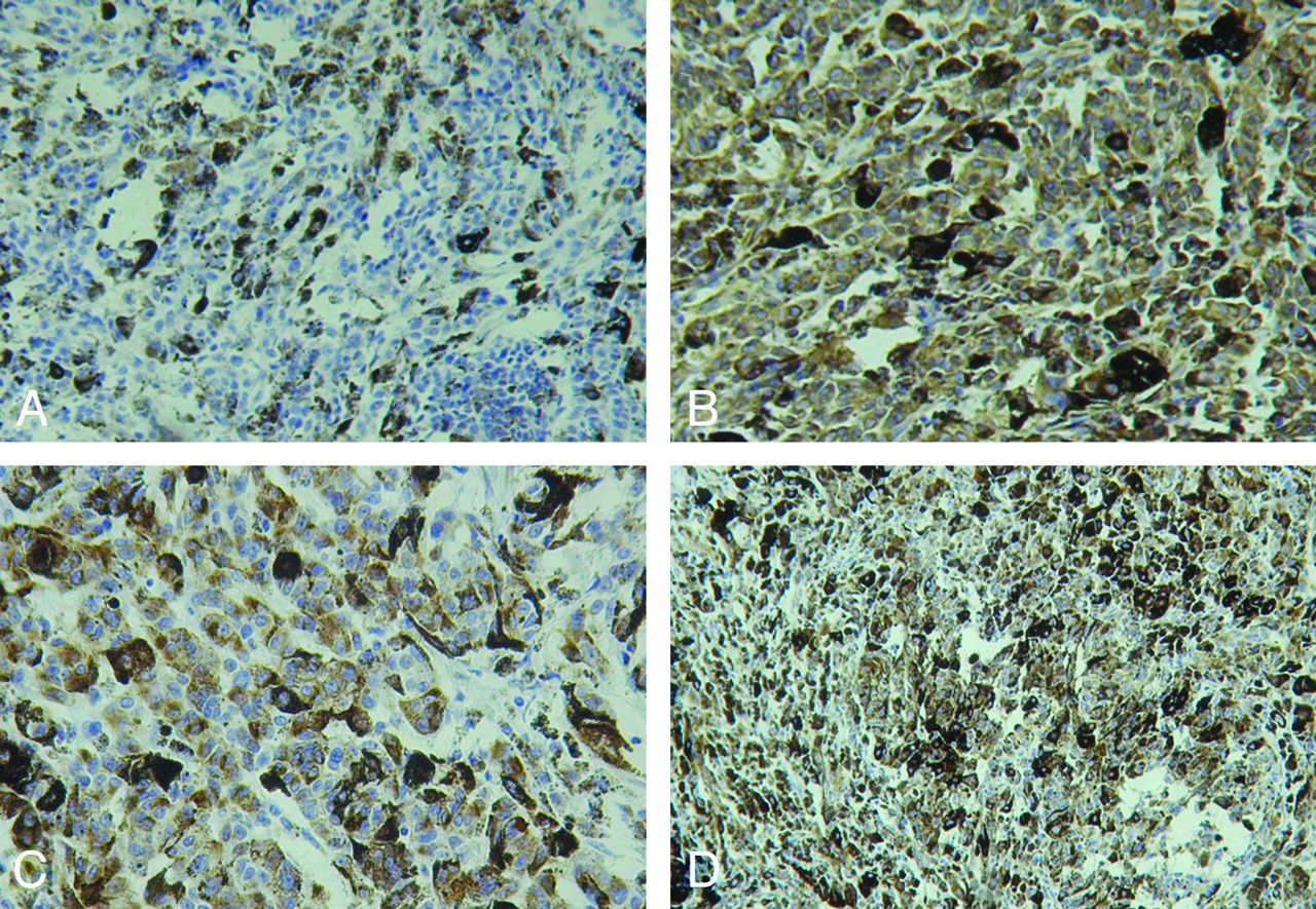
IHC detection of MM (DAB stain). The tumor cells have a negative reaction to the EMA antibody (A, original magnification ×200) but positive reactions to vimentin (B, ×200), HMB-45 (C, original magnification ×400), and S-100 (D, original magnification ×200).

Discussion

Melanocytes originate from the neural crest and are normally found in the leptomeninges.2,4 These cells are present in the leptomeninges of healthy adults and may result in primary leptomeningeal pigmented tumors, including pigmented meningioma, malignant melanoma, MM, melanotic schwannoma, and melanoblastosis.6,7 Malignant melanoma and MM are of the same origin and represent the malignant and benign ends of the tumor spectrum, respectively.5

MM was first coined by Limas and Tio3 in 1972. It is a very rare, pigmented, and slow-growing benign neoplasm.8 However, malignant transformation of melanocytoma to malignant melanoma has been reported.5,9 To date, approximately 112 cases of MM have been reported,5 and most were found in the cerebrum. The tumor occurs almost exclusively in the posterior fossa and spinal cord.6,7,10,11 The annual incidence of MM is estimated to be 1 case per 10 million people, and women are vulnerable.10 Our hospital has an extremely large collection area for patients from Guangdong province and 4 neighboring provinces. The orthopedics center in our hospital is very famous and is also a referral center for spinal tumors. Therefore, we have collected 5 cases of patients with MM in the past 7 years. The incidence is not affected by race or genetic risk factor, and there are no literature reports about MM, to our knowledge. MM may occur at any age but is most commonly seen in the fifth decade. The average age in our series is 49.2 years, which is consistent with that in other reports.10,11

Approximate 59 cases of intraspinal MM have been reported.5 Nineteen cases were founded in the intramedullary location, while others were located in the extramedullary area.5,9 The clinical signs and symptoms depend on its location and may present as myelopathy and seizures.10,12 The symptoms of our patients were nonspecific, mainly caused by mass compression.

Most MMs are commonly found as a solitary lesion, but they can also present as multifocal or disseminating along the arachnoid and dura mater.13,14 To date, approximately 4 cases with multifocal lesions have been reported.15,16 Ali et al6 considered multifocal MM as a distinct pathologic entity that seems to portend an aggressive clinical course and a poor prognosis, even when malignant pathologic features are absent.

In our study, 2 patients with multifocal lesions did not show malignant pathologic features compared with those with solitary lesions, but 1 patient with multifocal lesions developed malignant melanoma and died of metastasis 13 months after surgery. It has been reported that MM might transform into malignant melanoma9,17 and then cause distant metastasis.28 Our study also supports multifocal MM possibly predicting a poor outcome.

MR imaging is the first choice for spinal tumors. On MR imaging, MM is generally an attenuated extra-axial lesion attached to the dura, which may not have a distinct margin.5 It is typically hyperintense on T1WI and hypointense on T2WI and enhances homogeneously with gadolinium.10,16⇓⇓–19 The characteristic signal intensity of MM is thought to be caused by the paramagnetic susceptibility effect of the melanin pigment or hemorrhagic foci; hence, its radiologic signal intensity is variable and inconstant.16,19,20 However, differences in duration, different amounts of melanin, and hemorrhage in malignant melanoma may influence the signal intensity of tumor cells on MR imaging.9,14,19,20

Amelanotic melanoma and melanoma without hemorrhage are iso- or hypointense on T1WI and moderately hyperintense on T2WI.21⇓–23 Isiklar et al24 reviewed the MR images of 30 patients with intracerebral melanoma and sorted these into 4 groups: melanotic pattern (hyperintense on T1WI, hypointense on T2WI), amelanotic pattern (hypointense or isointense on T1WI and hyperintense or isontense on T2WI), indeterminate or mixed pattern (MR imaging characteristics not conforming to the first 2 categories), and hematoma pattern (MR imaging exhibiting only hematoma characteristics).27 In our study, cases with >10% of melanin-containing cells showed a melanotic pattern, while case 2 did not have typical MR imaging and was then proved to have <10% melanin-containing cells by histopathology.

Wadasadawala et al22 considered the following aspects useful for the differential diagnosis of MM: 1) Location: Pigmented meningioma is located in the extra-axial area; melanotic schwannoma is located in the cerebellopontine angle; MM and primary melanoma are often located in the high cervical cord or perimedullary area, whereas metastatic melanoma is located in the supratentorial or gray-white matter junction. 2) Number: The lesion of pigmented meningioma, MM, or primary melanoma is often single, while metastatic melanoma is multiple. 3) Hemorrhage: The pigmented meningioma and melanotic schwannoma do not have a hemorrhagic focus, while MM and primary and metastatic melanoma often have it.22 4) Dural involvement: Dural involvement and the “dural tail” sign are often observed in meningioma, MM, or meningeal melanocytosis, but metastatic melanoma seldom presents dural involvement.

Because the differential diagnosis of MM from other pigmented malignancies is difficult on the basis of only the imaging information, a final diagnosis is dependent on the results of pathologic examination. Grossly, MM usually presents as a solitary well-circumscribed black mass, while lesions in red, white, tan, brown, or blue have been reported.10,23,24 MMs may be mistaken for meningioma at gross inspection because they are attached to the meninges. MM cells tend to compress adjacent tissues instead of infiltrating them. However, local invasion of neural structures has been reported in some cases.6,25,26 The cytologic feature of MM is uniform, lacking in atypia and mitotic activity and showing different degrees of melanization.12,27⇓⇓–30

IHC staining is always indispensable for differentiating MM from other pigmented tumors. MM is characterized by a positive reaction to HMB-45, vimentin, and S-100 proteins12 and a negative reaction to EMA and Leu-7.7,20 Meningioma cells lack the expressions of HMB-45 and S-100, and a positive reaction to EMA differentiates pigmented meningioma from MM.7,12 The absence of a reaction to Leu-7 excludes the diagnosis of melanocytic schwannoma.7,28 Vimentin usually appears in MM but is only rarely present in malignant melanoma. Ultrastructurally, desmosomes are characteristic of meningioma but are generally absent in MM.3,5,19 However, a differential diagnosis between MM and malignant leptomeningeal melanoma may present some difficulties.20 Because the histologic characteristics of MM and melanoma are overlapping, thorough physical and radiologic examinations of the patient are required to exclude a primary cutaneous, ocular, or mucosal melanoma.5,10

Conclusions

We report 5 rare cases of intraspinal MM. To date, to our knowledge, this is the largest collection of intraspinal MM cases with MR imaging. MR imaging plays an important role in the detection and diagnosis of intraspinal MM. The different amounts of melanin-containing cells or hemorrhage may influence the MR imaging signal intensity of tumor cells. Final diagnosis should be based on histopathology and IHC examinations.

Acknowledgments

The authors thank Mou Ren-Qi for his assistance with the study.

Footnotes

  • The first 2 authors contributed equally to this work.

References

  1. 1.↵
    1. Kwon SC,
    2. Rhim SC,
    3. Lee DH,
    4. et al
    . Primary malignant melanoma of the cervical spinal nerve root. Yonsei Med J 2004; 45: 345– 48
    PubMed
  2. 2.↵
    1. El-Khashab M,
    2. Koral K,
    3. Bowers DC,
    4. et al
    . Intermediate grade meningeal melanocytoma of cervical spine. Childs Nerv Syst 2009; 25: 407– 10
    CrossRefPubMed
  3. 3.↵
    1. Limas C,
    2. Tio FO
    . Meningeal melanocytoma (“melanotic meningioma”): its melanocytic origin as revealed by electron microscopy. Cancer 1972; 30: 1286– 94
    CrossRefPubMed
  4. 4.↵
    1. Czarnecki EJ,
    2. Silbergleit R,
    3. Gutierrez JA
    . MR of spinal meningeal melanocytoma. AJNR Am J Neuroradiol 1997; 18: 180– 82
    Abstract
  5. 5.↵
    1. Liubinas SV,
    2. Maartens N,
    3. Drummond KJ
    . Primary melanocytic neoplasms of the central nervous system. J Clin Neurosci 2010; 17: 1227– 32
    CrossRefPubMed
  6. 6.↵
    1. Ali Y,
    2. Rahme R,
    3. Moussa R,
    4. et al
    . Multifocal meningeal melanocytoma: a new pathological entity or the result of leptomeningeal seeding? J Neurosurg 2009; 111: 488– 91
    CrossRefPubMed
  7. 7.↵
    1. Painter TJ,
    2. Chaljub G,
    3. Sethi R,
    4. et al
    . Intracranial and intraspinal meningeal melanocytosis. AJNR Am J Neuroradiol 2000; 21: 1349– 53
    Abstract/FREE Full Text
  8. 8.↵
    1. Karikari IO,
    2. Powers CJ,
    3. Bagley CA,
    4. et al
    . Primary intramedullary melanocytoma of the spinal cord: case report. Neurosurgery 2009; 64: E777– 78
    CrossRef
  9. 9.↵
    1. Rades D,
    2. Schild SE,
    3. Tatagiba M,
    4. et al
    . Therapy of meningeal melanocytomas. Cancer 2004; 100: 2442– 47
    CrossRefPubMed
  10. 10.↵
    1. Smith AB,
    2. Rushing EJ,
    3. Smirniotopoulos JG
    . Pigmented lesions of the central nervous system: radiologic-pathologic correlation. Radiographics 2009; 29: 1503– 24
    CrossRefPubMed
  11. 11.↵
    1. Chen CJ,
    2. Hsu YI,
    3. Ho YS,
    4. et al
    . Intracranial meningeal melanocytoma: CT and MRI. Neuroradiology 1997; 39: 811– 14
    CrossRefPubMed
  12. 12.↵
    1. Bydon A,
    2. Gutierrez JA,
    3. Mahmood A
    . Meningeal melanocytoma: an aggressive course for a benign tumor. J Neurooncol 2003; 64: 259– 63
    CrossRefPubMed
  13. 13.↵
    1. Horn EM,
    2. Nakaji P,
    3. Coons SW,
    4. et al
    . Surgical treatment for intramedullary spinal cord melanocytomas. J Neurosurg Spine 2008; 9: 48– 54
    CrossRefPubMed
  14. 14.↵
    1. Shownkeen HN,
    2. Harmath C,
    3. Thomas C
    . Multiform cervical melanocytoma: a case report. Neuroradiology 2002; 44: 1008– 10
    CrossRefPubMed
  15. 15.↵
    1. Iida M,
    2. Llena JF,
    3. Suarez MA,
    4. et al
    . Two cases of spinal meningeal melanocytoma. Brain Tumor Pathol 2002; 19: 41– 45
    CrossRefPubMed
  16. 16.↵
    1. O'Brien DF,
    2. Crooks D,
    3. Mallucci C,
    4. et al
    . Meningeal melanocytoma. Childs Nerv Syst 2006; 22: 556– 61
    CrossRefPubMed
  17. 17.↵
    1. Roser F,
    2. Nakamura M,
    3. Brandis A,
    4. et al
    . Transition from meningeal melanocytoma to primary cerebral melanoma. J Neurosurg 2004; 101: 528– 31
    CrossRefPubMed
  18. 18.↵
    1. Wang F,
    2. Li X,
    3. Chen L,
    4. et al
    . Malignant transformation of spinal meningeal melanocytoma: case report and review of the literature. J Neurosurg Spine 2007; 6: 451– 54
    CrossRefPubMed
  19. 19.↵
    1. Uematsu Y,
    2. Yukawa S,
    3. Yokote H,
    4. et al
    . Meningeal melanocytoma: magnetic resonance imaging characteristics and pathological features-case report. J Neurosurg 1992; 76: 705– 09
    CrossRefPubMed
  20. 20.↵
    1. Ruelle A,
    2. Tunesi G,
    3. Andrioli G
    . Spinal meningeal melanocytoma: case report and analysis of diagnostic criteria. Neurosurg Rev 1996; 19: 39– 42
    CrossRefPubMed
  21. 21.↵
    1. Farrokh D,
    2. Fransen P,
    3. Faverly D
    . MR findings of a primary intramedullary malignant melanoma: case report and literature review. AJNR Am J Neuroradiol 2001; 22: 1864– 66
    Abstract/FREE Full Text
  22. 22.↵
    1. Wadasadawala T,
    2. Trivedi S,
    3. Gupta T,
    4. et al
    . The diagnostic dilemma of primary central nervous system melanoma. J Clin Neurosci 2010; 17: 1014– 17
    CrossRefPubMed
  23. 23.↵
    1. Vanzieleghem BD,
    2. Lemmerling MM,
    3. Van Coster RN
    . Neurocutaneous melanosis presenting with intracranial amelanotic melanoma. AJNR Am J Neuroradiol 1999; 20: 457– 60
    Abstract/FREE Full Text
  24. 24.↵
    1. Isiklar I,
    2. Leeds NE,
    3. Fuller GN,
    4. et al
    . Intracranial metastatic melanoma: correlation between MR imaging characteristics and melanin content. AJR Am J Roentgenol 1995; 165: 1503– 12
    PubMed
  25. 25.↵
    1. Ahluwalia S,
    2. Ashkan K,
    3. Casey AT
    . Meningeal melanocytoma: clinical features and review of the literature. Br J Neurosurg 2003; 17: 347– 51
    CrossRefPubMed
  26. 26.↵
    1. Chow M,
    2. Clarke DB,
    3. Maloney WJ,
    4. et al
    . Meningeal melanocytoma of the planum sphenoidale: case report and review of the literature. J Neurosurg 2001; 94: 841– 45
    CrossRefPubMed
  27. 27.↵
    1. Czirjak S,
    2. Vitanovic D,
    3. Slowik F,
    4. et al
    . Primary meningeal melanocytoma of the pineal region. J Neurosurg 2000; 92: 461– 65
    CrossRefPubMed
  28. 28.↵
    1. Offiah CJ,
    2. Laitt RD
    . Intracranial meningeal melanocytoma: a cause of high signal on T1- and low signal on T2-weighted MRI. Clin Radiol 2006; 61: 294– 98
    CrossRefPubMed
  29. 29.↵
    1. Kleihues P,
    2. Cavenee WK
    1. Jellinger K,
    2. Chou P,
    3. Paulus W
    . Melanocytic lesions. In: Kleihues P, Cavenee WK eds. World Health Organization Classification of Tumours: Pathology and Genetics of Tumours of the Nervous System. Lyon, France: IARC Press: 2000: 193– 95
  30. 30.↵
    1. Prayson RA,
    2. Cohen ML
    1. Prayson RA,
    2. Cohen ML
    . Melanocytic lesions. In: Prayson RA, Cohen ML . Practical Differential Diagnosis in Surgical Neuropathology. Totowa, New Jersey: Humana Press; 2000: 137– 40
  • Received September 16, 2011.
  • Accepted after revision November 18, 2011.
  • © 2012 by American Journal of Neuroradiology
View Abstract
PreviousNext
Back to top

In this issue

American Journal of Neuroradiology: 33 (8)
American Journal of Neuroradiology
Vol. 33, Issue 8
1 Sep 2012
  • Table of Contents
  • Index by author
Advertisement
Print
Download PDF
Email Article

Thank you for your interest in spreading the word on American Journal of Neuroradiology.

NOTE: We only request your email address so that the person you are recommending the page to knows that you wanted them to see it, and that it is not junk mail. We do not capture any email address.

Enter multiple addresses on separate lines or separate them with commas.
MR Imaging Findings of the Intraspinal Meningeal Melanocytoma: Correlation with Histopathologic Findings
(Your Name) has sent you a message from American Journal of Neuroradiology
(Your Name) thought you would like to see the American Journal of Neuroradiology web site.
CAPTCHA
This question is for testing whether or not you are a human visitor and to prevent automated spam submissions.
Cite this article
G.Q. Hou, J.C. Sun, X.J. Zhang, B.X. Shen, X.J. Zhu, L. Liang, X.L. Zhang
MR Imaging Findings of the Intraspinal Meningeal Melanocytoma: Correlation with Histopathologic Findings
American Journal of Neuroradiology Sep 2012, 33 (8) 1525-1529; DOI: 10.3174/ajnr.A2987

Citation Manager Formats

  • BibTeX
  • Bookends
  • EasyBib
  • EndNote (tagged)
  • EndNote 8 (xml)
  • Medlars
  • Mendeley
  • Papers
  • RefWorks Tagged
  • Ref Manager
  • RIS
  • Zotero
0 Responses
Respond to this article
Share
Bookmark this article
MR Imaging Findings of the Intraspinal Meningeal Melanocytoma: Correlation with Histopathologic Findings
G.Q. Hou, J.C. Sun, X.J. Zhang, B.X. Shen, X.J. Zhu, L. Liang, X.L. Zhang
American Journal of Neuroradiology Sep 2012, 33 (8) 1525-1529; DOI: 10.3174/ajnr.A2987
del.icio.us logo Twitter logo Facebook logo Mendeley logo
  • Tweet Widget
  • Facebook Like
  • Google Plus One
Purchase

Jump to section

  • Article
    • Abstract
    • ABBREVIATIONS:
    • Materials and Methods
    • Results
    • Discussion
    • Conclusions
    • Acknowledgments
    • Footnotes
    • References
  • Figures & Data
  • Info & Metrics
  • Responses
  • References
  • PDF

Related Articles

  • No related articles found.
  • PubMed
  • Google Scholar

Cited By...

  • No citing articles found.
  • Crossref (11)
  • Google Scholar

This article has been cited by the following articles in journals that are participating in Crossref Cited-by Linking.

  • Spinal meningeal melanocytomas: clinical manifestations, radiological and pathological characteristics, and surgical outcomes
    Chenlong Yang, Jingyi Fang, Guang Li, Wenqing Jia, Hai Liu, Wei Qi, Yulun Xu
    Journal of Neuro-Oncology 2016 127 2
  • Diagnostic Clue of Meningeal Melanocytoma: Case Report and Review of Literature
    Jae Koo Lee, Young Joon Rho, Dong Mun Jeong, Seung Chul Rhim, Sang Joon Kim
    Yonsei Medical Journal 2017 58 2
  • How Should We Treat Meningeal Melanocytoma? A Retrospective Analysis of Potential Treatment Strategies
    Sarah Ricchizzi, Marco Gallus, Walter Stummer, Markus Holling
    Cancers 2022 14 23
  • Malignant Transformation and Metastatic Spread of Dumbbell-Shaped Meningeal Melanocytoma of the Cervical Spine: A Case Report and Literature Review
    Shuang-lin Deng, Yu-bo Wang, Dan-hua Wang, Shuang Zhan, Yi Jing, Yi Guan
    Frontiers in Surgery 2022 9
  • Spinal meningeal melanocytoma in a 5-year-old child: a case report and review of literature
    Ahmed M. Salah El-Din, Hashem M. Aboul-Ela, Mohamed F. Alsawy, Ahmed Koheil, Ahmed H. Ashry
    The Egyptian Journal of Neurology, Psychiatry and Neurosurgery 2018 54 1
  • Primary scattered multifocal melanocytomas in spinal canal mimicking neurofibromatosis
    Chenlong Yang, Jingyi Fang, Guang Li, Jun Yang, Yulun Xu
    The Spine Journal 2016 16 8
  • C6-T1 Intradural Extramedullary Ventral Meningeal Melanocytoma Resected Via Anterior Corpectomy with Reconstruction
    Mehmet Yigit Akgun, Cihan Isler, Mustafa Onur Ulu
    World Neurosurgery 2020 138
  • A rare case of multifocal craniospinal leptomeningeal melanocytoma: A case report and scoping review
    Daniel Lewis, Timothy P. Dawson, Rebecca Hyde, George Adrian Rata, Andrew F. Alalade, Kaushik Ghosh, Ahmed Elhabal
    Brain and Spine 2024 4
  • Meningeal melanocytoma of the central nervous system in children
    Lukasz Antkowiak, Jerzy Luszawski, Wieslawa Grajkowska, Joanna Trubicka, Marek Mandera
    Child's Nervous System 2025 41 1
  • Radiopaedia.org
    Jose Bacalla
    2020

More in this TOC Section

  • Bern Score Validity for SIH
  • MP2RAGE 7T in MS Lesions of the Cervical Spine
  • Deep Learning for STIR Spine MRI Quality
Show more Spine

Similar Articles

Advertisement

Indexed Content

  • Current Issue
  • Accepted Manuscripts
  • Article Preview
  • Past Issues
  • Editorials
  • Editors Choice
  • Fellow Journal Club
  • Letters to the Editor

Cases

  • Case Collection
  • Archive - Case of the Week
  • Archive - Case of the Month
  • Archive - Classic Case

Special Collections

  • Special Collections

Resources

  • News and Updates
  • Turn around Times
  • Submit a Manuscript
  • Author Policies
  • Manuscript Submission Guidelines
  • Evidence-Based Medicine Level Guide
  • Publishing Checklists
  • Graphical Abstract Preparation
  • Imaging Protocol Submission
  • Submit a Case
  • Become a Reviewer/Academy of Reviewers
  • Get Peer Review Credit from Publons

Multimedia

  • AJNR Podcast
  • AJNR SCANtastic
  • Video Articles

About Us

  • About AJNR
  • Editorial Board
  • Not an AJNR Subscriber? Join Now
  • Alerts
  • Feedback
  • Advertise with us
  • Librarian Resources
  • Permissions
  • Terms and Conditions

American Society of Neuroradiology

  • Not an ASNR Member? Join Now

© 2025 by the American Society of Neuroradiology All rights, including for text and data mining, AI training, and similar technologies, are reserved.
Print ISSN: 0195-6108 Online ISSN: 1936-959X

Powered by HighWire