Advertisement

Main menu

  • Home
  • Content
    • Current Issue
    • Accepted Manuscripts
    • Article Preview
    • Past Issue Archive
    • AJNR Case Collection
    • Case of the Week Archive
    • Classic Case Archive
    • Case of the Month Archive
  • Special Collections
    • Spinal CSF Leak Articles (Jan 2020-June 2024)
    • 2024 AJNR Journal Awards
    • Most Impactful AJNR Articles
  • Multimedia
    • AJNR Podcast
    • AJNR Scantastics
    • Video Articles
  • For Authors
    • Submit a Manuscript
    • Author Policies
    • Fast publishing of Accepted Manuscripts
    • Graphical Abstract Preparation
    • Manuscript Submission Guidelines
    • Imaging Protocol Submission
    • Submit a Case for the Case Collection
  • About Us
    • About AJNR
    • Editorial Board
  • More
    • Become a Reviewer/Academy of Reviewers
    • Subscribers
    • Permissions
    • Alerts
    • Feedback
    • Advertisers
    • ASNR Home
  • Other Publications
    • ajnr

User menu

  • Alerts
  • Log in

Search

  • Advanced search
American Journal of Neuroradiology

American Journal of Neuroradiology

ASHNR American Society of Functional Neuroradiology ASHNR American Society of Pediatric Neuroradiology ASSR
  • Alerts
  • Log in

Advanced Search

  • Home
  • Content
    • Current Issue
    • Accepted Manuscripts
    • Article Preview
    • Past Issue Archive
    • AJNR Case Collection
    • Case of the Week Archive
    • Classic Case Archive
    • Case of the Month Archive
  • Special Collections
    • Spinal CSF Leak Articles (Jan 2020-June 2024)
    • 2024 AJNR Journal Awards
    • Most Impactful AJNR Articles
  • Multimedia
    • AJNR Podcast
    • AJNR Scantastics
    • Video Articles
  • For Authors
    • Submit a Manuscript
    • Author Policies
    • Fast publishing of Accepted Manuscripts
    • Graphical Abstract Preparation
    • Manuscript Submission Guidelines
    • Imaging Protocol Submission
    • Submit a Case for the Case Collection
  • About Us
    • About AJNR
    • Editorial Board
  • More
    • Become a Reviewer/Academy of Reviewers
    • Subscribers
    • Permissions
    • Alerts
    • Feedback
    • Advertisers
    • ASNR Home
  • Follow AJNR on Twitter
  • Visit AJNR on Facebook
  • Follow AJNR on Instagram
  • Join AJNR on LinkedIn
  • RSS Feeds

Welcome to the new AJNR, Updated Hall of Fame, and more. Read the full announcements.


AJNR is seeking candidates for the position of Associate Section Editor, AJNR Case Collection. Read the full announcement.


Getting new auth cookie, if you see this message a lot, tell someone!
Research ArticleBrain

Diagnostic Accuracy of Dynamic Contrast-Enhanced MR Imaging Using a Phase-Derived Vascular Input Function in the Preoperative Grading of Gliomas

T.B. Nguyen, G.O. Cron, J.F. Mercier, C. Foottit, C.H. Torres, S. Chakraborty, J. Woulfe, G.H. Jansen, J.M. Caudrelier, J. Sinclair, M.J. Hogan, R.E. Thornhill and I.G. Cameron
American Journal of Neuroradiology September 2012, 33 (8) 1539-1545; DOI: https://doi.org/10.3174/ajnr.A3012
T.B. Nguyen
aFrom the Departments of Radiology (T.B.N., G.O.C., J.F.M., C.H.T., R.E.T., I.G.C., S.C., J.M.C.)
  • Find this author on Google Scholar
  • Find this author on PubMed
  • Search for this author on this site
G.O. Cron
aFrom the Departments of Radiology (T.B.N., G.O.C., J.F.M., C.H.T., R.E.T., I.G.C., S.C., J.M.C.)
  • Find this author on Google Scholar
  • Find this author on PubMed
  • Search for this author on this site
J.F. Mercier
aFrom the Departments of Radiology (T.B.N., G.O.C., J.F.M., C.H.T., R.E.T., I.G.C., S.C., J.M.C.)
  • Find this author on Google Scholar
  • Find this author on PubMed
  • Search for this author on this site
C. Foottit
bMedical Physics (C.F., I.G.C.)
  • Find this author on Google Scholar
  • Find this author on PubMed
  • Search for this author on this site
C.H. Torres
aFrom the Departments of Radiology (T.B.N., G.O.C., J.F.M., C.H.T., R.E.T., I.G.C., S.C., J.M.C.)
  • Find this author on Google Scholar
  • Find this author on PubMed
  • Search for this author on this site
S. Chakraborty
aFrom the Departments of Radiology (T.B.N., G.O.C., J.F.M., C.H.T., R.E.T., I.G.C., S.C., J.M.C.)
  • Find this author on Google Scholar
  • Find this author on PubMed
  • Search for this author on this site
J. Woulfe
cPathology (G.H.J., J.W.)
  • Find this author on Google Scholar
  • Find this author on PubMed
  • Search for this author on this site
G.H. Jansen
cPathology (G.H.J., J.W.)
  • Find this author on Google Scholar
  • Find this author on PubMed
  • Search for this author on this site
J.M. Caudrelier
aFrom the Departments of Radiology (T.B.N., G.O.C., J.F.M., C.H.T., R.E.T., I.G.C., S.C., J.M.C.)
  • Find this author on Google Scholar
  • Find this author on PubMed
  • Search for this author on this site
J. Sinclair
dSurgery, Division of Neurosurgery (J.S.)
  • Find this author on Google Scholar
  • Find this author on PubMed
  • Search for this author on this site
M.J. Hogan
eMedicine, Division of Neurology (M.J.H.); The Ottawa Hospital, University of Ottawa, Ottawa, Ontario, Canada.
  • Find this author on Google Scholar
  • Find this author on PubMed
  • Search for this author on this site
R.E. Thornhill
aFrom the Departments of Radiology (T.B.N., G.O.C., J.F.M., C.H.T., R.E.T., I.G.C., S.C., J.M.C.)
  • Find this author on Google Scholar
  • Find this author on PubMed
  • Search for this author on this site
I.G. Cameron
aFrom the Departments of Radiology (T.B.N., G.O.C., J.F.M., C.H.T., R.E.T., I.G.C., S.C., J.M.C.)
bMedical Physics (C.F., I.G.C.)
  • Find this author on Google Scholar
  • Find this author on PubMed
  • Search for this author on this site
  • Article
  • Figures & Data
  • Info & Metrics
  • Responses
  • References
  • PDF
Loading

Article Figures & Data

Figures

  • Tables
  • Fig 1.
    • Download figure
    • Open in new tab
    • Download powerpoint
    Fig 1.

    A, Phase-derived VIF and tumor CT(t) from a patient with confirmed grade IV glioma obtained at 3T. B and C, Ktrans_ϕ (B) and Vp_ϕ (C) maps. D, Magnitude-derived VIF and tumor signal-intensity changes from the same patient for comparison. E and F, Ktrans_SI (E) and Vp_SI (F) maps. Maximal Ktrans_ϕ and Vp_ϕ values in tumor are 0.099 minutes−1 and 5.7 mL/100 g compared with 0.15 minutes−1 and 18 mL/100 g for Ktrans_SI and Vp_SI.
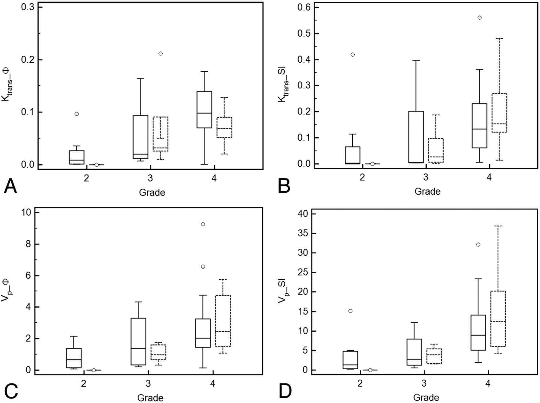
  • Fig 2.
    • Download figure
    • Open in new tab
    • Download powerpoint
    Fig 2.

    Boxplots showing ability of parameters Ktrans_ϕ (A), Ktrans_SI (B), Vp_ϕ (C), and Vp_SI (D) in differentiating various glioma grades for 2D (solid line) and 3D data (dashed line).

  • Fig 3.
    • Download figure
    • Open in new tab
    • Download powerpoint
    Fig 3.

    A, ROC for comparison of maximal Ktrans_ϕ and Ktrans_SI for differentiation of low-grade from high-grade gliomas. B, ROC for comparison of Vp_ϕ and Vp_SI in differentiating low-grade and high-grade gliomas.

  • Fig 4.
    • Download figure
    • Open in new tab
    • Download powerpoint
    Fig 4.

    Agreement between Ktrans_ϕ and Ktrans_SI (A), Vp_ϕ, and Vp_SI (B). A, Bland-Altman plot of difference between Ktrans_ϕ and Ktrans_SI against a mean of Ktrans_ϕ and Ktrans_SI, with a mean absolute difference (bias) (solid line) and 95% confidence interval of the mean difference (limits of agreement) (dashed lines). B, Bland-Altman plot of difference between Vp_ϕ and Vp_SI_ϕ against a mean of Vp_ϕ and Vp_SI.

Tables

  • Figures
    • View popup
    Table 1:

    Median values of the maximal perfusion parameters and coefficient of variation for different grades of gliomas using a 2D gradient-recalled echo sequence on a 1.5T MR imaging scanner

    No.Ktrans_ϕ(min−1) (95% CI)CV(%)Ktrans_SI(min−1) (95% CI)CV(%)Vp_ϕ (mL/100 g) (95% CI)CV(%)Vp_SI (ml/100 g) (95% CI)CV(%)
    Grade 280.0086 (0.0010–0.047)1530.0032 (0.00056–0.17)2100.66 (0.091–1.55)911.35 (0.22–6.9)143
    Grade 340.0201420.00521911.371052.8114
    Not definedNot definedNot definedNot defined
    Grade 4190.10 (0.072–0.13)460.13 (0.073–0.23)852.0 (1.7–3.1)788.8 (5.3–14)76
    • View popup
    Table 2:

    Median values of the maximal perfusion parameters and coefficient of variation for different grades of gliomas using a 3D gradient-recalled echo sequence on a 3T MR scanner

    No.Ktrans_ϕ(min−1) (95% CI)CV(%)Ktrans_SI(min−1) (95% CI)CV(%)Vp_ϕ (mL/100 g) (95% CI)CV(%)Vp_SI (mL/100 g) (95% CI)CV(%)
    Grade 210000
    Not defined Not definedNot definedNot defined
    Grade 350.0321220.0261310.98543.957
    Not defined Not definedNot definedNot defined
    Grade 490.069 (0.047–0.096)450.15 (0.082–0.31)722.4 (1.3–5.6)5912.5 (5.5–25)70
    • View popup
    Table 3:

    Median values of the maximal perfusion parameters and coefficient of variation for different grades of gliomas combining 2D and 3D acquisition methods

    No.Ktrans_ϕ(min−1) (95% CI)CV(%)Ktrans_SI(min−1) (95% CI)CV(%)Vp_ϕ (mL/100 g) (95% CI)CV(%)Vp_SI (mL/100 g) (95% CI)CV(%)
    Grade 290.0041 (0.00062–0.033)1650.0024 (0.00050–0.10)2240.64 (0.063–1.4)1031.12 (0.18–4.9)155
    Grade 2 excluding biopsy60.0014 (0.00010–0.011)1440.0016 (0.00011–0.014)1560.15 (0.011–0.67)1070.81 (0.032–4.3)133
    Grade 390.031 (0.011–0.15)1220.0093 (0.0030–0.17)1720.98 (0.34–2.2)923.7 (1.6–6.4)86
    Grade 3 excluding biopsy70.031 (0.0083–0.19)1160.0093 (0.0017–0.29)1690.79 (0.26–2.9)1161.9 (1.0–8.6)103
    Grade 4280.088 (0.069–0.11)480.15 (0.093–0.21)792.16 (1.8–3.1)719.4 (5.8–14)75
    • View popup
    Table 4:

    The statistical significance (P Values) of differences in median perfusion parameter values for various grades of gliomas and imaging techniques

    P ValueKtrans_ϕKtrans_SIVp_ϕVp_SI
    Grade II vs III.050.17.15.14
    Grade II vs III excluding biopsy.0047.070.051.070
    Grade III vs IV.040.014.015.0026
    Grade III 2D vs 3D.32.62.80.80
    Grade IV 2D vs 3D.058.50.570.21
    • View popup
    Table 5:

    AUC values for various ROC curves

    AUC for Ktrans_ϕ (95% CI)AUC for Ktrans_SI (95% CI)AUC for Vp_ϕ (95% CI)AUC for Vp_SI (95% CI)
    Low-grade from high-grade gliomas0.87 (0.73–1)0.81 (0.59–1)0.84 (0.69–0.98)0.84 (0.66–0.91)
    Grade III from grade IV gliomas0.73 (0.46–1)0.77 (0.54–1)0.77 (0.58–0.97)0.84 (0.68–0.99)
PreviousNext
Back to top

In this issue

American Journal of Neuroradiology: 33 (8)
American Journal of Neuroradiology
Vol. 33, Issue 8
1 Sep 2012
  • Table of Contents
  • Index by author
Advertisement
Print
Download PDF
Email Article

Thank you for your interest in spreading the word on American Journal of Neuroradiology.

NOTE: We only request your email address so that the person you are recommending the page to knows that you wanted them to see it, and that it is not junk mail. We do not capture any email address.

Enter multiple addresses on separate lines or separate them with commas.
Diagnostic Accuracy of Dynamic Contrast-Enhanced MR Imaging Using a Phase-Derived Vascular Input Function in the Preoperative Grading of Gliomas
(Your Name) has sent you a message from American Journal of Neuroradiology
(Your Name) thought you would like to see the American Journal of Neuroradiology web site.
CAPTCHA
This question is for testing whether or not you are a human visitor and to prevent automated spam submissions.
Cite this article
T.B. Nguyen, G.O. Cron, J.F. Mercier, C. Foottit, C.H. Torres, S. Chakraborty, J. Woulfe, G.H. Jansen, J.M. Caudrelier, J. Sinclair, M.J. Hogan, R.E. Thornhill, I.G. Cameron
Diagnostic Accuracy of Dynamic Contrast-Enhanced MR Imaging Using a Phase-Derived Vascular Input Function in the Preoperative Grading of Gliomas
American Journal of Neuroradiology Sep 2012, 33 (8) 1539-1545; DOI: 10.3174/ajnr.A3012

Citation Manager Formats

  • BibTeX
  • Bookends
  • EasyBib
  • EndNote (tagged)
  • EndNote 8 (xml)
  • Medlars
  • Mendeley
  • Papers
  • RefWorks Tagged
  • Ref Manager
  • RIS
  • Zotero
0 Responses
Respond to this article
Share
Bookmark this article
Diagnostic Accuracy of Dynamic Contrast-Enhanced MR Imaging Using a Phase-Derived Vascular Input Function in the Preoperative Grading of Gliomas
T.B. Nguyen, G.O. Cron, J.F. Mercier, C. Foottit, C.H. Torres, S. Chakraborty, J. Woulfe, G.H. Jansen, J.M. Caudrelier, J. Sinclair, M.J. Hogan, R.E. Thornhill, I.G. Cameron
American Journal of Neuroradiology Sep 2012, 33 (8) 1539-1545; DOI: 10.3174/ajnr.A3012
del.icio.us logo Twitter logo Facebook logo Mendeley logo
  • Tweet Widget
  • Facebook Like
  • Google Plus One
Purchase

Jump to section

  • Article
    • Abstract
    • ABBREVIATIONS:
    • Materials and Methods
    • Results
    • Discussion
    • Conclusions
    • Appendix
    • Footnotes
    • References
  • Figures & Data
  • Info & Metrics
  • Responses
  • References
  • PDF

Related Articles

  • No related articles found.
  • PubMed
  • Google Scholar

Cited By...

  • Comparison of Dynamic Contrast-Enhancement Parameters between Gadobutrol and Gadoterate Meglumine in Posttreatment Glioma: A Prospective Intraindividual Study
  • Improved Brain Tumor Classification by Sodium MR Imaging: Prediction of IDH Mutation Status and Tumor Progression
  • Comparison of the Diagnostic Accuracy of DSC- and Dynamic Contrast-Enhanced MRI in the Preoperative Grading of Astrocytomas
  • ASFNR Recommendations for Clinical Performance of MR Dynamic Susceptibility Contrast Perfusion Imaging of the Brain
  • Preoperative Prognostic Value of Dynamic Contrast-Enhanced MRI-Derived Contrast Transfer Coefficient and Plasma Volume in Patients with Cerebral Gliomas
  • Evaluation of Microvascular Permeability with Dynamic Contrast-Enhanced MRI for the Differentiation of Primary CNS Lymphoma and Glioblastoma: Radiologic-Pathologic Correlation
  • Crossref (34)
  • Google Scholar

This article has been cited by the following articles in journals that are participating in Crossref Cited-by Linking.

  • Perfusion MRI: The Five Most Frequently Asked Technical Questions
    Marco Essig, Mark S. Shiroishi, Thanh Binh Nguyen, Marc Saake, James M. Provenzale, David Enterline, Nicoletta Anzalone, Arnd Dörfler, Àlex Rovira, Max Wintermark, Meng Law
    American Journal of Roentgenology 2013 200 1
  • ASFNR Recommendations for Clinical Performance of MR Dynamic Susceptibility Contrast Perfusion Imaging of the Brain
    K. Welker, J. Boxerman, A. Kalnin, T. Kaufmann, M. Shiroishi, M. Wintermark
    American Journal of Neuroradiology 2015 36 6
  • Diagnostic performance of texture analysis on MRI in grading cerebral gliomas
    Karoline Skogen, Anselm Schulz, Johann Baptist Dormagen, Balaji Ganeshan, Eirik Helseth, Andrès Server
    European Journal of Radiology 2016 85 4
  • Dynamic contrast-enhanced and dynamic susceptibility contrast perfusion MR imaging for glioma grading: Preliminary comparison of vessel compartment and permeability parameters using hotspot and histogram analysis
    Corrado Santarosa, Antonella Castellano, Gian Marco Conte, Marcello Cadioli, Antonella Iadanza, Maria Rosa Terreni, Alberto Franzin, Lorenzo Bello, Massimo Caulo, Andrea Falini, Nicoletta Anzalone
    European Journal of Radiology 2016 85 6
  • Perfusion MRI: The Five Most Frequently Asked Clinical Questions
    Marco Essig, Thanh Binh Nguyen, Mark S. Shiroishi, Marc Saake, James M. Provenzale, David S. Enterline, Nicoletta Anzalone, Arnd Dörfler, Àlex Rovira, Max Wintermark, Meng Law
    American Journal of Roentgenology 2013 201 3
  • Evaluation of Microvascular Permeability with Dynamic Contrast-Enhanced MRI for the Differentiation of Primary CNS Lymphoma and Glioblastoma: Radiologic-Pathologic Correlation
    P. Kickingereder, F. Sahm, B. Wiestler, M. Roethke, S. Heiland, H.- P. Schlemmer, W. Wick, A. von Deimling, M. Bendszus, A. Radbruch
    American Journal of Neuroradiology 2014 35 8
  • The role of imaging in the management of adults with diffuse low grade glioma
    Sarah Jost Fouke, Tammie Benzinger, Daniel Gibson, Timothy C. Ryken, Steven N. Kalkanis, Jeffrey J. Olson
    Journal of Neuro-Oncology 2015 125 3
  • Clinical Applications of Contrast-Enhanced Perfusion MRI Techniques in Gliomas: Recent Advances and Current Challenges
    Junfeng Zhang, Heng Liu, Haipeng Tong, Sumei Wang, Yizeng Yang, Gang Liu, Weiguo Zhang
    Contrast Media & Molecular Imaging 2017 2017
  • Improved Brain Tumor Classification by Sodium MR Imaging: Prediction ofIDHMutation Status and Tumor Progression
    A. Biller, S. Badde, A. Nagel, J.-O. Neumann, W. Wick, A. Hertenstein, M. Bendszus, F. Sahm, N. Benkhedah, J. Kleesiek
    American Journal of Neuroradiology 2016 37 1
  • Comparison of the Diagnostic Accuracy of DSC- and Dynamic Contrast-Enhanced MRI in the Preoperative Grading of Astrocytomas
    T.B. Nguyen, G.O. Cron, K. Perdrizet, K. Bezzina, C.H. Torres, S. Chakraborty, J. Woulfe, G.H. Jansen, J. Sinclair, R.E. Thornhill, C. Foottit, B. Zanette, I.G. Cameron
    American Journal of Neuroradiology 2015 36 11

More in this TOC Section

  • Evaluating the Effects of White Matter Multiple Sclerosis Lesions on the Volume Estimation of 6 Brain Tissue Segmentation Methods
  • White Matter Alterations in the Brains of Patients with Active, Remitted, and Cured Cushing Syndrome: A DTI Study
  • Enhanced Axonal Metabolism during Early Natalizumab Treatment in Relapsing-Remitting Multiple Sclerosis
Show more Brain

Similar Articles

Advertisement

Indexed Content

  • Current Issue
  • Accepted Manuscripts
  • Article Preview
  • Past Issues
  • Editorials
  • Editors Choice
  • Fellow Journal Club
  • Letters to the Editor

Cases

  • Case Collection
  • Archive - Case of the Week
  • Archive - Case of the Month
  • Archive - Classic Case

Special Collections

  • Special Collections

Resources

  • News and Updates
  • Turn around Times
  • Submit a Manuscript
  • Author Policies
  • Manuscript Submission Guidelines
  • Evidence-Based Medicine Level Guide
  • Publishing Checklists
  • Graphical Abstract Preparation
  • Imaging Protocol Submission
  • Submit a Case
  • Become a Reviewer/Academy of Reviewers
  • Get Peer Review Credit from Publons

Multimedia

  • AJNR Podcast
  • AJNR SCANtastic
  • Video Articles

About Us

  • About AJNR
  • Editorial Board
  • Not an AJNR Subscriber? Join Now
  • Alerts
  • Feedback
  • Advertise with us
  • Librarian Resources
  • Permissions
  • Terms and Conditions

American Society of Neuroradiology

  • Not an ASNR Member? Join Now

© 2025 by the American Society of Neuroradiology All rights, including for text and data mining, AI training, and similar technologies, are reserved.
Print ISSN: 0195-6108 Online ISSN: 1936-959X

Powered by HighWire