Advertisement

Main menu

  • Home
  • Content
    • Current Issue
    • Accepted Manuscripts
    • Article Preview
    • Past Issue Archive
    • Video Articles
    • AJNR Case Collection
    • Case of the Week Archive
    • Case of the Month Archive
    • Classic Case Archive
  • Special Collections
    • AJNR Awards
    • Low-Field MRI
    • Alzheimer Disease
    • ASNR Foundation Special Collection
    • Photon-Counting CT
    • View All
  • Multimedia
    • AJNR Podcasts
    • AJNR SCANtastic
    • Trainee Corner
    • MRI Safety Corner
    • Imaging Protocols
  • For Authors
    • Submit a Manuscript
    • Submit a Video Article
    • Submit an eLetter to the Editor/Response
    • Manuscript Submission Guidelines
    • Statistical Tips
    • Fast Publishing of Accepted Manuscripts
    • Graphical Abstract Preparation
    • Imaging Protocol Submission
    • Author Policies
  • About Us
    • About AJNR
    • Editorial Board
    • Editorial Board Alumni
  • More
    • Become a Reviewer/Academy of Reviewers
    • Subscribers
    • Permissions
    • Alerts
    • Feedback
    • Advertisers
    • ASNR Home

User menu

  • Alerts
  • Log in

Search

  • Advanced search
American Journal of Neuroradiology

American Journal of Neuroradiology

ASHNR American Society of Functional Neuroradiology ASHNR American Society of Pediatric Neuroradiology ASSR
  • Alerts
  • Log in

Advanced Search

  • Home
  • Content
    • Current Issue
    • Accepted Manuscripts
    • Article Preview
    • Past Issue Archive
    • Video Articles
    • AJNR Case Collection
    • Case of the Week Archive
    • Case of the Month Archive
    • Classic Case Archive
  • Special Collections
    • AJNR Awards
    • Low-Field MRI
    • Alzheimer Disease
    • ASNR Foundation Special Collection
    • Photon-Counting CT
    • View All
  • Multimedia
    • AJNR Podcasts
    • AJNR SCANtastic
    • Trainee Corner
    • MRI Safety Corner
    • Imaging Protocols
  • For Authors
    • Submit a Manuscript
    • Submit a Video Article
    • Submit an eLetter to the Editor/Response
    • Manuscript Submission Guidelines
    • Statistical Tips
    • Fast Publishing of Accepted Manuscripts
    • Graphical Abstract Preparation
    • Imaging Protocol Submission
    • Author Policies
  • About Us
    • About AJNR
    • Editorial Board
    • Editorial Board Alumni
  • More
    • Become a Reviewer/Academy of Reviewers
    • Subscribers
    • Permissions
    • Alerts
    • Feedback
    • Advertisers
    • ASNR Home
  • Follow AJNR on Twitter
  • Visit AJNR on Facebook
  • Follow AJNR on Instagram
  • Join AJNR on LinkedIn
  • RSS Feeds

AJNR Awards, New Junior Editors, and more. Read the latest AJNR updates

Research ArticleNeurointervention
Open Access

The Effect of Intracranial Stent Implantation on the Curvature of the Cerebrovasculature

R.M. King, J.-Y. Chueh, I.M.J. van der Bom, C.F. Silva, S.L. Carniato, G. Spilberg, A.K. Wakhloo and M.J. Gounis
American Journal of Neuroradiology October 2012, 33 (9) 1657-1662; DOI: https://doi.org/10.3174/ajnr.A3062
R.M. King
aFrom the Department of Radiology (R.M.K., J.-Y.C., I.M.J.v.d.B., C.F.S., G.S., A.K.W., M.J.G.), New England Center for Stroke Research, University of Massachusetts Medical School, Worcester, Massachusetts
  • Find this author on Google Scholar
  • Find this author on PubMed
  • Search for this author on this site
J.-Y. Chueh
aFrom the Department of Radiology (R.M.K., J.-Y.C., I.M.J.v.d.B., C.F.S., G.S., A.K.W., M.J.G.), New England Center for Stroke Research, University of Massachusetts Medical School, Worcester, Massachusetts
  • Find this author on Google Scholar
  • Find this author on PubMed
  • Search for this author on this site
I.M.J. van der Bom
aFrom the Department of Radiology (R.M.K., J.-Y.C., I.M.J.v.d.B., C.F.S., G.S., A.K.W., M.J.G.), New England Center for Stroke Research, University of Massachusetts Medical School, Worcester, Massachusetts
  • Find this author on Google Scholar
  • Find this author on PubMed
  • Search for this author on this site
C.F. Silva
aFrom the Department of Radiology (R.M.K., J.-Y.C., I.M.J.v.d.B., C.F.S., G.S., A.K.W., M.J.G.), New England Center for Stroke Research, University of Massachusetts Medical School, Worcester, Massachusetts
  • Find this author on Google Scholar
  • Find this author on PubMed
  • Search for this author on this site
S.L. Carniato
bResearch and Development (S.L.C.), Stryker Neurovascular, Fremont, California
  • Find this author on Google Scholar
  • Find this author on PubMed
  • Search for this author on this site
G. Spilberg
aFrom the Department of Radiology (R.M.K., J.-Y.C., I.M.J.v.d.B., C.F.S., G.S., A.K.W., M.J.G.), New England Center for Stroke Research, University of Massachusetts Medical School, Worcester, Massachusetts
  • Find this author on Google Scholar
  • Find this author on PubMed
  • Search for this author on this site
A.K. Wakhloo
aFrom the Department of Radiology (R.M.K., J.-Y.C., I.M.J.v.d.B., C.F.S., G.S., A.K.W., M.J.G.), New England Center for Stroke Research, University of Massachusetts Medical School, Worcester, Massachusetts
  • Find this author on Google Scholar
  • Find this author on PubMed
  • Search for this author on this site
M.J. Gounis
aFrom the Department of Radiology (R.M.K., J.-Y.C., I.M.J.v.d.B., C.F.S., G.S., A.K.W., M.J.G.), New England Center for Stroke Research, University of Massachusetts Medical School, Worcester, Massachusetts
  • Find this author on Google Scholar
  • Find this author on PubMed
  • Search for this author on this site
  • Article
  • Figures & Data
  • Info & Metrics
  • Responses
  • References
  • PDF
Loading

Article Figures & Data

Figures

  • Tables
  • Fig 1.
    • Download figure
    • Open in new tab
    • Download powerpoint
    Fig 1.

    Illustrative case of a 61-year-old female with family history of aneurysmal subarachnoid hemorrhage. MR angiography revealed a left ICA terminus aneurysm. Catheter angiography demonstrated a 2.5 mm aneurysm having a 2 mm neck (A, frontal oblique projection) that subsequently underwent stent assisted coil embolization (B, frontal oblique projection). Pre-embolization 3DRA (C) and post-embolization CBCT (D) are used to isolate a 3D model of the stented artery (pre-embolization, E; post-embolization, F). The centerlines of the stented vascular segment pre and post stent-assisted coiling are extracted in G and H, respectively.

  • Fig 2.
    • Download figure
    • Open in new tab
    • Download powerpoint
    Fig 2.

    The best-fit polynomial (green line) of the resampled centerline data (blue triangles) is used to calculate the radii of curvature before (top) and after (bottom) stent implantation.

  • Fig 3.
    • Download figure
    • Open in new tab
    • Download powerpoint
    Fig 3.

    Rotational angiography (A) and contrast-enhanced CBCT (B) of 1 of the vascular phantoms. Bland-Altman plot (C) of the differences in the radii of curvature measured based on data obtained from each imaging technique (bold dashed line is the mean difference; dotted lines are the limits of the agreement).

  • Fig 4.
    • Download figure
    • Open in new tab
    • Download powerpoint
    Fig 4.

    A, Box-and-whisker plots showing the radii of curvature before and after stent implantation (***, P < .0001, paired t test). B, Histogram of the increase of the radii of curvature from pre- to post-stent implantation.

  • Fig 5.
    • Download figure
    • Open in new tab
    • Download powerpoint
    Fig 5.

    Box-and-whisker plot of the change in the radius of curvature due to stent implantation based on the anatomic location of the aneurysm.
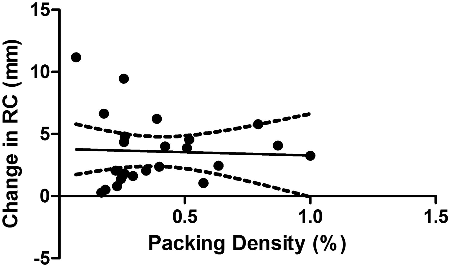
  • Fig 6.
    • Download figure
    • Open in new tab
    • Download powerpoint
    Fig 6.

    Linear regression analysis of the effect of packing attenuation on changes in the radius of the curvature of the stented segment (P > 0.05; R2 = 0.002).

Tables

  • Figures
  • Patient demographics and aneurysm information

    Demographics/Aneurysms
    Patients24
    Aneurysms24
    Females18 (75.0%)
    Mean age (yr)49.5
    Age range (yr)18–68
    Presentation/indications for treatment
        Headache/incidental15 (62.5%)
        Recurrent after coiling7 (29.2%)
        Stroke/transient ischemic attack2 (8.3%)
    Aneurysm dimensions
        Mean aneurysm dome size (SEM) (mm)5.6 (0.8)
        Mean aneurysm neck size (SEM) (mm)4.8 (0.7)
        Dome/neck ratio (SEM) (%)1.2 (0.09)
    Aneurysm location
        ACA2 (8.3%)
        AcomA3 (12.5%)
        ICA16 (67.7%)
        ICA terminus3 (12.5%)
    • Note:—SEM indicates standard error of the mean; AcomA, anterior communicating artery.

PreviousNext
Back to top

In this issue

American Journal of Neuroradiology: 33 (9)
American Journal of Neuroradiology
Vol. 33, Issue 9
1 Oct 2012
  • Table of Contents
  • Index by author
Advertisement
Print
Download PDF
Email Article

Thank you for your interest in spreading the word on American Journal of Neuroradiology.

NOTE: We only request your email address so that the person you are recommending the page to knows that you wanted them to see it, and that it is not junk mail. We do not capture any email address.

Enter multiple addresses on separate lines or separate them with commas.
The Effect of Intracranial Stent Implantation on the Curvature of the Cerebrovasculature
(Your Name) has sent you a message from American Journal of Neuroradiology
(Your Name) thought you would like to see the American Journal of Neuroradiology web site.
CAPTCHA
This question is for testing whether or not you are a human visitor and to prevent automated spam submissions.
Cite this article
R.M. King, J.-Y. Chueh, I.M.J. van der Bom, C.F. Silva, S.L. Carniato, G. Spilberg, A.K. Wakhloo, M.J. Gounis
The Effect of Intracranial Stent Implantation on the Curvature of the Cerebrovasculature
American Journal of Neuroradiology Oct 2012, 33 (9) 1657-1662; DOI: 10.3174/ajnr.A3062

Citation Manager Formats

  • BibTeX
  • Bookends
  • EasyBib
  • EndNote (tagged)
  • EndNote 8 (xml)
  • Medlars
  • Mendeley
  • Papers
  • RefWorks Tagged
  • Ref Manager
  • RIS
  • Zotero
0 Responses
Respond to this article
Share
Bookmark this article
The Effect of Intracranial Stent Implantation on the Curvature of the Cerebrovasculature
R.M. King, J.-Y. Chueh, I.M.J. van der Bom, C.F. Silva, S.L. Carniato, G. Spilberg, A.K. Wakhloo, M.J. Gounis
American Journal of Neuroradiology Oct 2012, 33 (9) 1657-1662; DOI: 10.3174/ajnr.A3062
del.icio.us logo Twitter logo Facebook logo Mendeley logo
  • Tweet Widget
  • Facebook Like
  • Google Plus One
Purchase

Jump to section

  • Article
    • Abstract
    • ABBREVIATIONS:
    • Materials and Methods
    • Results
    • Discussion
    • Conclusions
    • Footnotes
    • References
  • Figures & Data
  • Info & Metrics
  • Responses
  • References
  • PDF

Related Articles

  • No related articles found.
  • PubMed
  • Google Scholar

Cited By...

  • Parent Artery Straightening after Flow-Diverter Stenting Improves the Odds of Aneurysm Occlusion
  • Increased focal internal carotid artery angulation in patients with posterior communicating artery aneurysms
  • Selection of helical braided flow diverter stents based on hemodynamic performance and mechanical properties
  • Virtual-versus-Real Implantation of Flow Diverters: Clinical Potential and Influence of Vascular Geometry
  • High-Resolution C-Arm CT and Metal Artifact Reduction Software: A Novel Imaging Modality for Analyzing Aneurysms Treated with Stent-Assisted Coil Embolization
  • Effect of Structural Remodeling (Retraction and Recoil) of the Pipeline Embolization Device on Aneurysm Occlusion Rate
  • Crossref
  • Google Scholar

This article has not yet been cited by articles in journals that are participating in Crossref Cited-by Linking.

More in this TOC Section

  • A Retrospective Study in Tentorial DAVFs
  • Proximal Protection Devices for Carotid Stenting
  • Rescue Reentry in Carotid Near-Occlusion
Show more NEUROINTERVENTION

Similar Articles

Advertisement

Indexed Content

  • Current Issue
  • Accepted Manuscripts
  • Article Preview
  • Past Issues
  • Editorials
  • Editor's Choice
  • Fellows' Journal Club
  • Letters to the Editor
  • Video Articles

Cases

  • Case Collection
  • Archive - Case of the Week
  • Archive - Case of the Month
  • Archive - Classic Case

More from AJNR

  • Trainee Corner
  • Imaging Protocols
  • MRI Safety Corner
  • Book Reviews

Multimedia

  • AJNR Podcasts
  • AJNR Scantastics

Resources

  • Turnaround Time
  • Submit a Manuscript
  • Submit a Video Article
  • Submit an eLetter to the Editor/Response
  • Manuscript Submission Guidelines
  • Statistical Tips
  • Fast Publishing of Accepted Manuscripts
  • Graphical Abstract Preparation
  • Imaging Protocol Submission
  • Evidence-Based Medicine Level Guide
  • Publishing Checklists
  • Author Policies
  • Become a Reviewer/Academy of Reviewers
  • News and Updates

About Us

  • About AJNR
  • Editorial Board
  • Editorial Board Alumni
  • Alerts
  • Permissions
  • Not an AJNR Subscriber? Join Now
  • Advertise with Us
  • Librarian Resources
  • Feedback
  • Terms and Conditions
  • AJNR Editorial Board Alumni

American Society of Neuroradiology

  • Not an ASNR Member? Join Now

© 2025 by the American Society of Neuroradiology All rights, including for text and data mining, AI training, and similar technologies, are reserved.
Print ISSN: 0195-6108 Online ISSN: 1936-959X

Powered by HighWire