Advertisement

Main menu

  • Home
  • Content
    • Current Issue
    • Accepted Manuscripts
    • Article Preview
    • Past Issue Archive
    • AJNR Case Collection
    • Case of the Week Archive
    • Classic Case Archive
    • Case of the Month Archive
  • Special Collections
    • Spinal CSF Leak Articles (Jan 2020-June 2024)
    • 2024 AJNR Journal Awards
    • Most Impactful AJNR Articles
  • Multimedia
    • AJNR Podcast
    • AJNR Scantastics
    • Video Articles
  • For Authors
    • Submit a Manuscript
    • Author Policies
    • Fast publishing of Accepted Manuscripts
    • Graphical Abstract Preparation
    • Manuscript Submission Guidelines
    • Imaging Protocol Submission
    • Submit a Case for the Case Collection
  • About Us
    • About AJNR
    • Editorial Board
  • More
    • Become a Reviewer/Academy of Reviewers
    • Subscribers
    • Permissions
    • Alerts
    • Feedback
    • Advertisers
    • ASNR Home
  • Other Publications
    • ajnr

User menu

  • Alerts
  • Log in

Search

  • Advanced search
American Journal of Neuroradiology

American Journal of Neuroradiology

ASHNR American Society of Functional Neuroradiology ASHNR American Society of Pediatric Neuroradiology ASSR
  • Alerts
  • Log in

Advanced Search

  • Home
  • Content
    • Current Issue
    • Accepted Manuscripts
    • Article Preview
    • Past Issue Archive
    • AJNR Case Collection
    • Case of the Week Archive
    • Classic Case Archive
    • Case of the Month Archive
  • Special Collections
    • Spinal CSF Leak Articles (Jan 2020-June 2024)
    • 2024 AJNR Journal Awards
    • Most Impactful AJNR Articles
  • Multimedia
    • AJNR Podcast
    • AJNR Scantastics
    • Video Articles
  • For Authors
    • Submit a Manuscript
    • Author Policies
    • Fast publishing of Accepted Manuscripts
    • Graphical Abstract Preparation
    • Manuscript Submission Guidelines
    • Imaging Protocol Submission
    • Submit a Case for the Case Collection
  • About Us
    • About AJNR
    • Editorial Board
  • More
    • Become a Reviewer/Academy of Reviewers
    • Subscribers
    • Permissions
    • Alerts
    • Feedback
    • Advertisers
    • ASNR Home
  • Follow AJNR on Twitter
  • Visit AJNR on Facebook
  • Follow AJNR on Instagram
  • Join AJNR on LinkedIn
  • RSS Feeds

Welcome to the new AJNR, Updated Hall of Fame, and more. Read the full announcements.


AJNR is seeking candidates for the position of Associate Section Editor, AJNR Case Collection. Read the full announcement.


Research ArticleInterventional

Intracranial Aneurysm Neck Size Overestimation with 3D Rotational Angiography: The Impact on Intra-Aneurysmal Hemodynamics Simulated with Computational Fluid Dynamics

J.J. Schneiders, H.A. Marquering, L. Antiga, R. van den Berg, E. VanBavel and C.B. Majoie
American Journal of Neuroradiology January 2013, 34 (1) 121-128; DOI: https://doi.org/10.3174/ajnr.A3179
J.J. Schneiders
aFrom the Departments of Radiology (J.J.S., R.v.d.B., H.A.M., C.B.M.)
  • Find this author on Google Scholar
  • Find this author on PubMed
  • Search for this author on this site
H.A. Marquering
aFrom the Departments of Radiology (J.J.S., R.v.d.B., H.A.M., C.B.M.)
bBiomedical Engineering and Physics (H.A.M., E.v.B.), Academic Medical Center, University of Amsterdam, the Netherlands
  • Find this author on Google Scholar
  • Find this author on PubMed
  • Search for this author on this site
L. Antiga
cOrobix Srl. (L.A.), Bergamo, Italy.
  • Find this author on Google Scholar
  • Find this author on PubMed
  • Search for this author on this site
R. van den Berg
aFrom the Departments of Radiology (J.J.S., R.v.d.B., H.A.M., C.B.M.)
  • Find this author on Google Scholar
  • Find this author on PubMed
  • Search for this author on this site
E. VanBavel
bBiomedical Engineering and Physics (H.A.M., E.v.B.), Academic Medical Center, University of Amsterdam, the Netherlands
  • Find this author on Google Scholar
  • Find this author on PubMed
  • Search for this author on this site
C.B. Majoie
aFrom the Departments of Radiology (J.J.S., R.v.d.B., H.A.M., C.B.M.)
  • Find this author on Google Scholar
  • Find this author on PubMed
  • Search for this author on this site
  • Article
  • Figures & Data
  • Info & Metrics
  • Responses
  • References
  • PDF
Loading

Article Figures & Data

Figures

  • Tables
  • Fig 1.
    • Download figure
    • Open in new tab
    • Download powerpoint
    Fig 1.

    Comparison of the aneurysm neck as imaged by 3DRA (A) and 2D DSA (B). The 3DRA image suggests a wider neck than 2D DSA, which is expressed in the original 3DRA-derived vascular model (C). D, The vascular model after modification.
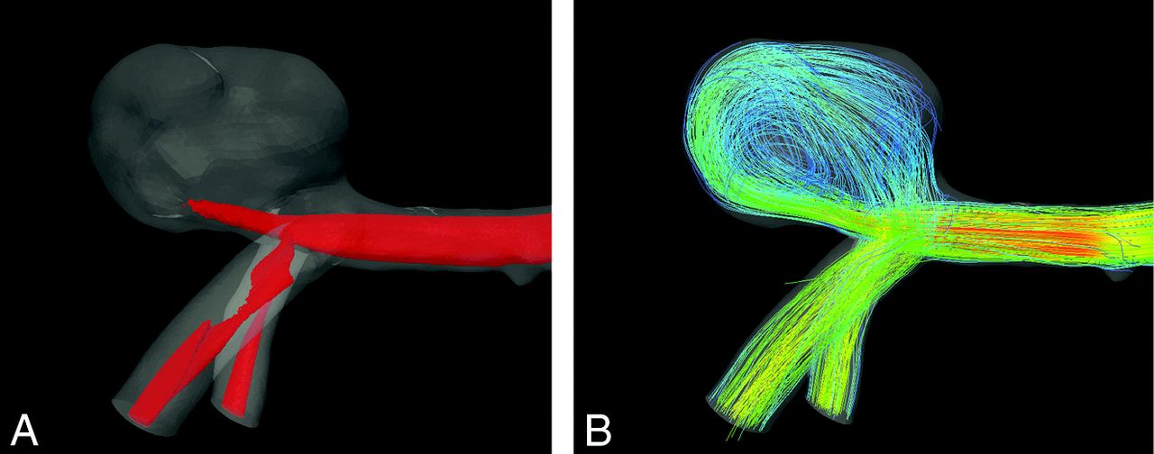
  • Fig 2.
    • Download figure
    • Open in new tab
    • Download powerpoint
    Fig 2.

    Illustration of the hemodynamic features displayed for comparison of case 7. A, Isosurface of the maximal 25% of the flow magnitude within the aneurysm as used in the assessment of the inflow jet. B, Streamlines of the flow used in the assessment of the main vortices.

  • Fig 3.
    • Download figure
    • Open in new tab
    • Download powerpoint
    Fig 3.

    Eight cases in which the 3DRA-derived vascular models were considered to disagree with 2D DSA. From left to right: the original vascular model; the working projection DSA; the modified 3DRA-derived vascular model.

  • Fig 4.
    • Download figure
    • Open in new tab
    • Download powerpoint
    Fig 4.

    Hemodynamic features in original (left) and modified vascular models (right). Case 1 shows a similar impingement zone and WSS distribution, flow velocity, and direction of the inflow jet. Case 2 shows an increase in maximal WSS, unchanged flow velocity, and direction of the inflow jet. Case 4 shows an increased maximal WSS, an increased impingement zone, an increased maximal flow velocity, and a change in inflow jet position. Case 6 shows a decrease in maximal WSS, a change in impingement zone, and a change in the maximal flow velocity and the position of the inflow jet.

  • Fig 5.
    • Download figure
    • Open in new tab
    • Download powerpoint
    Fig 5.

    Inflow jet in the original model (A) and modified model (B) showing a considerable difference in the inflow jet position of case 6. Both inflow jets are shown in superposition in image C.

  • Fig 6.
    • Download figure
    • Open in new tab
    • Download powerpoint
    Fig 6.

    A change in position of the impingement zone (IZ) between the original (A) and modified (B) vascular model after modification of case 5.

  • Fig 7.
    • Download figure
    • Open in new tab
    • Download powerpoint
    Fig 7.

    Schematic front view (left), bottom view (middle), and side view (right) of an idealized aneurysm and its parent artery. The front view would be chosen as the working projection. This figure shows that the parent artery is closer to the artery in the front view (area A) than perpendicular to the working projection (area B), suggesting that the blurring of the neck is more severe in this direction.

Tables

  • Figures
    • View popup
    Table 1:

    Demographic and clinical data of the 8 patients with aneurysm neck size overestimation in the 3DRA-derived vascular models

    CaseLocationAge/SexAneurysm Size (mm)Original Neck Diameter (mm)Absolute Neck Diameter Reduction (mm)Relative Neck Diameter Reduction
    1AcomA56/F5.95.240.7715%
    2AcomA27/M6.94.280.348%
    3BA53/F11.06.860.548%
    4MCA41/M4.52.860.8229%
    5PcomA57/F17.96.661.2018%
    6BA50/F6.03.861.1931%
    7MCA58/M12.86.790.7010%
    8PA39/F6.13.761.2032%
    Average488.95.040.8519%
    Minimum275.92.860.348%
    Maximum5817.96.861.2032%
    Standard deviation114.61.580.3210%
    • Note:—Aneurysm size is the largest aneurysm diameter. Neck diameters were measured in the working projection. AcomA indicates anterior communicating artery; BA, basilar artery; PA, pericallosal artery; PcomA: posterior communicating artery.

    • View popup
    Table 2:

    Consensus measurements of hemodynamic changes as a result of a correction of the 3DRA surface neck

    CaseInflow JetImpingement ZoneLow WSS ZoneVortices Change
    Change in PositionChange in SizeChange in PositionChange in SizeChange in PositionChange in Size
    1NoNarrower (2)NoSmaller (3)NoLarger (1)No
    2NoNarrower (2)NoSmaller (5)NoLarger (4)No
    3NoWider (2)Yes (3)Larger (3)Yes (4)NoNo
    4Yes (2)Wider (2)NoLarger (5)Yes (4)Smaller (5)No
    5NoNarrower (2)Yes (5)Smaller (5)NoLarger (5)No
    6Yes (2)Narrower (3)Yes (5)Larger (5)Yes (4)Larger (5)No
    7NoNarrower (5)NoSmaller (2)Yes (5)Larger (5)No
    8NoWider (1)NoSmaller (2)NoLarger (3)No
    • Note:—The significance scoring is given in brackets (1 = little significance; 5 = great significance).

    • View popup
    Table 3:

    Quantitative differences in hemodynamic patterns of the original and modified vascular models

    CaseRelative Increase in Maximal Inflow VelocityRelative Increase in Maximal WSSDistance Between Impingement Zones (mm)Region Classification of Impingement Zone (Original/Modified)
    1−2.7%+0.7%0.25Body/Body
    2−5.4%−17.7%0.27Neck/Neck
    32.6%+22.0%1.14Dome/Lobulation
    48.3%+98.0%0.46Body/Body
    5−0.3%+1.5%6.51Body/Dome
    69.3%−18.4%0.47Neck/Neck
    7−1.6%−4.5%0.27Neck/Neck
    83.8%+6.3%0.23Dome/Dome
    • View popup
    Table 4:

    Interobserver agreement on the scoring of the hemodynamic patterns

    κWeighted κ
    Inflow jet position0.63 (0.36)0.16 (0.11)
    Inflow jet sizeNA0.67 (0.22)
    Impingement zone position10.89 (0.06)
    Impingement zone size10.62 (0.12)
    Visible change in low WSS1NA
    Change in low WSS position0.67 (0.20)0.50 (0.16)
    Change in low WSS size10.63 (0.23)
    Number of vortices1NA
    • Note:—The weighted interobserver agreement is measured including the significance of a reported change in a hemodynamic feature. Standard error is given between parentheses. NA indicates not available.

PreviousNext
Back to top

In this issue

American Journal of Neuroradiology: 34 (1)
American Journal of Neuroradiology
Vol. 34, Issue 1
1 Jan 2013
  • Table of Contents
  • Index by author
Advertisement
Print
Download PDF
Email Article

Thank you for your interest in spreading the word on American Journal of Neuroradiology.

NOTE: We only request your email address so that the person you are recommending the page to knows that you wanted them to see it, and that it is not junk mail. We do not capture any email address.

Enter multiple addresses on separate lines or separate them with commas.
Intracranial Aneurysm Neck Size Overestimation with 3D Rotational Angiography: The Impact on Intra-Aneurysmal Hemodynamics Simulated with Computational Fluid Dynamics
(Your Name) has sent you a message from American Journal of Neuroradiology
(Your Name) thought you would like to see the American Journal of Neuroradiology web site.
CAPTCHA
This question is for testing whether or not you are a human visitor and to prevent automated spam submissions.
Cite this article
J.J. Schneiders, H.A. Marquering, L. Antiga, R. van den Berg, E. VanBavel, C.B. Majoie
Intracranial Aneurysm Neck Size Overestimation with 3D Rotational Angiography: The Impact on Intra-Aneurysmal Hemodynamics Simulated with Computational Fluid Dynamics
American Journal of Neuroradiology Jan 2013, 34 (1) 121-128; DOI: 10.3174/ajnr.A3179

Citation Manager Formats

  • BibTeX
  • Bookends
  • EasyBib
  • EndNote (tagged)
  • EndNote 8 (xml)
  • Medlars
  • Mendeley
  • Papers
  • RefWorks Tagged
  • Ref Manager
  • RIS
  • Zotero
0 Responses
Respond to this article
Share
Bookmark this article
Intracranial Aneurysm Neck Size Overestimation with 3D Rotational Angiography: The Impact on Intra-Aneurysmal Hemodynamics Simulated with Computational Fluid Dynamics
J.J. Schneiders, H.A. Marquering, L. Antiga, R. van den Berg, E. VanBavel, C.B. Majoie
American Journal of Neuroradiology Jan 2013, 34 (1) 121-128; DOI: 10.3174/ajnr.A3179
del.icio.us logo Twitter logo Facebook logo Mendeley logo
  • Tweet Widget
  • Facebook Like
  • Google Plus One
Purchase

Jump to section

  • Article
    • Abstract
    • ABBREVIATIONS:
    • Materials and Methods
    • Results
    • Discussion
    • Conclusions
    • Footnotes
    • References
  • Figures & Data
  • Info & Metrics
  • Responses
  • References
  • PDF

Related Articles

  • No related articles found.
  • PubMed
  • Google Scholar

Cited By...

  • Improving visualization of three-dimensional aneurysm features via segmentation with upsampled resolution and gradient enhancement (SURGE)
  • Geometric uncertainty in intracranial aneurysm rupture status discrimination: a two-site retrospective study
  • Comparing Morphology and Hemodynamics of Stable-versus-Growing and Grown Intracranial Aneurysms
  • Critical role of angiographic acquisition modality and reconstruction on morphometric and haemodynamic analysis of intracranial aneurysms
  • Does the DSA reconstruction kernel affect hemodynamic predictions in intracranial aneurysms? An analysis of geometry and blood flow variations
  • Better Than Nothing: A Rational Approach for Minimizing the Impact of Outflow Strategy on Cerebrovascular Simulations
  • Inflow Jet Patterns of Unruptured Cerebral Aneurysms Based on the Flow Velocity in the Parent Artery: Evaluation Using 4D Flow MRI
  • Additional Value of Intra-Aneurysmal Hemodynamics in Discriminating Ruptured versus Unruptured Intracranial Aneurysms
  • Hemodynamic Differences in Intracranial Aneurysms before and after Rupture
  • Rupture-Associated Changes of Cerebral Aneurysm Geometry: High-Resolution 3D Imaging before and after Rupture
  • Mind the Gap: Impact of Computational Fluid Dynamics Solution Strategy on Prediction of Intracranial Aneurysm Hemodynamics and Rupture Status Indicators
  • 3D Cine Phase-Contrast MRI at 3T in Intracranial Aneurysms Compared with Patient-Specific Computational Fluid Dynamics
  • Crossref (41)
  • Google Scholar

This article has been cited by the following articles in journals that are participating in Crossref Cited-by Linking.

  • Mind the Gap: Impact of Computational Fluid Dynamics Solution Strategy on Prediction of Intracranial Aneurysm Hemodynamics and Rupture Status Indicators
    K. Valen-Sendstad, D.A. Steinman
    American Journal of Neuroradiology 2014 35 3
  • CFD for Evaluation and Treatment Planning of Aneurysms: Review of Proposed Clinical Uses and Their Challenges
    Bongjae Chung, Juan Raul Cebral
    Annals of Biomedical Engineering 2015 43 1
  • Better Than Nothing: A Rational Approach for Minimizing the Impact of Outflow Strategy on Cerebrovascular Simulations
    C. Chnafa, O. Brina, V.M. Pereira, D.A. Steinman
    American Journal of Neuroradiology 2018 39 2
  • A review on the reliability of hemodynamic modeling in intracranial aneurysms: why computational fluid dynamics alone cannot solve the equation
    Philipp Berg, Sylvia Saalfeld, Samuel Voß, Oliver Beuing, Gábor Janiga
    Neurosurgical Focus 2019 47 1
  • How patient specific are patient-specific computational models of cerebral aneurysms? An overview of sources of error and variability
    David A. Steinman, Vitor M. Pereira
    Neurosurgical Focus 2019 47 1
  • Rupture-Associated Changes of Cerebral Aneurysm Geometry: High-Resolution 3D Imaging before and after Rupture
    J.J. Schneiders, H.A. Marquering, R. van den Berg, E. VanBavel, B. Velthuis, G.J.E. Rinkel, C.B. Majoie
    American Journal of Neuroradiology 2014 35 7
  • Ultrahigh-resolution CT scan of the temporal bone
    Koji Yamashita, Akio Hiwatashi, Osamu Togao, Kazufumi Kikuchi, Nozomu Matsumoto, Daichi Momosaka, Hiroshi Nakatake, Yuki Sakai, Hiroshi Honda
    European Archives of Oto-Rhino-Laryngology 2018 275 11
  • 3D Cine Phase-Contrast MRI at 3T in Intracranial Aneurysms Compared with Patient-Specific Computational Fluid Dynamics
    P. van Ooij, J.J. Schneiders, H.A. Marquering, C.B. Majoie, E. van Bavel, A.J. Nederveen
    American Journal of Neuroradiology 2013 34 9
  • Reproducibility of image-based computational models of intracranial aneurysm: a comparison between 3D rotational angiography, CT angiography and MR angiography
    Yuan Ren, Guo-Zhong Chen, Zhen Liu, Yan Cai, Guang-Ming Lu, Zhi-Yong Li
    BioMedical Engineering OnLine 2016 15 1
  • Implantation of 3D-Printed Patient-Specific Aneurysm Models into Cadaveric Specimens: A New Training Paradigm to Allow for Improvements in Cerebrovascular Surgery and Research
    Arnau Benet, Julio Plata-Bello, Adib A. Abla, Gabriel Acevedo-Bolton, David Saloner, Michael T. Lawton
    BioMed Research International 2015 2015

More in this TOC Section

  • SAVE vs. Solumbra Techniques for Thrombectomy
  • CT Perfusion&Reperfusion in Acute Ischemic Stroke
  • Delayed Reperfusion Post-Thrombectomy&Thrombolysis
Show more Interventional

Similar Articles

Advertisement

Indexed Content

  • Current Issue
  • Accepted Manuscripts
  • Article Preview
  • Past Issues
  • Editorials
  • Editors Choice
  • Fellow Journal Club
  • Letters to the Editor

Cases

  • Case Collection
  • Archive - Case of the Week
  • Archive - Case of the Month
  • Archive - Classic Case

Special Collections

  • Special Collections

Resources

  • News and Updates
  • Turn around Times
  • Submit a Manuscript
  • Author Policies
  • Manuscript Submission Guidelines
  • Evidence-Based Medicine Level Guide
  • Publishing Checklists
  • Graphical Abstract Preparation
  • Imaging Protocol Submission
  • Submit a Case
  • Become a Reviewer/Academy of Reviewers
  • Get Peer Review Credit from Publons

Multimedia

  • AJNR Podcast
  • AJNR SCANtastic
  • Video Articles

About Us

  • About AJNR
  • Editorial Board
  • Not an AJNR Subscriber? Join Now
  • Alerts
  • Feedback
  • Advertise with us
  • Librarian Resources
  • Permissions
  • Terms and Conditions

American Society of Neuroradiology

  • Not an ASNR Member? Join Now

© 2025 by the American Society of Neuroradiology All rights, including for text and data mining, AI training, and similar technologies, are reserved.
Print ISSN: 0195-6108 Online ISSN: 1936-959X

Powered by HighWire