Advertisement

Main menu

  • Home
  • Content
    • Current Issue
    • Accepted Manuscripts
    • Article Preview
    • Past Issue Archive
    • AJNR Case Collection
    • Case of the Week Archive
    • Classic Case Archive
    • Case of the Month Archive
  • Special Collections
    • Spinal CSF Leak Articles (Jan 2020-June 2024)
    • 2024 AJNR Journal Awards
    • Most Impactful AJNR Articles
  • Multimedia
    • AJNR Podcast
    • AJNR Scantastics
    • Video Articles
  • For Authors
    • Submit a Manuscript
    • Author Policies
    • Fast publishing of Accepted Manuscripts
    • Graphical Abstract Preparation
    • Manuscript Submission Guidelines
    • Imaging Protocol Submission
    • Submit a Case for the Case Collection
  • About Us
    • About AJNR
    • Editorial Board
  • More
    • Become a Reviewer/Academy of Reviewers
    • Subscribers
    • Permissions
    • Alerts
    • Feedback
    • Advertisers
    • ASNR Home
  • Other Publications
    • ajnr

User menu

  • Alerts
  • Log in

Search

  • Advanced search
American Journal of Neuroradiology

American Journal of Neuroradiology

ASHNR American Society of Functional Neuroradiology ASHNR American Society of Pediatric Neuroradiology ASSR
  • Alerts
  • Log in

Advanced Search

  • Home
  • Content
    • Current Issue
    • Accepted Manuscripts
    • Article Preview
    • Past Issue Archive
    • AJNR Case Collection
    • Case of the Week Archive
    • Classic Case Archive
    • Case of the Month Archive
  • Special Collections
    • Spinal CSF Leak Articles (Jan 2020-June 2024)
    • 2024 AJNR Journal Awards
    • Most Impactful AJNR Articles
  • Multimedia
    • AJNR Podcast
    • AJNR Scantastics
    • Video Articles
  • For Authors
    • Submit a Manuscript
    • Author Policies
    • Fast publishing of Accepted Manuscripts
    • Graphical Abstract Preparation
    • Manuscript Submission Guidelines
    • Imaging Protocol Submission
    • Submit a Case for the Case Collection
  • About Us
    • About AJNR
    • Editorial Board
  • More
    • Become a Reviewer/Academy of Reviewers
    • Subscribers
    • Permissions
    • Alerts
    • Feedback
    • Advertisers
    • ASNR Home
  • Follow AJNR on Twitter
  • Visit AJNR on Facebook
  • Follow AJNR on Instagram
  • Join AJNR on LinkedIn
  • RSS Feeds

Welcome to the new AJNR, Updated Hall of Fame, and more. Read the full announcements.


AJNR is seeking candidates for the position of Associate Section Editor, AJNR Case Collection. Read the full announcement.


Research ArticleBrain
Open Access

Intracranial 4D Flow MRI: Toward Individualized Assessment of Arteriovenous Malformation Hemodynamics and Treatment-Induced Changes

S.A. Ansari, S. Schnell, T. Carroll, P. Vakil, M.C. Hurley, C. Wu, J. Carr, B.R. Bendok, H. Batjer and M. Markl
American Journal of Neuroradiology October 2013, 34 (10) 1922-1928; DOI: https://doi.org/10.3174/ajnr.A3537
S.A. Ansari
aFrom the Department of Radiology (S.A.A., S.S., T.C., P.V., M.C.H., J.C., B.R.B., M.M.)
bDepartment of Neurological Surgery (S.A.A., M.C.H., B.R.B., H.B.), Feinberg School of Medicine
  • Find this author on Google Scholar
  • Find this author on PubMed
  • Search for this author on this site
S. Schnell
aFrom the Department of Radiology (S.A.A., S.S., T.C., P.V., M.C.H., J.C., B.R.B., M.M.)
  • Find this author on Google Scholar
  • Find this author on PubMed
  • Search for this author on this site
T. Carroll
aFrom the Department of Radiology (S.A.A., S.S., T.C., P.V., M.C.H., J.C., B.R.B., M.M.)
cDepartment of Biomedical Engineering (T.C., P.V., C.W., M.M.), McCormick School of Engineering, Northwestern University, Chicago, Illinois.
  • Find this author on Google Scholar
  • Find this author on PubMed
  • Search for this author on this site
P. Vakil
aFrom the Department of Radiology (S.A.A., S.S., T.C., P.V., M.C.H., J.C., B.R.B., M.M.)
cDepartment of Biomedical Engineering (T.C., P.V., C.W., M.M.), McCormick School of Engineering, Northwestern University, Chicago, Illinois.
  • Find this author on Google Scholar
  • Find this author on PubMed
  • Search for this author on this site
M.C. Hurley
aFrom the Department of Radiology (S.A.A., S.S., T.C., P.V., M.C.H., J.C., B.R.B., M.M.)
bDepartment of Neurological Surgery (S.A.A., M.C.H., B.R.B., H.B.), Feinberg School of Medicine
  • Find this author on Google Scholar
  • Find this author on PubMed
  • Search for this author on this site
C. Wu
cDepartment of Biomedical Engineering (T.C., P.V., C.W., M.M.), McCormick School of Engineering, Northwestern University, Chicago, Illinois.
  • Find this author on Google Scholar
  • Find this author on PubMed
  • Search for this author on this site
J. Carr
aFrom the Department of Radiology (S.A.A., S.S., T.C., P.V., M.C.H., J.C., B.R.B., M.M.)
  • Find this author on Google Scholar
  • Find this author on PubMed
  • Search for this author on this site
B.R. Bendok
aFrom the Department of Radiology (S.A.A., S.S., T.C., P.V., M.C.H., J.C., B.R.B., M.M.)
bDepartment of Neurological Surgery (S.A.A., M.C.H., B.R.B., H.B.), Feinberg School of Medicine
  • Find this author on Google Scholar
  • Find this author on PubMed
  • Search for this author on this site
H. Batjer
bDepartment of Neurological Surgery (S.A.A., M.C.H., B.R.B., H.B.), Feinberg School of Medicine
  • Find this author on Google Scholar
  • Find this author on PubMed
  • Search for this author on this site
M. Markl
aFrom the Department of Radiology (S.A.A., S.S., T.C., P.V., M.C.H., J.C., B.R.B., M.M.)
cDepartment of Biomedical Engineering (T.C., P.V., C.W., M.M.), McCormick School of Engineering, Northwestern University, Chicago, Illinois.
  • Find this author on Google Scholar
  • Find this author on PubMed
  • Search for this author on this site
  • Article
  • Figures & Data
  • Info & Metrics
  • Responses
  • References
  • PDF
Loading

Article Figures & Data

Figures

  • Tables
  • Fig 1.
    • Download figure
    • Open in new tab
    • Download powerpoint
    Fig 1.

    AVM hemodynamics visualized by time-integrated 3D pathlines in 6 of 20 patients. Color-coding shows the local velocity of blood traveling along the traces during 1 cardiac cycle. Different AVM sizes as well as feeding artery (solid white arrows) and draining vein patterns (white open arrows) can clearly be appreciated. AVM 4, 5, and 10: Deep AVM location and deep venous drainage resulting in with high SMG. AVM 17, 19, and 20: Superficial AVM location with superficial and deep venous drainage.
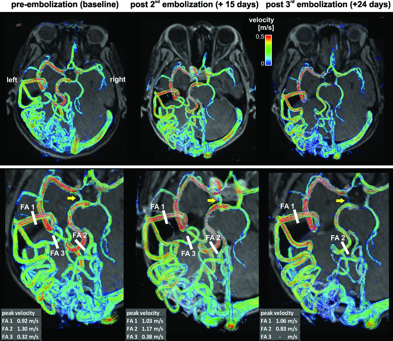
  • Fig 2.
    • Download figure
    • Open in new tab
    • Download powerpoint
    Fig 2.

    Three-dimensional blood flow visualization and quantification in a patient with a large unruptured temporo-occipital AVM (AVM 13, SMG = 4). Complex arterial feeding and convoluted hemodynamics as well as differences in pre-embolization and postembolization vascularization and hemodynamics are clearly visible. Flow quantification in 3 feeding arteries (FA 1–3) revealed substantial changes and redistribution of peak velocities during the course of staged embolization. Note that FA 3 was embolized during the third procedure. Note that embolization resulted in reduced flow in the right posterior communicating artery (yellow arrows). As evident from the changes in peak velocity in feeding arteries FA1 and FA2, the final embolization stage resulted in a substantial decrease in peak velocity in the FA2 systems. As a result, shunting to the FA2 system through the right posterior communicating artery was reduced, which resulted in its diminished appearance.
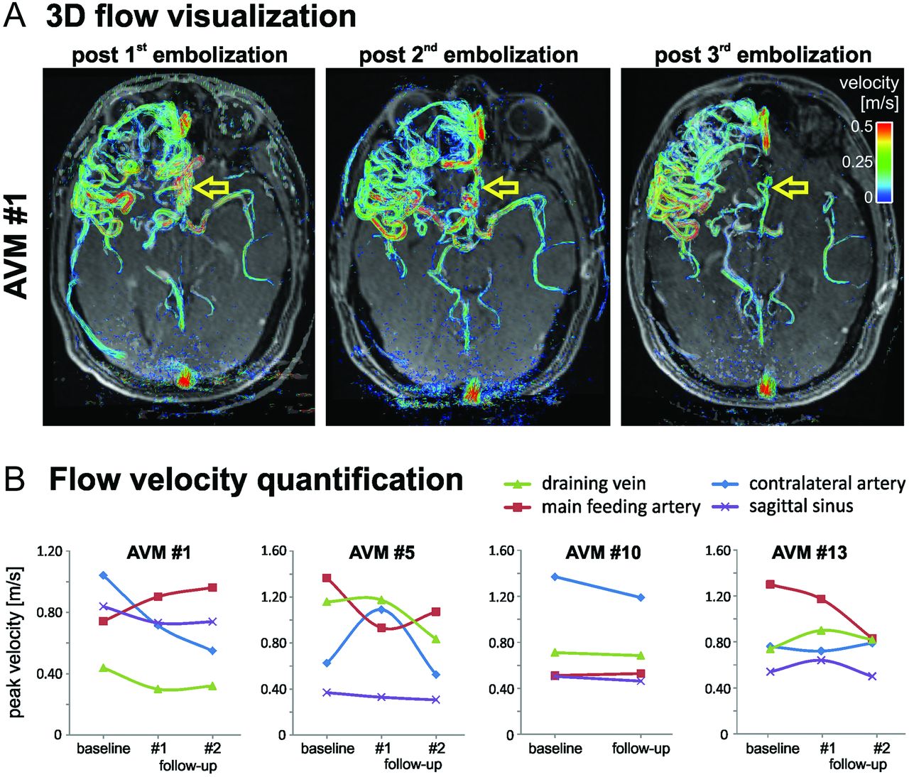
  • Fig 3.
    • Download figure
    • Open in new tab
    • Download powerpoint
    Fig 3.

    A, Three-dimensional blood flow in a large frontal AVM (AVM 1, SMG = 3). Staged embolization resulted in compaction of the AVM with reduced blood flow velocities (yellow arrows). B, Quantification of postinterventional changes in AVM hemodynamics on the basis of 4D flow MR imaging after multiple embolization procedures in all 4 patients who underwent MR imaging follow-up.

  • Fig 4.
    • Download figure
    • Open in new tab
    • Download powerpoint
    Fig 4.

    Flow connectivity mapping of AVM feeding and draining patterns in 2 large AVMs with complex vascularization (both SMG = 4). The gray shaded iso-surfaces show 3D-PC-MRA that was calculated from the 4D flow MR data. FA = feeding artery; DV = draining vein.

Tables

  • Figures
    • View popup
    Table 1:

    Demographics of 20 patients with AVM included in the study

    AVM No.Age, ySexHistoryVenous DrainageLocationAVM Size, cmSMG
    Deep/SuperficialEloquentSuperficial/Deep
    1a43Md+s0s5.6 × 4.1 × 5.13
    222MRupturedd+s0s2.5 × 2.3 × 2.42
    368Fd+s1d4.0 × 3.0 × 2.54
    444Md+s1d1.0 × 1.0 × 1.03
    5a29Md+s0d4.0 × 3.5 × 3.43
    648Fs0s3.5 × 3.5 × 3.02
    734MRuptureds0s3.3 × 2.3 × 2.51
    822Fs1s1.3 × 1.6 × 1.32
    940Fd+s0s3.0 × 2.0 × 4.02
    10a43MRupturedd+s1d2.7 × 3.1 × 3.13
    1135Fd+s0s3.1 × 1.6 × 1.02
    1225Fd+s0s2.3 × 2.3 × 1.72
    13a21Fd+s1s4.0 × 3.4 × 3.84
    1452Fs0s2.4 × 2.1 × 2.61
    1516Fs1d2.3 × 2.8 × 2.82
    1666Ms0s0.8 × 1.0 × 0.81
    1749Ms0s1.9 × 2.4 × 2.01
    1822Fs0s1.0 × 2.5 × 0.61
    1941Fd+s1s6.6 × 3.7 × 5.24
    2055Ms0s2.1 × 3.6 × 2.62
    • ↵a Patients for whom follow-up 4D flow MRI was obtained during staged embolization.

    • Note:—d indicates deep; s, superficial.

    • View popup
    Table 2:

    Summary of the results of visual flow pattern grading in patients with AVM (n = 18)

    SMG 1 (n = 4)SMG 2 (n = 7)SMG ≥3 (n = 7)
    Arterial feeders
    No. of arteriesSingleSingle and multiple (1–3)Single and multiple (1–4)
    Flow across arteriesHomogeneousHeterogeneous n = 4Heterogeneous n = 3
    Velocity grading1.7 ± 0.61.4 ± 0.51.5 ± 0.6
    Range = 1–2Range = 1–2Range = 0–2
    Median = 2Median = 1Median = 1
    Venous drainage
    No. of veinsSuperficial only (1–2)Superficial/deep (1–3)Superficial/deep (1–3)
    Flow across veinsHeterogeneous n = 2Heterogeneous n = 3Heterogeneous n = 4
    Velocity grading0.5 ± 0.60.8 ± 0.81.1 ± 0.6
    Range = 0–1Range = 0–2Range = 0–2
    Median = 0.5Median = 1Median = 1
PreviousNext
Back to top

In this issue

American Journal of Neuroradiology: 34 (10)
American Journal of Neuroradiology
Vol. 34, Issue 10
1 Oct 2013
  • Table of Contents
  • Index by author
  • Complete Issue (PDF)
Advertisement
Print
Download PDF
Email Article

Thank you for your interest in spreading the word on American Journal of Neuroradiology.

NOTE: We only request your email address so that the person you are recommending the page to knows that you wanted them to see it, and that it is not junk mail. We do not capture any email address.

Enter multiple addresses on separate lines or separate them with commas.
Intracranial 4D Flow MRI: Toward Individualized Assessment of Arteriovenous Malformation Hemodynamics and Treatment-Induced Changes
(Your Name) has sent you a message from American Journal of Neuroradiology
(Your Name) thought you would like to see the American Journal of Neuroradiology web site.
CAPTCHA
This question is for testing whether or not you are a human visitor and to prevent automated spam submissions.
Cite this article
S.A. Ansari, S. Schnell, T. Carroll, P. Vakil, M.C. Hurley, C. Wu, J. Carr, B.R. Bendok, H. Batjer, M. Markl
Intracranial 4D Flow MRI: Toward Individualized Assessment of Arteriovenous Malformation Hemodynamics and Treatment-Induced Changes
American Journal of Neuroradiology Oct 2013, 34 (10) 1922-1928; DOI: 10.3174/ajnr.A3537

Citation Manager Formats

  • BibTeX
  • Bookends
  • EasyBib
  • EndNote (tagged)
  • EndNote 8 (xml)
  • Medlars
  • Mendeley
  • Papers
  • RefWorks Tagged
  • Ref Manager
  • RIS
  • Zotero
0 Responses
Respond to this article
Share
Bookmark this article
Intracranial 4D Flow MRI: Toward Individualized Assessment of Arteriovenous Malformation Hemodynamics and Treatment-Induced Changes
S.A. Ansari, S. Schnell, T. Carroll, P. Vakil, M.C. Hurley, C. Wu, J. Carr, B.R. Bendok, H. Batjer, M. Markl
American Journal of Neuroradiology Oct 2013, 34 (10) 1922-1928; DOI: 10.3174/ajnr.A3537
del.icio.us logo Twitter logo Facebook logo Mendeley logo
  • Tweet Widget
  • Facebook Like
  • Google Plus One
Purchase

Jump to section

  • Article
    • Abstract
    • ABBREVIATIONS:
    • Materials and Methods
    • Results
    • Discussion
    • Conclusions
    • Footnotes
    • REFERENCES
  • Figures & Data
  • Info & Metrics
  • Responses
  • References
  • PDF

Related Articles

  • No related articles found.
  • PubMed
  • Google Scholar

Cited By...

  • Hemodynamic Analysis of Cerebral AVMs with 3D Phase-Contrast MR Imaging
  • In-room assessment of intravascular velocity from time-resolved rotational angiography in patients with arteriovenous malformation: a pilot study
  • Early Hemodynamic Response Assessment of Stereotactic Radiosurgery for a Cerebral Arteriovenous Malformation Using 4D Flow MRI
  • Validation of cerebral arteriovenous malformation hemodynamics assessed by DSA using quantitative magnetic resonance angiography: preliminary study
  • In Vivo Assessment of the Impact of Regional Intracranial Atherosclerotic Lesions on Brain Arterial 3D Hemodynamics
  • Three-dimensional printing of anatomically accurate, patient specific intracranial aneurysm models
  • Application of a Novel Brain Arteriovenous Malformation Endovascular Grading Scale for Transarterial Embolization
  • Fast Contrast-Enhanced 4D MRA and 4D Flow MRI Using Constrained Reconstruction (HYPRFlow): Potential Applications for Brain Arteriovenous Malformations
  • Evaluation of 4D Vascular Flow and Tissue Perfusion in Cerebral Arteriovenous Malformations: Influence of Spetzler-Martin Grade, Clinical Presentation, and AVM Risk Factors
  • Quantitative Assessment of Changes in Cerebral Arteriovenous Malformation Hemodynamics After Embolization
  • Value of 4D MR Angiography at 3T Compared with DSA for the Follow-Up of Treated Brain Arteriovenous Malformation
  • Hemodynamic Quantification in Brain Arteriovenous Malformations With Time-Resolved Spin-Labeled Magnetic Resonance Angiography
  • Crossref (92)
  • Google Scholar

This article has been cited by the following articles in journals that are participating in Crossref Cited-by Linking.

  • Phase-Contrast MRI: Physics, Techniques, and Clinical Applications
    David T. Wymer, Kunal P. Patel, William F. Burke, Vinay K. Bhatia
    RadioGraphics 2020 40 1
  • Advanced flow MRI: emerging techniques and applications
    M. Markl, S. Schnell, C. Wu, E. Bollache, K. Jarvis, A.J. Barker, J.D. Robinson, C.K. Rigsby
    Clinical Radiology 2016 71 8
  • Three-dimensional printing of anatomically accurate, patient specific intracranial aneurysm models
    Jeff R Anderson, Walker L Thompson, Abdulaziz K Alkattan, Orlando Diaz, Richard Klucznik, Yi J Zhang, Gavin W Britz, Robert G Grossman, Christof Karmonik
    Journal of NeuroInterventional Surgery 2016 8 5
  • Advances and Innovations in Brain Arteriovenous Malformation Surgery
    Bernard R. Bendok, Najib E. El Tecle, Tarek Y. El Ahmadieh, Antoun Koht, Thomas A. Gallagher, Timothy J. Carroll, Michael Markl, Randa Sabbagha, Asma Sabbagha, David Cella, Cindy Nowinski, Julius P.A. Dewald, Thomas J. Meade, Duke Samson, H. Hunt Batjer
    Neurosurgery 2014 74 Supplement 1
  • Four-dimensional MRI flow examinations in cerebral and extracerebral vessels – ready for clinical routine?
    Susanne Schnell, Can Wu, Sameer A. Ansari
    Current Opinion in Neurology 2016 29 4
  • Risk factors for hemorrhage of brain arteriovenous malformation
    Sonali S. Shaligram, Ethan Winkler, Daniel Cooke, Hua Su
    CNS Neuroscience & Therapeutics 2019 25 10
  • Influence of eddy current, Maxwell and gradient field corrections on 3D flow visualization of 3D CINE PC-MRI data
    Ramona Lorenz, Jelena Bock, Jeff Snyder, Jan G. Korvink, Bernd A. Jung, Michael Markl
    Magnetic Resonance in Medicine 2014 72 1
  • Quantitative Assessment of Changes in Cerebral Arteriovenous Malformation Hemodynamics After Embolization
    Ali Alaraj, Sepideh Amin-Hanjani, Sophia F. Shakur, Victor A. Aletich, Alexander Ivanov, Andrew P. Carlson, Gerald Oh, Fady T. Charbel
    Stroke 2015 46 4
  • Evaluation of 4D Vascular Flow and Tissue Perfusion in Cerebral Arteriovenous Malformations: Influence of Spetzler-Martin Grade, Clinical Presentation, and AVM Risk Factors
    C. Wu, S.A. Ansari, A.R. Honarmand, P. Vakil, M.C. Hurley, B.R. Bendok, J. Carr, T.J. Carroll, M. Markl
    American Journal of Neuroradiology 2015 36 6
  • In vivo analysis of physiological 3D blood flow of cerebral veins
    Florian Schuchardt, Laure Schroeder, Constantin Anastasopoulos, Michael Markl, Jochen Bäuerle, Anja Hennemuth, Johann Drexl, José M. Valdueza, Irina Mader, Andreas Harloff
    European Radiology 2015 25 8

More in this TOC Section

  • Qualitative and Quantitative Analysis of MR Imaging Findings in Patients with Middle Cerebral Artery Stroke Implanted with Mesenchymal Stem Cells
  • Multimodal CT Provides Improved Performance for Lacunar Infarct Detection
  • Statin Therapy Does Not Affect the Radiographic and Clinical Profile of Patients with TIA and Minor Stroke
Show more Brain

Similar Articles

Advertisement

Indexed Content

  • Current Issue
  • Accepted Manuscripts
  • Article Preview
  • Past Issues
  • Editorials
  • Editors Choice
  • Fellow Journal Club
  • Letters to the Editor

Cases

  • Case Collection
  • Archive - Case of the Week
  • Archive - Case of the Month
  • Archive - Classic Case

Special Collections

  • Special Collections

Resources

  • News and Updates
  • Turn around Times
  • Submit a Manuscript
  • Author Policies
  • Manuscript Submission Guidelines
  • Evidence-Based Medicine Level Guide
  • Publishing Checklists
  • Graphical Abstract Preparation
  • Imaging Protocol Submission
  • Submit a Case
  • Become a Reviewer/Academy of Reviewers
  • Get Peer Review Credit from Publons

Multimedia

  • AJNR Podcast
  • AJNR SCANtastic
  • Video Articles

About Us

  • About AJNR
  • Editorial Board
  • Not an AJNR Subscriber? Join Now
  • Alerts
  • Feedback
  • Advertise with us
  • Librarian Resources
  • Permissions
  • Terms and Conditions

American Society of Neuroradiology

  • Not an ASNR Member? Join Now

© 2025 by the American Society of Neuroradiology All rights, including for text and data mining, AI training, and similar technologies, are reserved.
Print ISSN: 0195-6108 Online ISSN: 1936-959X

Powered by HighWire