
Article Figures & Data
Figures
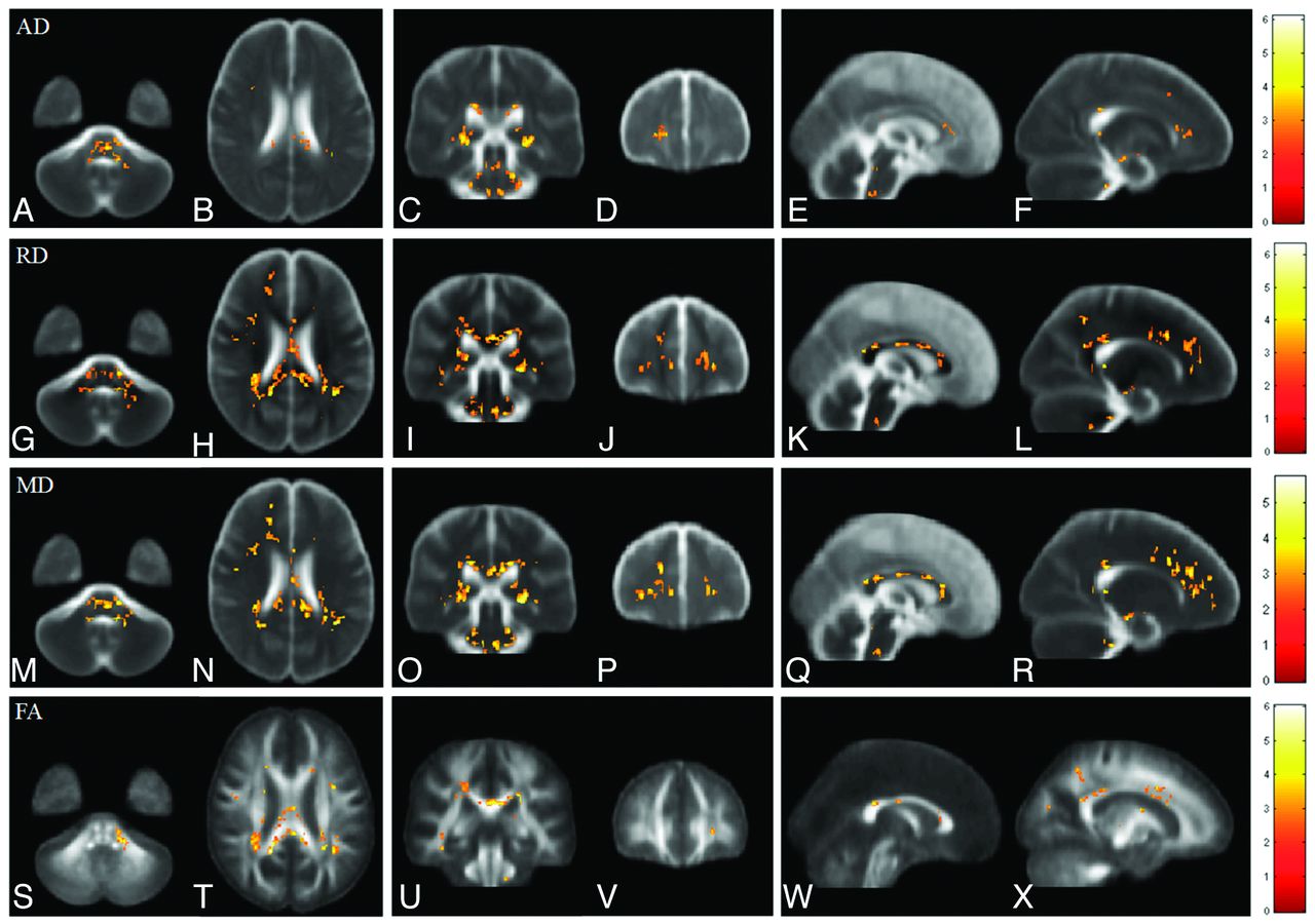
- Fig 1.
Voxelwise comparisons of AD, RD, MD, and FA between patients with ESRD and healthy subjects. Red-yellow colors indicate significant increases of AD (A–F), RD (G–L), and MD (M–R) and significant decreases of FA (S–X) in patients with ESRD. Color bars show the ranges of t-scores.
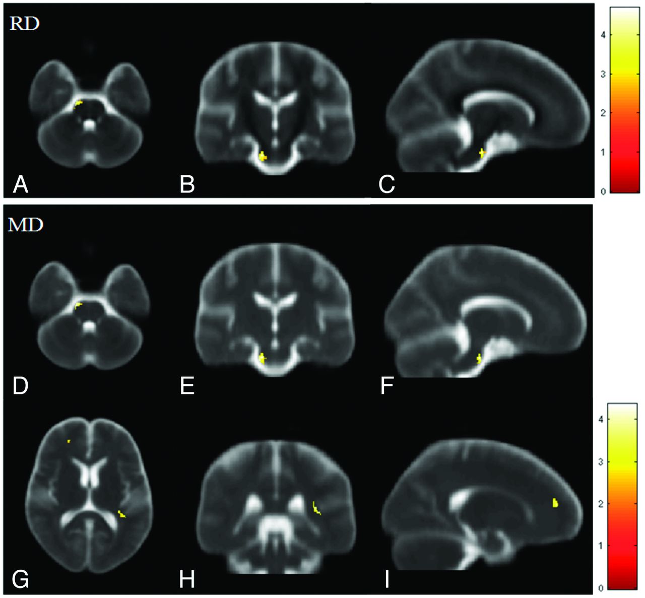
- Fig 2.
Voxelwise multiple regression analysis of DTI indices and the duration of hemodialysis in patients with ESRD. Red-yellow colors indicate the significant positive correlation in RD (A–C) and MD (D–I). Color bars show the ranges of t-scores.
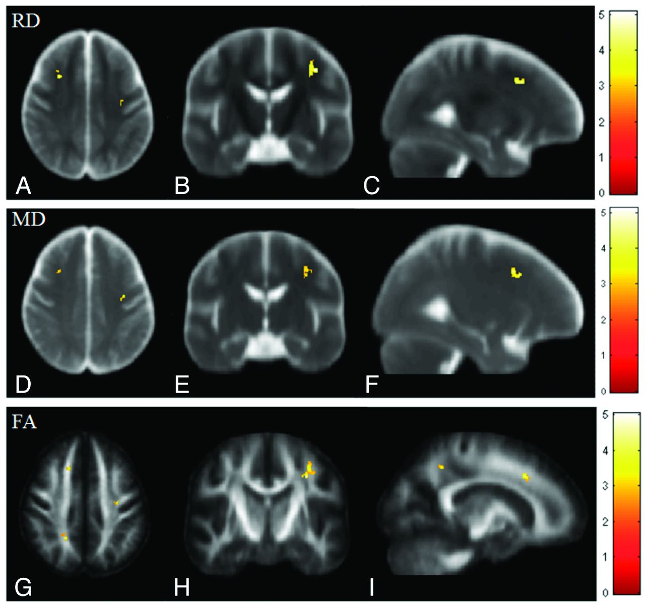
- Fig 3.
Voxelwise multiple regression analysis of DTI indices and CASI scores in patients with ESRD. Red-yellow colors indicate the significant negative correlations in RD (A–C) and MD (D–F) and a positive correlation in FA (G–I). Color bars show the ranges of t-scores.


Tables
- Table 2:
MNI coordinates of major areas with significant AD differences along with means of AD values
Brain Regions MNI Coordinate (mm) AD (mean) × 10−3 mm2/s X Y Z ESRD Healthy Rt RLIC 25 −16 −12 1.43 ± 0.17 1.39 ± 0.14 Lt RLIC −27 −14 −14 1.66 ± 0.21 1.57 ± 0.13 Lt Fminor −21 52 −12 1.43 ± 0.07 1.33 ± 0.07 Pons 1 −14 −56 1.85 ± 0.18 1.73 ± 0.11 Note:—Fminor indicates forceps minor; RLIC, retrolenticular part of the internal capsule; MNI, Montreal Neurological Institute; Rt, right; Lt, left.
- Table 3:
MNI coordinates of major areas with significant RD differences along with means of RD values
Brain Regions MNI Coordinate (mm) RD (mean) × 10−3 mm2/s X Y Z ESRD Healthy GCC 1 39 −1 0.94 ± 0.2 0.86 ± 0.14 SCC 1 −14 5 0.65 ± 0.15 0.56 ± 0.08 Rt. Fmajor 24 −54 −8 0.66 ± 0.11 0.62 ± 0.09 Lt. Fmajor −28 −42 −8 0.67 ± 0.09 0.61 ± 0.11 Rt. PCR 25 −23 7 0.64 ± 0.09 0.63 ± 0.04 Lt. PCR −28 −20 7 0.66 ± 0.09 0.65 ± 0.04 Rt. ACR 17 51 −6 0.64 ± 0.06 0.62 ± 0.05 Lt. ACR −20 56 −8 0.63 ± 0.06 0.59 ± 0.05 Pons 3 −13 −54 0.82 ± 0.19 0.71 ± 0.12 Note:—ACR indicates anterior corona radiata; Fmajor, forceps major; PCR, posterior corona radiata; SCC, splenium of the corpus callosum; MNI, Montreal Neurological Institute; GCC, genu of the corpus callosum; Rt, right; Lt, left.
- Table 4:
MNI coordinates of major areas with significant MD differences along with means of MD values
Brain Regions MNI Coordinate (mm) MD (mean) × 10−3 mm2/s X Y Z ESRD Healthy GCC 1 39 −8 1.16 ± 0.07 1.08 ± 0.05 SCC 1 −13 5 1.12 ± 0.13 1.02 ± 0.06 Rt. SS 31 −31 −9 1.27 ± 0.22 1.21 ± 0.15 Lt. SS −39 −24 −15 1.07 ± 0.12 1.04 ± 0.08 Rt. Fmajor 25 −42 −3 0.96 ± 0.11 0.89 ± 0.04 Lt. Fmajor −30 −34 −3 1.15 ± 0.11 0.99 ± 0.09 Rt. PCR 27 −25 4 1.07 ± 0.12 0.92 ± 0.06 Lt. PCR −29 −25 1 1.21 ± 0.18 1.11 ± 0.15 Pons 2 −14 −56 1.28 ± 0.13 1.13 ± 0.09 Note:—Fmajor indicates forceps major; PCR, posterior corona radiata; SCC, splenium of the corpus callosum; SS, sagittal stratum; MNI, Montreal Neurological Institute; GCC, genu of the corpus callosum; Rt, right; Lt, left.
- Table 5:
MNI coordinates of major areas with significant FA differences along with means of FA values
Brain Regions MNI Coordinate (mm) FA (mean) X Y Z ESRD Healthy GCC 3 40 −8 0.6 ± 0.06 0.67 ± 0.04 SCC 3 −14 5 0.65 ± 0.05 0.72 ± 0.05 Rt. SS 31 −28 −6 0.50 ± 0.04 0.56 ± 0.04 Lt. SS −27 −39 −6 0.46 ± 0.05 0.56 ± 0.07 Rt. MCP 13 −18 −58 0.45 ± 0.03 0.53 ± 0.04 Note:—MCP indicates middle cerebellar peduncle; SCC, splenium of the corpus callosum; SS, sagittal stratum; MNI, Montreal Neurological Institute; GCC, genu of the corpus callosum; Rt, right; Lt, left.
- Table 6:
MNI coordinates of areas with significant correlations along with their correlation coefficients
Brain Regions MNI Coordinate (mm) Correlation Coefficient X Y Z RD and duration Lt. Pons −16 −3 −46 0.7093 RD and CASI Lt. ACR −28 33 19 −0.7442 Rt. SCR 31 7 21 −0.6154 MD and duration Rt. RLIC 33 −20 −6 0.6009 Lt. FWM −21 63 −5 0.6287 Lt. Pons −12 −3 −46 0.6675 MD and CASI Lt. ACR −28 33 19 −0.7657 Rt. SCR 31 7 21 −0.5463 FA and CASI Lt. ACR −17 40 18 0.7067 Lt. PCR −20 −29 24 0.6117 Rt. SCR 33 6 26 0.6815 Note:—ACR indicates anterior corona radiata; PCR, posterior corona radiata; RLIC, retrolenticular part of the internal capsule; SCR, superior corona radiata; MNI, Montreal Neurological Institute; FWM, frontal white matter; Rt, right; Lt, left.


Jump to section
Related Articles
Cited By...
- Normalization of Cerebral Blood Flow, Neurochemicals, and White Matter Integrity after Kidney Transplantation
- Normalization of Cerebral Blood Flow, Neurochemicals, and White Matter Integrity After Kidney Transplantation
- Hemodialysis Induces an Acute Decline in Cerebral Blood Flow in Elderly Patients
- Relationship between Hypotension and Cerebral Ischemia during Hemodialysis
- Randomized Clinical Trial of Dialysate Cooling and Effects on Brain White Matter