
Article Figures & Data
Figures
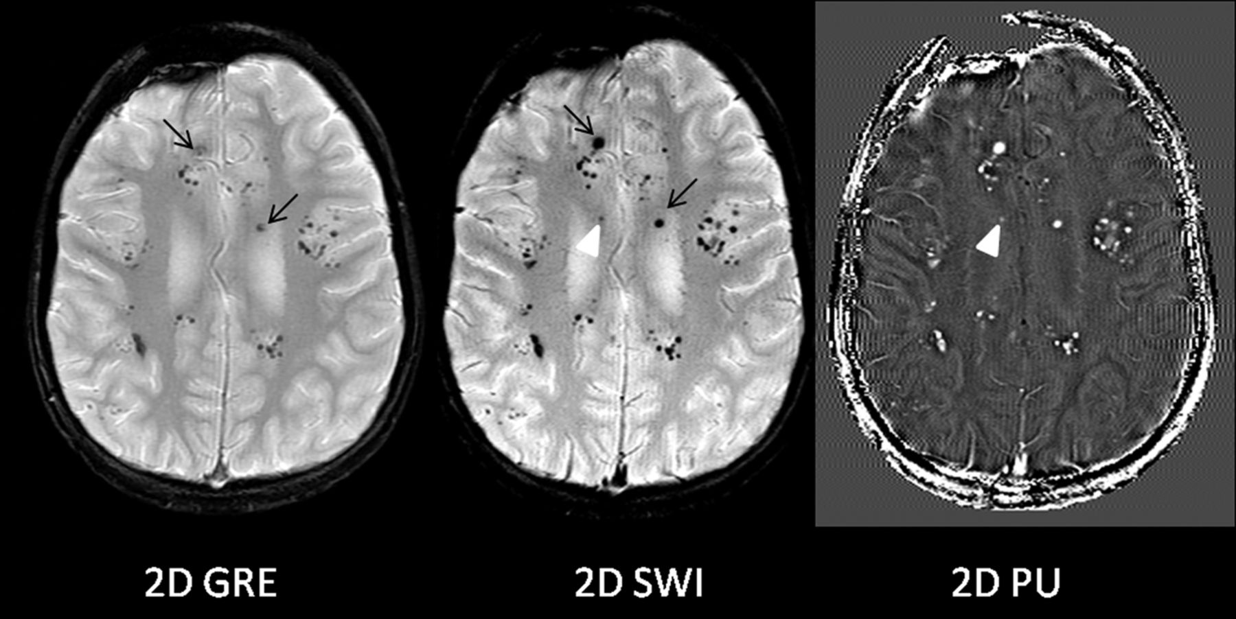
- Fig 1.
Improved T2* lesion conspicuity on 2D SWI compared with 2D GRE. Multiple T2* lesions are seen throughout the brain of a 14-year-old boy previously irradiated for medulloblastoma and presumed to have radiation-induced cavernous malformations/telangiectasias. Note improved visualization of these lesions (black arrows) on 2D SWI compared with corresponding lesions on 2D GRE (black arrows). Note a new T2* lesion on 2D SWI and PU images (arrowheads) not previously seen on 2D GRE.
- Fig 2.
Potential pitfalls of 2D SWI/PU. Focal venous thrombosis (black arrow) and an adjacent area suspicious for focal venous ischemia (black arrowhead) on DWI are identified in a 5-month-old boy with seizures. The T2* lesion corresponding to the thrombosis is well-visualized on both 2D GRE and 2D SWI (black arrows) and is, in fact, even more prominent on 2D SWI images. However, prominent adjacent calvarial bony artifacts (arrowhead) and heightened visualization of deoxygenated and slow-flow cortical veins on 2D SWI (white arrows) were thought to be distracting.
- Fig 3.
New T2* abnormality identified by 2D SWI. A, No T2* abnormality is seen on this baseline 2D GRE image in a 10-year-old boy with tectal glioma and associated ventriculomegaly. B, 2D SWI shows punctuate low-signal foci suggestive of T2* abnormality (arrow, arrowhead). C, This is shown as calcification/mineralization (arrows) by CT. D, 2D PU image shows a punctate low intensity (arrows) suggestive of calcification and foci of high intensity that may represent blood products (arrowhead) within the tumor.
- Fig 4.
Other comparison features of 2D GRE and 2D SWI/PU. A, Dental braces artifacts. Note increased artifacts on 2D SWI (arrow) and 2D PU images resulting from dental braces in a 15-year-old boy. B, Calcification versus hemorrhage. A T2* lesion (black arrows) is detected on both 2D GRE and 2D SWI in a 12-year-old boy previously treated for pineal germinoma. Corresponding 2D PU image shows dark signal (arrowhead), opposite of the signal expected for hemorrhage. An adjacent fluid-level lesion demonstrates high intensity typical of hemorrhage (white arrow). The presence of calcification is confirmed by prior CT. C, Catheter detail. The side holes of the catheter are better delineated on 2D SWI/PU images (arrows), particularly on the 2D PU image in an 8-year-old girl with periventricular cyst or encystment. While patency or functionality cannot be assessed by this method, improved catheter detail could be helpful when the position of the side hole relative to the cyst or ventricular system is clinically queried.
- Fig 5.
Sample case of 2D GRE, 2D SWI, and 3D SWI acquired in a 3-year-old boy with Sturge-Weber syndrome after surgical resection of the affected brain region. Axial images of the brain at the corona radiata (A) and basal ganglia (B) are shown. Note on 3D SWI, improved resolution and increased visualization of the venous structures, including prominent deep medullary venous drainage (white arrows) commonly seen in this condition. Note pulsation artifacts (small black arrow) associated with a large vessel (large black arrow) on 3D SWI. Longer scan time of 3D SWI (7 minutes, 40 seconds) compared with 2D SWI (1 minute, 50 seconds) has resulted in motion artifacts despite administration of general anesthesia in this child.


Jump to section
Related Articles
Cited By...
- Brain Injury Lesion Imaging Using Preconditioned Quantitative Susceptibility Mapping without Skull Stripping
- Diagnostic Performance of a 10-Minute Gadolinium-Enhanced Brain MRI Protocol Compared with the Standard Clinical Protocol for Detection of Intracranial Enhancing Lesions
- Iron and Non-Iron-Related Characteristics of Multiple Sclerosis and Neuromyelitis Optica Lesions at 7T MRI