
Article Figures & Data
Figures
- Fig 1.
Anterior oblique drawing (A) of a 5-week embryo showing the appearance of the nasal (olfactory) placode. B, Drawing shows the development of the medial and lateral nasal processes forming a downward-facing “horseshoe” around the sinking nasal placode, which forms the nasal pit.
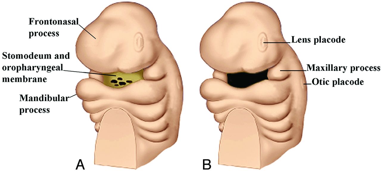
- Fig 2.
Anterior oblique drawing of an embryo in the late fourth week (A) shows the stomodeum with the oropharyngeal membrane surrounded by the further development of the frontonasal process and the maxillary and mandibular processes. B, Drawing shows the breakdown of the oropharyngeal membrane by the fifth week. The early appearance of the lens and otic placodes is also seen.
- Fig 3.
Drawing (A) of a ventral view of a 3- to 4-week embryo shows the anterior neuropore and the early formation of the maxillary processes. Drawing (B) in the late fourth week shows closure of the anterior neuropore and the location of the future frontonasal process. (Modified with permission from Netter's Atlas of Human Embryology. Edited by Cochard, L.R., PhD. 2002. Icon Learning Systems, Teterboro, New Jersey, Figures 9.5. Netter Illustrations from www.netterimages.com, © Elsevier Inc, All rights reserved).
- Fig 4.
Anterior oblique drawing of a 5-week embryo (A) shows the further growth of the medial and lateral nasal processes and the development of the nasal sac. The bucconasal groove is shown. B, Anterior oblique drawing of a 6-week embryo shows closure of bucconasal groove completing the floor of the nasal cavity and progressive flattening of the nasal sac openings, mainly as a result of ventrolateral growth of the medial nasal processes. The nasal sacs are also pushed toward the midline as the maxillary processes grow. Anterior oblique drawing of a 7-week embryo (C) and a 10-week (D) embryo shows the progressive medial movement of the nasal sacs and the resulting progressive pushing upwards of the frontonasal process.
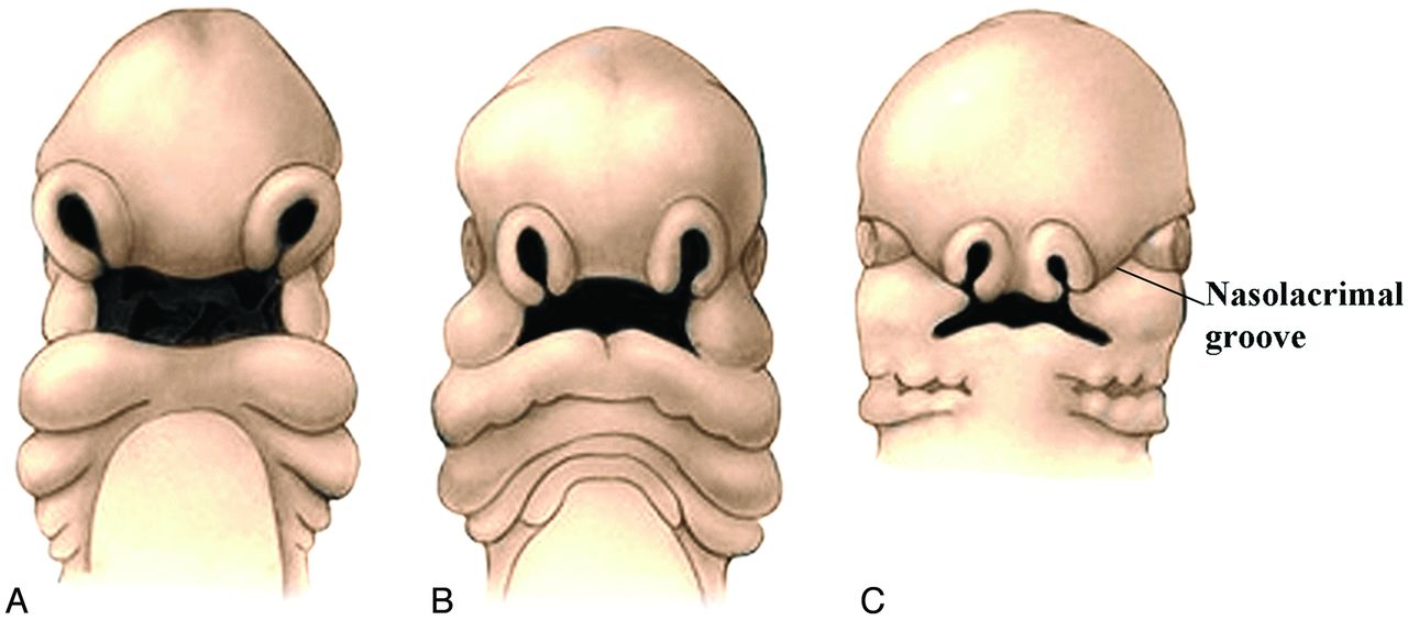
- Fig 5.
Frontal drawings of 4- to 5-week (A) and 5- to 6-week (B) embryos illustrate the progressive displacement of the nasal sacs toward the midline as a result of medial growth of the maxillary processes. Frontal view of a 6- to 7-week embryo (C) shows the nasolacrimal groove. Closure of this groove establishes continuity between the side of the nose formed by the lateral nasal process and the cheek formed by the maxillary process. (Modified with permission from Levine HL, Clemente MP, eds. Chapter 1, Surgical Anatomy of the Paranasal Sinus. China: 2005. Sinus Surgery Endoscopic and Microscopic Approaches. Figures 1–2. Thieme Medical Publishers Inc., Georg Thieme Verlag Stuttgart).
- Fig 6.
Drawing from below of a 6-week fetus showing the emergence of the intermaxillary segment below the medial nasal processes. This segment may actually arise from the globular processes of His. Also shown is the maxillary process and, arising from its medial surface, the lateral palatine process, which will form the secondary palate. (Modified with permission from Levine HL, Clemente MP, eds. Chapter 1, Surgical Anatomy of the Paranasal Sinus. China: 2005. Sinus Surgery Endoscopic and Microscopic Approaches. Figures 1–3. Thieme Medical Publishers Inc., Georg Thieme Verlag Stuttgart).
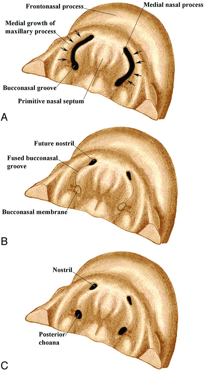
- Fig 7.
Drawings of the inferior view of an embryo from the sixth (A) to seventh weeks (C) show the bucconasal groove starting to close because of medial growth of the maxillary processes (A). B, There is fusion along most of the course of the bucconasal groove and membranous closure posteriorly by the bucconasal membrane. C, The oronasal membrane (thinned bucconasal membrane) has ruptured, creating an opening for communication between the primitive nasal and oral cavities (posterior choana).
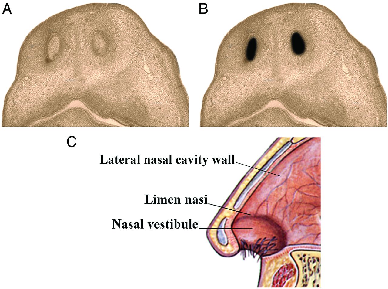
- Fig 8.
Drawings from below of a 7-week embryo (A) and a 13- to 15-week fetus (B) show epithelial plugs closing the nostrils (A) and then dissolving (B). If a plug does not dissolve, there will be atresia of the nostril. Lateral drawing of the anterior nose (C) shows the nasal vestibule and limen nasi. These areas mark the location of the nasal epithelial plugs.
- Fig 9.
Frontal drawings of the upper lip region in embryos at 5 (A), 6 (B) and 7 (C) weeks show the progressive medial growth of the maxillary processes toward the midline. However, the actual midline of the upper lip (the philtrum) is formed by the medial nasal processes. (Modified with permission from Levine HL, Clemente MP, eds. Chapter 1, Surgical Anatomy of the Paranasal Sinus. China: 2005. Sinus Surgery Endoscopic and Microscopic Approaches. Figures 1–3. Thieme Medical Publishers Inc., Georg Thieme Verlag Stuttgart).
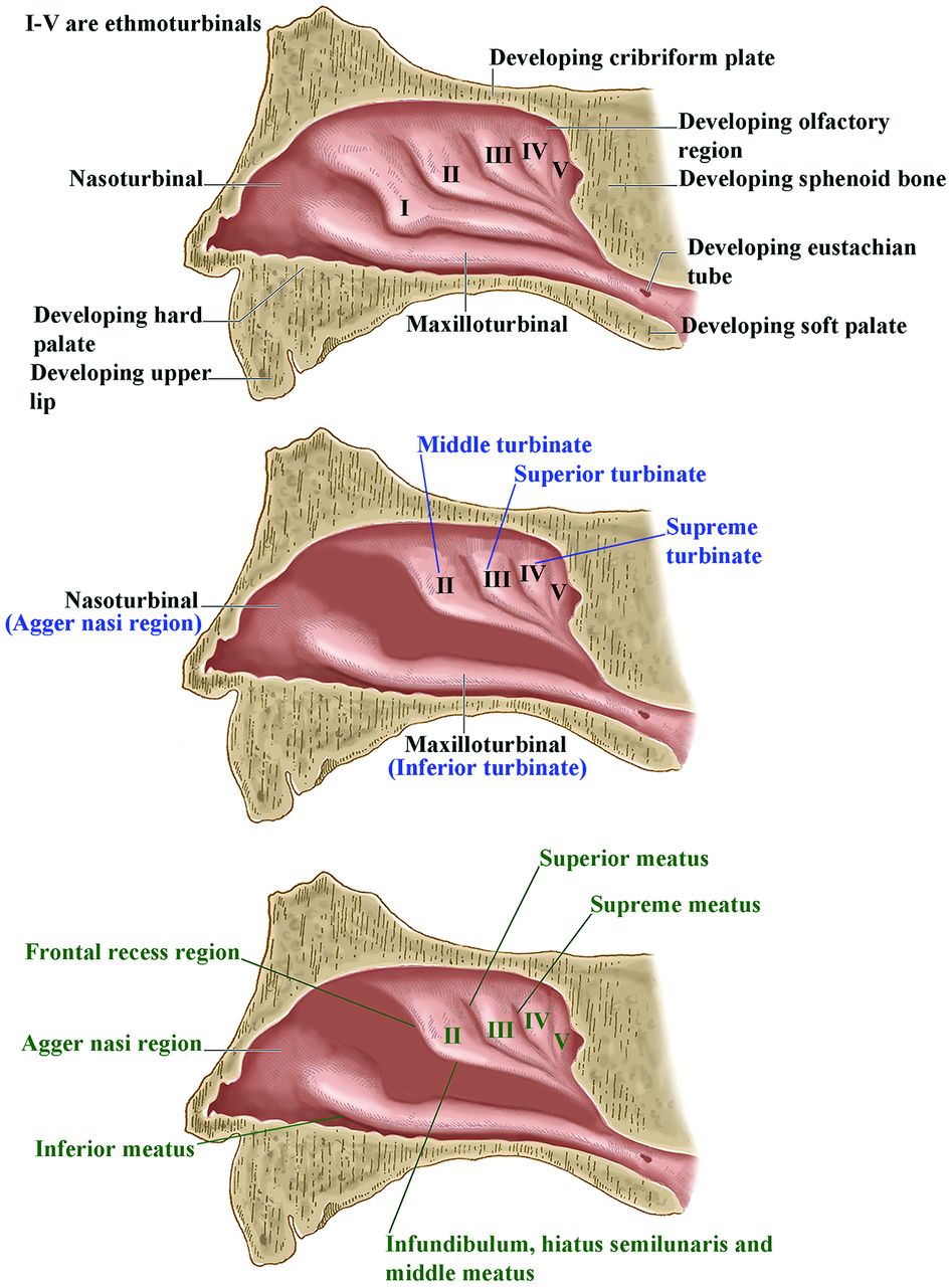
- Fig 10.
Lateral drawings of the developing lateral nasal wall showing the appearance of the nasal turbinals and their eventual development into the nasal turbinates and meati.


Tables
Summary of facial developmenta
Week Description Late 3rd to early 4th Anterior neuropore closes; oropharyngeal membrane appears Early 4th Frontonasal process appears; early development of maxillary and mandibular processes creates stomodeum Late 4th Oropharyngeal membrane disintegrates; nasal (olfactory) placodes appear Early 5th Lateral nasal processes and then the medial nasal processes appear, forming nasal pit; mandibular processes meet in midline; olfactory epithelium starts to form in upper nasal cavities Late 5th Nasal sacs form; medial nasal processes fuse with maxillary processes and form nasal fins, separating primitive nasal and oral cavities; nasal sacs migrate more medially and become more slit-like; olfactory nerves form; Meckel cartilage forms Early 6th Oronasal membrane forms and disintegrates opening posterior nasal choana; medial nasal processes start to form primitive nasal septum and primary palate Late 6th Lateral palatine processes develop; they are initially directed caudally alongside and above the tongue; naso-optic furrow develops and nasolacrimal duct forms (will become patent at birth); external ear develops; labiogingival laminae appear; lips and gums separate; dental lamina appears; maxilloturbinal (future inferior turbinate) starts to form; remaining nasal turbinals form; eyelids start to form Early 7th Philtrum and upper lip complete; nasal septum further develops; external olfactory epithelium confined to upper nasal cavities and further develops Late 7th External ear fully developed; nasal plugs close nostrils (will open again in 16th week); cartilaginous nasal capsule develops; palatal shelves elevate and fuse, forming secondary palate; future nostril now complete; eyes move more to midline; fusion of maxillary and mandibular processes narrows the width of the mouth and completes lower cheeks 8th Lateral nasal wall well-developed; facial muscle primordia appear (muscles develop by 9th week) 9th–10th Nasal septum starts to fuse with palate (will be complete by 12th week); ossification of maxillas occurs 10th–11th Uncinate process arises; infundibulum then develops; tooth buds become cup-shaped (teeth will be near complete in 6th month); eyelids fuse (will open in 26th–28th weeks) 12th Ossification centers of all facial bones are present ↵a The timeline of development differs slightly in the references quoted in Parts 1 and 2 of these articles. The timeline in this Table represents a compilation of the various dates in these references, and because some normal variation is to be expected, this Table represents the best averaging of any differences in developmental dates that we could make.


Related Articles
Cited By...
- Cell Cycle Arrest of a 'Zippering Epithelial Cell Cluster Shapes the Face and is Disrupted in Craniofacial Disorders
- Identification of novel genes regulating the development of the palate
- Cultured Mesenchymal Cells from Nasal Turbinate as a Cellular Model of the Neurodevelopmental Component of Schizophrenia Etiology
- Interfacial energy constraints are sufficient to align cells over large distances
- Investigating the shared genetics of non-syndromic cleft lip/palate and facial morphology