
Abstract
BACKGROUND AND PURPOSE: The anterior diskoligamentous complex is important for cervical spinal stability. Subjective widening of the disk space after trauma has been used to gauge disruption of the anterior diskoligamentous complex on CT scanning, but no quantitative CT measurements exist to evaluate injury. The purpose of our study was to evaluate if an increased intervertebral disk angle could serve as a more sensitive, reproducible indicator of disruption of the anterior diskoligamentous complex compared with subjective assessment.
MATERIALS AND METHODS: The intervertebral disk angle was retrospectively measured on CT scanning for 122 disk levels with disruption of the anterior diskoligamentous complex by MR imaging and 1095 disk levels with an intact anterior diskoligamentous complex by MR imaging. The intervertebral disk angle was measured between the anterior superior endplate and anterior inferior endplate, with angle apex at the midposterior disk. Area under the receiver operating characteristic curves for subjective disk widening and specific angle values were obtained. Intervertebral disk angle reproducibility was also evaluated.
RESULTS: Intervertebral disk angle measurements were “substantially reproducible.” No disk with an intact anterior diskoligamentous complex had an intervertebral disk angle greater than 18° or 2 standard deviations from the average intervertebral disk angle of the remaining disks. The area under the receiver operating characteristic curve for a criterion of subjective disk widening was 0.58. The area under the receiver operating characteristic curve for objective criteria, an intervertebral disk angle greater than 13 or above 1 standard deviation from normal values, was 0.85. The maximal area under the receiver operating characteristic curve was achieved if an intervertebral disk angle greater than 2 SD from the average angle of the other disks was used (0.86).
CONCLUSIONS: Subjective disk widening does not accurately detect disruption of the anterior diskoligamentous complex on CT scanning; an elevated intervertebral disk angle provides a more sensitive and objective measurement to help direct further imaging in trauma patients.
ABBREVIATIONS:
- ADL
- anterior diskoligamentous complex
- IDA
- intervertebral disk angle
- ALL
- anterior longitudinal ligament
- AUC
- area under the receiver operating characteristic curve
- ROC
- receiver operating characteristic
Acute cervical diskoligamentous injury is difficult to detect with standard trauma screening protocols, with the incidence of occult cervical diskoligamentous injury, in patients with persistent midline tenderness and a negative result on cervical spine CT scan, estimated to be as high as 44%.1 Timely diagnosis of these injuries is imperative, as the risk for neurologic sequelae is 10 times higher in patients with cervical injury missed on initial screening.2 The anterior diskoligamentous complex (ADL), composed of the anterior longitudinal ligament (ALL) and the intervertebral disk, is a key component of anterior cervical spine stability. Catastrophic injury to the ADL may result in cervical instability and acute disability, whereas subcatastrophic injury may lead to chronic pathologic conditions including disk degeneration, facet osteoarthritis, and chronic instability.3 Instability with ADL disruption may result in pain by compressing neural structures or muscle fatigue from increased reliance on the spinal musculature for stability.4 ADL injuries heal poorly, and a missed ADL injury is thought to contribute to chronic neck pain.5 Even in patients with other known cervical spine injuries, an unrecognized disk injury can be devastating, as potentially a higher number of columns may be injured. Identifying disruption of ligamentous integrity even in the setting of known fractures is critical, as knowledge of the extent of cervical column compromise is important in alerting clinicians to the potential for delayed instability.6
Although MR imaging is the standard method to analyze spinal soft tissue injuries, CT scan remains the first-line screening technique in cervical spine trauma, which relies heavily on vertebral body alignment to evaluate for ligamentous injury. Assessment of alignment is based on subjective evaluation of CT images because of a lack of clinically validated and reproducible criteria for alignment on CT scan. An overly sensitive assessment may lead to unnecessary MR imaging and its associated risk for transport in severely injured patients. Conversely, diminished sensitivity may result in missed injuries and the associated costs of both acute and delayed morbidity. Uniform measurements to detect ADL injury easily obtained on existing digitally based PACS systems could improve patient care by decreasing missed injuries and allowing for more judicious use of MR imaging.
The purpose of this study was to evaluate if an increased intervertebral disk angle (IDA) could serve as a more sensitive, reproducible indicator of ADL disruption compared with subjective assessment of disk space widening.
Materials and Methods
Patient Selection and Image Acquisition
Our institutional review board approved this study with waiver of informed consent. All CT and MR examinations included were performed during the clinical care of patients, and results were retrospectively reviewed.
We searched our enterprise-wide electronic medical record, encompassing 20 academic and community hospitals, to identify patients with ADL disruption on MR imaging examinations. We searched for radiology reports of MR imaging examinations performed from July 1, 2007, to May 1, 2012, on a PACS-capable search system, by using the following individual keywords: anterior longitudinal ligament, discoligamentous, ligament injury, ligament tear, and ligament disruption. MR images were reviewed in consensus by 2 fellowship-trained neuroradiologists (L.M.A., S.F.), without taking into account the initial diagnostic interpretation, to confirm the findings of ADL disruption.
Multiple prior studies have demonstrated the usefulness and accuracy of MR imaging of ligamentous injury in the cervical spine.7⇓⇓⇓⇓–12 For our study, the ADL was considered torn if a focal area of ALL/disk discontinuity could be identified on sagittal T2 or inversion recovery sequences. To further improve our specificity, we excluded patients if a focal point of ligament/disk discontinuity could not be definitively identified or if there was not agreement between the 2 neuroradiologists regarding the presence of ADL disruption. Prevertebral soft tissue swelling, ALL edema, or disk edema were not considered sufficient indicators of ADL disruption. Patients were also excluded if they did not have a comparison cervical spine CT examination performed within 7 days before the MR examination.
We identified control participants by searching the electronic medical record for cervical spine MR reports by using the keywords cervicalgia, stenosis, pain, and disk disease. Control participants were excluded if they had evidence of ADL disruption or edema on MR imaging, a history of trauma within the last 6 months, or no cervical spine CT examination performed within 1 month of the MR examination. Demographic data collected included age and sex. Clinical and imaging data collected included clinical history, initial CT findings/interpretation, reasons for MR imaging examination, level of ADL disruption, levels of disk degeneration, and final clinical management.
CT examinations were performed on 16- or 64-MDCT scanners (LightSpeed VCT; GE Healthcare, Milwaukee, Wisconsin). CT acquisitions were obtained from the infraorbital rim to the level of T1–T2 by use of the axial technique, 0.5 pitch, 1.2-mm collimation, 350 maximal mA, 120 kVp, and 18-cm FOV, in bone and standard algorithms, with 2.5-mm sagittal and coronal reconstructions. Patients were immobilized in a cervical collar during CT image acquisition.
MR imaging examinations were performed on 1.5T Optima 450W and 3T Discovery 750 systems (GE Healthcare) with neutral positioning by use of a standard spine coil. Sagittal sequences were obtained with 24-cm FOV and 256 × 192 matrix as follows: sagittal spin-echo T1-weighted (TR, 500 ms; TE, minimal; section thickness, 3 mm; NEX, 3), sagittal inversion recovery (TR, 9000–10000 ms; TE, 68 ms; section thickness, 3 mm; TI, 2200 ms), and sagittal gradient-echo (TR, 800 ms; TE, 25 ms; flip angle 20°; section thickness, 3 mm; NEX, 2). Additional axial 3D gradient-echo images (TR, 35 ms; TE, 13 ms; flip angle, 5°; section thickness, 2 mm; NEX, 1) were obtained with a 22-cm FOV and 256 × 192 matrix. Sagittal diffusion images (single-shot echo-planar; TR, 10,000 ms; TE, minimal; section thickness, 5 mm; matrix, 128 × 128) were also performed.
IDA Measurements
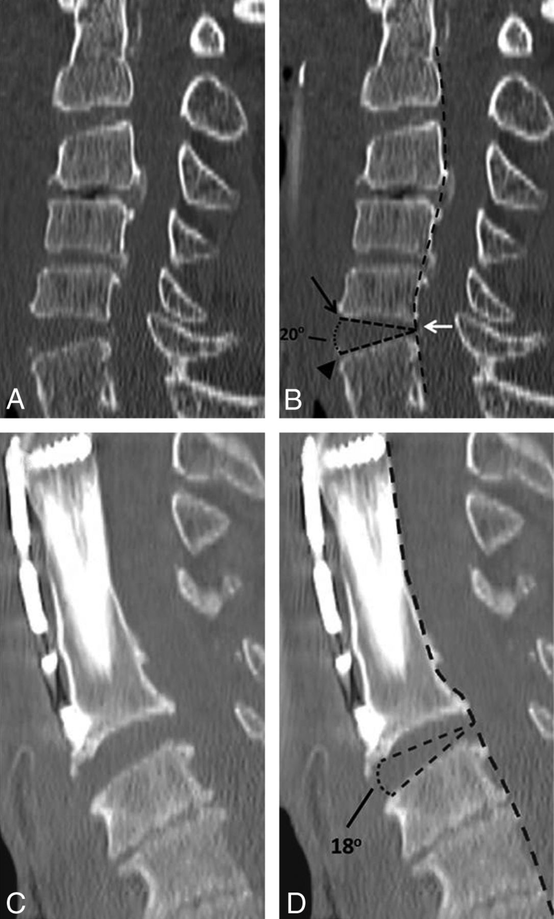
For measurement of the IDA, 3 points are first identified: 1) the first point at the midpoint of the disk space at its most posterior margin, 2) a second point at the anterior aspect of the endplate of the upper vertebral body, and 3) a third point at the anterior aspect of the endplate of the lower vertebral body. A line is drawn between the first and second points and the first and third points. The IDA is defined as the angle formed between these 2 lines.
IDA was measured on midline sagittal CT images by use of the angle measurement tool on our PACS. If there were anterior osteophytes, the angle excluded the osteophytes (Fig 1A,-B). Vertebral body distraction, or relative parallel configuration of vertebral body endplates, did not affect angle measurement, as the angle apex is placed at the middle of the distracted disk at the posterior vertebral body (Fig 1C,-D). IDA was measured for all disk spaces from C2–3 to C7–T1 in both patients with ADL disruption and in control participants.

A, Sagittal CT reconstruction of the cervical spine in a 65-year-old man with blunt trauma demonstrating (B) measurement of the IDA in a level with ADL disruption, measured between the anterior superior endplate (black arrow) and anterior inferior endplate (arrowhead), with the apex of the angle at the midpoint of the posterior disk (white arrow) at the posterior vertebral body margin (broken black line). C, Sagittal CT reconstruction of the cervical spine in an 82-year-old man with a history of fall demonstrating measurement of an IDA in the presence of anterior osteophytes and vertebral body distraction, resulting in parallel endplates, at a level with ADL disruption. D, The IDA measurement excludes the osteophytes and is still measured at the midpoint of the distracted posterior disk.
Reproducibility of the IDA was assessed by having 2 neuroradiologists independently measure the IDA for all disk spaces from C2–3 to C7–T1 in 17 randomly selected patients from both control and trauma groups (102 total disk spaces), with 10 of the tested disk spaces demonstrating ADL disruption on MR imaging. Radiologists were blinded to both each other's measurements and MR imaging findings. IDAs were then measured in all patients and control participants, in random order, at all levels from C2–3 to C7–T1 by 1 of the 2 neuroradiologists blinded to the clinical history and MR imaging findings.
Evaluation of subjective disk widening was performed with use of the prospective interpretation of the CT study by the initial interpreting radiologist. At our institution, cervical spine CT examinations are interpreted by fellowship-trained neuroradiologists and musculoskeletal radiologists. A true-positive finding for subjective disk widening was considered an initial interpretation of the CT scan, suggesting disk space widening at a level of injury seen on MR imaging.
Data Analysis
Confidence intervals for proportions used a continuity correction.13 IDA reproducibility was assessed by the Lin concordance correlation coefficient,14 interpreted according to McBride,15 that is, poor agreement (< 0.90), moderate agreement (0.90 to < 0.95), substantial agreement (0.95–0.99), and almost perfect agreement (> 0.99). Sensitivity and specificity for subjective disk widening and IDA measurements were obtained, with confidence intervals obtained without a continuity correction.13 Sensitivity and specificity for the various tests were compared with subjective assessment by use of the extended McNemar test with 2-tailed P values.16 Positive predictive value and negative predicative value of subjective assessment of disk widening, IDA measurements, and IDA variation were calculated. Receiver operating characteristic (ROC) curves were created for IDA measurements. Areas under the ROC curve (AUC) for subjective disk widening and specific angle values and angle variation were obtained17 and were interpreted according to Hosmer and Lemeshow,18 that is, no discrimination (AUC = 0.5), acceptable discrimination (0.7 ≤ AUC ≤ 0.8), excellent discrimination (0.8 ≤ AUC ≤ 0.9), and outstanding discrimination (AUC ≥ 0.9). Standard errors of the AUC values were calculated for each test.19 P values comparing the diagnostic performance of subjective disk widening and specific angle values were obtained by paired analysis.20 P values of < .05 were considered statistically significant.
Results
Patient Selection and Image Acquisition
A total of 139 cervical spine MR imaging studies with ADL disruption were evaluated based on our search of the electronic medical record. A total of 172 MR imaging studies of control patients were reviewed. Demographic data are summarized in Table 1.
Patient demographics
Among trauma patients, the most common history was either fall (41.7%; 95% CI, 32.2%–51.9%) or motor vehicle collision (33.0%; 95% CI, 24.3%–43.1%). Most of patients received an MR imaging examination for fractures on CT (62.1%; 95% CI, 52.0%–71.4%), whereas additional studies were obtained for persistent cervical pain (11.7%; 95% CI, 6.4%–19.8%) or continued neurologic deficit (12.6%; 95% CI, 7.2%–21.0%) in the absence of positive CT findings. MR imaging examinations in the remaining patients were obtained because of concern for ligamentous injury on the part of the radiologist interpreting the preceding CT study without evidence of bony fracture (11.7%; 95% CI, 6.4%–19.8%). Most patients required surgical stabilization for their injuries (53.4%, 95% CI, 43.3%–63.2%), whereas the remaining patients were either discharged from the hospital wearing a cervical collar (38.8%; 95% CI, 29.5%–49.0%) or died from either spinal or other injuries before treatment (5.8%; 95% CI, 2.4%–12.8%).
Measurements of IDA
The Lin correlation coefficient (ρc) for IDA measurements on test disk spaces, used to assess IDA reproducibility, was 0.9576 (95% CI, 0.9417–0.9692), indicating substantial agreement among observers. Concordance results are shown in Fig 2.

Concordance correlation coefficient plot measuring both precision and accuracy to determine how far the measured IDAs from the 2 different observers deviate from the line of perfect concordance (the line at 45° on a square scatterplot). The Lin coefficient increases in value as a function of the nearness of the data's reduced major axis to the line of perfect concordance (the accuracy of the data) and of the tightness of the data about its reduced major axis (the precision of the data).
Among trauma patients, 122 levels of ADL disruption were identified. IDAs for the 122 disk levels with ADL disruption as well as the remaining 487 uninjured disk levels were measured. Nine disk levels were excluded because of solid bony fusion. Among control patients, the IDAs for 608 uninjured disk spaces were evaluated, with 16 levels excluded for solid bony fusion. This provided 122 IDA measurements at levels with ADL disruption and 1095 IDA measurements at uninjured levels (487 IDA measurements at uninjured levels in trauma patients and 608 IDA measurements at uninjured levels in nontrauma patients) for analysis. Average uninjured IDA values for each disk level were calculated (Table 2). The distribution of angles for uninjured and injured disks is shown in Fig 3.
IDA measurements in 1095 uninjured disks

Distribution of disk angles among injured and uninjured disks, showing a Gaussian-like distribution of angles among uninjured disks, somewhat overlapping with angles in injured disks predominantly at 14° and below. Above an angle of 14°, there is minimal overlap.
Subjective disk widening had a low sensitivity (16.4%; 95% CI, 10.5%–24.3%) but a high specificity (99.4%; 95% CI, 98.0%–99.8%). The sensitivity increased with smaller IDA values as low as 13 (82.0%; 95% CI, 73.7%–88.1%) with a mild loss of specificity (89.1%, 95% CI, 87.1%–90.9%). When an angle of 12 was used, the sensitivity increased only marginally (85.2%; 95% CI, 77.4%–90.8%) with significant decrease in specificity (75.8%; 95% CI, 73.1%–78.2%). Maximal sensitivity was achieved by use of an IDA greater than 1 SD from normal values (86.1%; 95% CI, 78.3%–91.4%) or the average IDA of the remaining disks (86.1%; 95% CI, 74.7%–88.8%).
The highest diagnostic accuracy for a single IDA measurement was achieved with an IDA of 13 (0.884; 95% CI, 0.869–0.896). Using variation from normal values, we obtained the highest diagnostic accuracy by using an IDA greater than 1 SD from normal values (0.849; 95% CI, 0.834–0.859). The overall highest diagnostic accuracy was achieved by use of an IDA greater than 2 SDs from the remaining disks (0.972; 95% CI, 0.965–0.972). Diagnostic performance for subjective disk widening, an IDA of 13, as well as IDA measurement variations are compared in Table 3.
Comparison of diagnostic performance among subjective disk angle measurements and IDA measurements
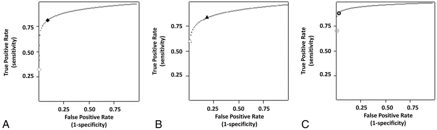
ROC curves for progressively smaller IDA measurements (Fig 4A) as well as for IDA variation (Fig 4B,-C) were obtained. The AUC for subjective widening of the disk was 0.58 (95% CI, 0.52–0.64), improving to 0.85 (95% CI, 0.81–0.90) if the criterion was an IDA of 13. Maximal AUC was achieved with an IDA greater than 2 SDs from the average IDA of the other disks (0.86; 95% CI, 0.82–0.90).

ROC curves for IDA measurements. A, ROC curve for progressively smaller IDAs, demonstrating marked increase in sensitivity with only minor loss in specificity as the angle is decreased from 20° (white diamond) to 13° (black diamond). B, ROC curve for deviation of the IDA from normal values, demonstrating an increase in sensitivity with only mild loss in specificity as the range is changed from 2 SDs from normal values (white triangle) to 1 SD (black triangle). C, ROC curve for deviation of IDA from the average IDA of the remaining disks also demonstrating an increase in sensitivity with only mild loss in specificity as the range is changed from 2 SDs from the average (white open circle) to 1 SD (black open circle).
Discussion
CT scanning has excellent usefulness in the evaluation of the bony integrity of the cervical spine; however, diskoligamentous injury, particularly in the absence of listhesis, can be difficult to detect. Although flexion/extension radiography is useful in the gauging of potential cervical spine instability, it can be difficult to perform in the setting of cervical spine trauma and has been shown to have low usefulness in the evaluation of potential diskoligamentous injury.21⇓⇓⇓–25 Normative data have been published regarding the upper limits of acceptable prevertebral soft tissue thickness on CT scan,26 and spinous process widening on plain film27; however, neither normative data regarding disk widening nor objective criteria for evaluation of potential ADL disruption have been published. Therefore, when evaluating the ADL on CT scan, radiologists are forced to rely on a subjective assessment of disk widening.
The purpose of our study was to evaluate the diagnostic performance of subjective disk space widening on CT scan for determination of ADL disruption and to evaluate if objective criteria based on the IDA can more accurately predict ADL disruption. Our results indicate that subjective disk space widening on CT scan is not adequate for evaluation of ADL disruption—achieving an AUC of only 0.58 (95% CI, 0.52–0.64) and a sensitivity of only 16.4% (95% CI, 10.5%–24.3%)—and that an elevated IDA provides a more objective, reproducible criterion to evaluate potential ADL disruption and guide further imaging.
Of all of the criteria evaluated for the detection of ADL disruption on CT scan, an IDA greater than 2 SDs from the average of the remaining disks offered the best diagnostic accuracy, 0.972 (95% CI, 0.965–0.972), and an AUC of 0.860 (95% CI, 0.817–0.903), with 72.1% sensitivity (95% CI, 63.2%–79.7%) and 100% specificity (95% CI, 99.6%–100%). However, we realize that a test requiring calculation of an SD is impractical for everyday use. Our results, however, also indicate that more practical tests relying on only a single angle measurement, easily performed on most PACS systems, can reliably predict ADL disruption.
Of 1095 total disks with an intact ADL evaluated (in both control patients and in those with ADL disruption at other levels), none had an IDA 18 or greater. We believe that an IDA of 18 or greater should always be considered abnormal and worthy of further evaluation with MR imaging. In our study, subjective assessment of disk widening on CT scan only detected ADL disruption when the IDA was 22 or greater; below this angle, subjective assessment failed to detect any of the abnormal levels. Considering an angle of 18 or greater as always being abnormal will improve detection of ADL disruption beyond subjective evaluation (P < .01).
Although an angle of 18 was always abnormal in our study, as a criterion for ADL disruption, it still failed to detect a significant number of abnormal levels (sensitivity, 41.8%; 95% CI, 33.0%–51.1%). An angle of 13 or greater had a similar AUC to an IDA 2 SDs from the average of the remaining disks (0.854; 95% CI, 0.832–0.876), with a sensitivity of 82.0% (95% CI, 73.7%–88.1%) and a specificity of 89.1% (95% CI, 87.1%–90.9%), with significantly greater ease of use. An IDA of 13 or greater may serve as a screening tool for ADL disruption, detecting significantly more injured levels than subjective widening alone (P < .01), with knowledge that this will result in some false-positive results.
IDA measurements were less sensitive in patients with cervical kyphosis, because an injured disk compresses with flexion, possibly even more easily than a normal disk, and an injured disk may decompress through the disrupted anterior margin. However, in our study, injured disks with normal or decreased IDA in kyphosis usually demonstrated some degree of listhesis related to the ADL injury in combination with other ligamentous or bony injury. Therefore, although the IDA measurement alone did not detect these injured levels, in clinical practice, these abnormal levels may be detected by the overall constellation of findings. Therefore, our study underestimates the overall sensitivity provided by the IDA measurement in clinical practice, as the IDA should never be used in isolation from other imaging or clinical findings suggestive of injury.
In our study, approximately one-quarter of patients with ADL disruption underwent MR imaging because of clinical criteria of persistent pain or neurologic deficit. In consideration of the significant number of trauma patients with possible cervical injury who are obtunded or are otherwise unable to cooperate with a full clinical examination, an objective, highly sensitive criterion for ADL disruption on CT scanning would be a vital tool to prevent missed injuries in obtunded patients unable to compensate for CT oversights by providing physical examination clues to their injury.
Furthermore, multiple studies have demonstrated that the force required to disrupt the ADL are similar to, if not greater than, the forces required to disrupt the posterior longitudinal ligament and ligamentum flavum.28,29 These findings suggest that patients with ADL disruption have received a significant blunt force, and if the force is sufficient to disrupt the ADL, they are at higher risk for additional ligamentous injury, making an MR imaging examination of these injuries even more important.
There were limitations to our study. First, as our study was retrospective, some images were evaluated by the same reader who performed the initial diagnostic evaluation. To eliminate potential recall bias, readers were blinded to patient information, and no study was re-evaluated by the same reader within 4 months of the initial reading. Second, we used MR imaging as our reference standard to identify ADL injuries. Although intraoperative correlation would have been ideal, most patients in our study who underwent intraoperative stabilization did not undergo an anterior fusion procedure, rendering direct operative correlation impossible. However, as prior studies have shown the high diagnostic accuracy of MR imaging in the detection of ALL and disk injury,7⇓⇓⇓⇓–12 we do not believe that this affects our overall conclusions. Furthermore, we attempted to imbue our reference standard with a high specificity by setting stringent imaging criteria for the diagnosis of ADL disruption and requiring concordance between 2 neuroradiologists. A further limitation of the IDA measurement was that although an elevated IDA is associated with ADL disruption, the reverse does not hold true; namely, a normal IDA does not exclude ADL disruption and should not be used to do so. Finally, although our data strongly indicate a useful role for the IDA in cervical spine trauma, further validation of this through a prospective trial would be ideal.
Conclusions
In trauma patients, subjective disk widening on CT scan does not accurately detect ADL disruption. An elevated IDA provides a more sensitive, objective, and reproducible measurement to help direct further evaluation with MR imaging. We recommend that an IDA greater then 13 warrants an MR imaging examination to exclude ADL disruption, noting that an IDA greater than 18 is always associated with ADL disruption.
REFERENCES
- Received December 18, 2012.
- Accepted after revision February 25, 2013.
- © 2013 by American Journal of Neuroradiology


Jump to section
Related Articles
Cited By...
- No citing articles found.