Advertisement

Main menu

  • Home
  • Content
    • Current Issue
    • Accepted Manuscripts
    • Article Preview
    • Past Issue Archive
    • AJNR Case Collection
    • Case of the Week Archive
    • Classic Case Archive
    • Case of the Month Archive
  • Special Collections
    • Spinal CSF Leak Articles (Jan 2020-June 2024)
    • 2024 AJNR Journal Awards
    • Most Impactful AJNR Articles
  • Multimedia
    • AJNR Podcast
    • AJNR Scantastics
    • Video Articles
  • For Authors
    • Submit a Manuscript
    • Author Policies
    • Fast publishing of Accepted Manuscripts
    • Graphical Abstract Preparation
    • Manuscript Submission Guidelines
    • Imaging Protocol Submission
    • Submit a Case for the Case Collection
  • About Us
    • About AJNR
    • Editorial Board
  • More
    • Become a Reviewer/Academy of Reviewers
    • Subscribers
    • Permissions
    • Alerts
    • Feedback
    • Advertisers
    • ASNR Home
  • Other Publications
    • ajnr

User menu

  • Alerts
  • Log in

Search

  • Advanced search
American Journal of Neuroradiology

American Journal of Neuroradiology

ASHNR American Society of Functional Neuroradiology ASHNR American Society of Pediatric Neuroradiology ASSR
  • Alerts
  • Log in

Advanced Search

  • Home
  • Content
    • Current Issue
    • Accepted Manuscripts
    • Article Preview
    • Past Issue Archive
    • AJNR Case Collection
    • Case of the Week Archive
    • Classic Case Archive
    • Case of the Month Archive
  • Special Collections
    • Spinal CSF Leak Articles (Jan 2020-June 2024)
    • 2024 AJNR Journal Awards
    • Most Impactful AJNR Articles
  • Multimedia
    • AJNR Podcast
    • AJNR Scantastics
    • Video Articles
  • For Authors
    • Submit a Manuscript
    • Author Policies
    • Fast publishing of Accepted Manuscripts
    • Graphical Abstract Preparation
    • Manuscript Submission Guidelines
    • Imaging Protocol Submission
    • Submit a Case for the Case Collection
  • About Us
    • About AJNR
    • Editorial Board
  • More
    • Become a Reviewer/Academy of Reviewers
    • Subscribers
    • Permissions
    • Alerts
    • Feedback
    • Advertisers
    • ASNR Home
  • Follow AJNR on Twitter
  • Visit AJNR on Facebook
  • Follow AJNR on Instagram
  • Join AJNR on LinkedIn
  • RSS Feeds

Welcome to the new AJNR, Updated Hall of Fame, and more. Read the full announcements.


AJNR is seeking candidates for the position of Associate Section Editor, AJNR Case Collection. Read the full announcement.


Research ArticlePediatrics

MR Spectroscopy of the Fetal Brain: Is It Possible without Sedation?

V. Berger-Kulemann, P.C. Brugger, D. Pugash, M. Krssak, M. Weber, A. Wielandner and D. Prayer
American Journal of Neuroradiology February 2013, 34 (2) 424-431; DOI: https://doi.org/10.3174/ajnr.A3196
V. Berger-Kulemann
aFrom the Departments of Radiology (V.B.-K., M.K., M.W., A.W., D.P.)
  • Find this author on Google Scholar
  • Find this author on PubMed
  • Search for this author on this site
P.C. Brugger
bAnatomy and Cell Biology (P.C.B.), Medical University of Vienna, Vienna, Austria
  • Find this author on Google Scholar
  • Find this author on PubMed
  • Search for this author on this site
D. Pugash
aFrom the Departments of Radiology (V.B.-K., M.K., M.W., A.W., D.P.)
cDepartment of Gynecology and Obstetrics (D.P.), Medical University of British Columbia, BC Women's Hospital, Vancouver, British Columbia, Canada.
  • Find this author on Google Scholar
  • Find this author on PubMed
  • Search for this author on this site
M. Krssak
aFrom the Departments of Radiology (V.B.-K., M.K., M.W., A.W., D.P.)
  • Find this author on Google Scholar
  • Find this author on PubMed
  • Search for this author on this site
M. Weber
aFrom the Departments of Radiology (V.B.-K., M.K., M.W., A.W., D.P.)
  • Find this author on Google Scholar
  • Find this author on PubMed
  • Search for this author on this site
A. Wielandner
aFrom the Departments of Radiology (V.B.-K., M.K., M.W., A.W., D.P.)
  • Find this author on Google Scholar
  • Find this author on PubMed
  • Search for this author on this site
D. Prayer
aFrom the Departments of Radiology (V.B.-K., M.K., M.W., A.W., D.P.)
  • Find this author on Google Scholar
  • Find this author on PubMed
  • Search for this author on this site
  • Article
  • Figures & Data
  • Info & Metrics
  • Responses
  • References
  • PDF
Loading

Article Figures & Data

Figures

  • Tables
  • Fig 1.
    • Download figure
    • Open in new tab
    • Download powerpoint
    Fig 1.

    GW 29 + 6, single-voxel MR spectroscopy of the brain. PRESS with a long TE (144 ms). VOI 2.7 cm3. Cephalic presentation. Normal age-related spectrum.

  • Fig 2.
    • Download figure
    • Open in new tab
    • Download powerpoint
    Fig 2.

    GW 31 + 3, single-voxel MR spectroscopy of the brain. PRESS with a short TE (35 ms). VOI 2.7 cm3. Cephalic presentation. Normal age-related spectrum.
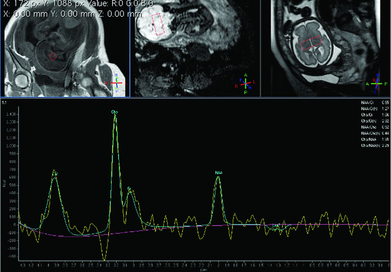
  • Fig 3.
    • Download figure
    • Open in new tab
    • Download powerpoint
    Fig 3.

    GW 24 + 0, single-voxel MR spectroscopy of the brain. PRESS with a long TE (144 ms). VOI 2.7 cm3. Cephalic presentation. Distinct peak in the lactate region in a fetus with a brain tumor.
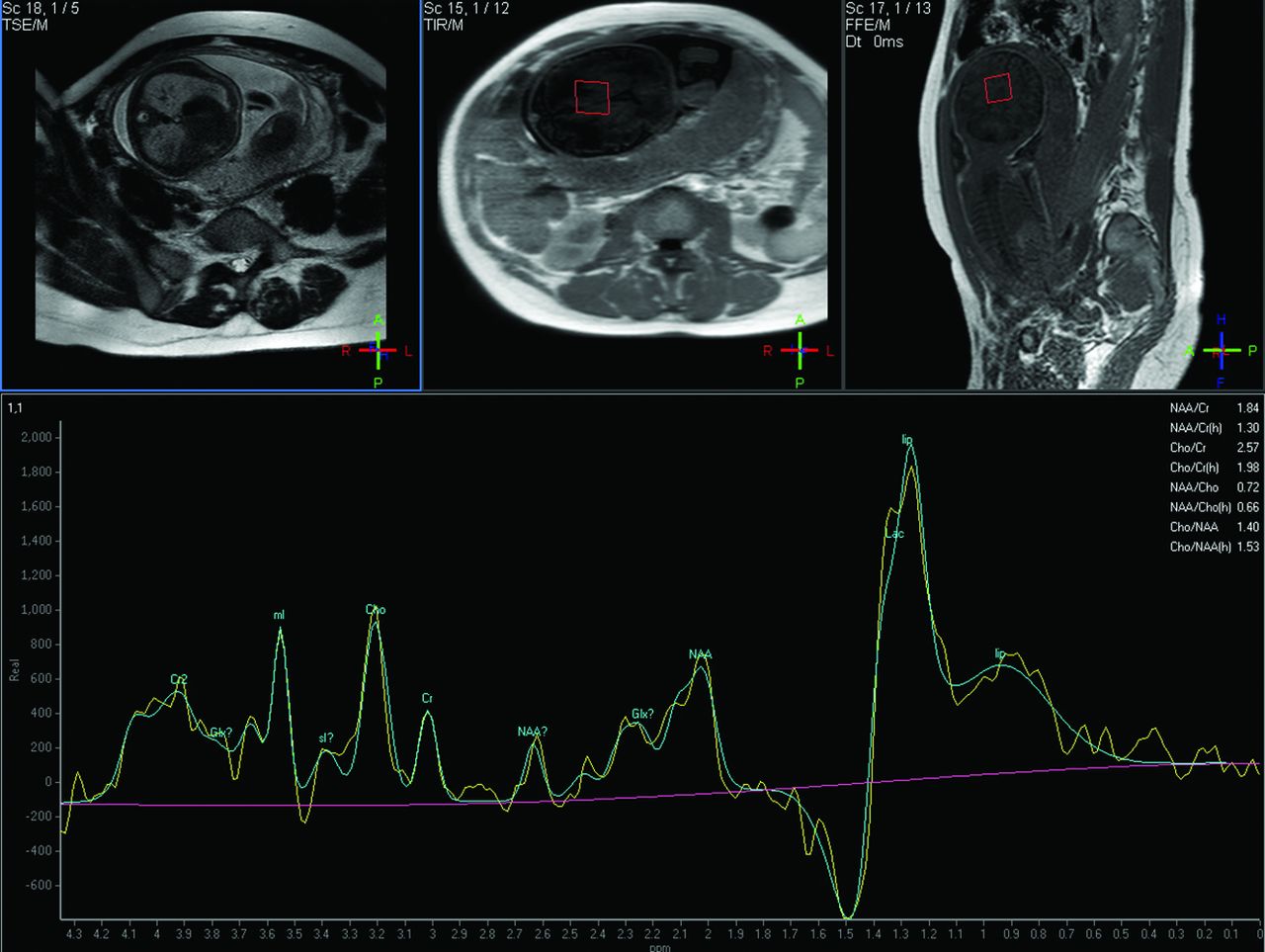
  • Fig 4.
    • Download figure
    • Open in new tab
    • Download powerpoint
    Fig 4.

    GW 31 + 3, single-voxel MR spectroscopy of the brain. PRESS with a short TE (35 ms). VOI 2.7 cm3. Breech presentation. Peak in the lipid/Lac region. No fetal pathology was found. In this case, a premature rupture of the membranes was present and indications of maternal infection, which confirmed the decision to perform a cesarean delivery. Notice the reduced amniotic fluid on the scout scans.
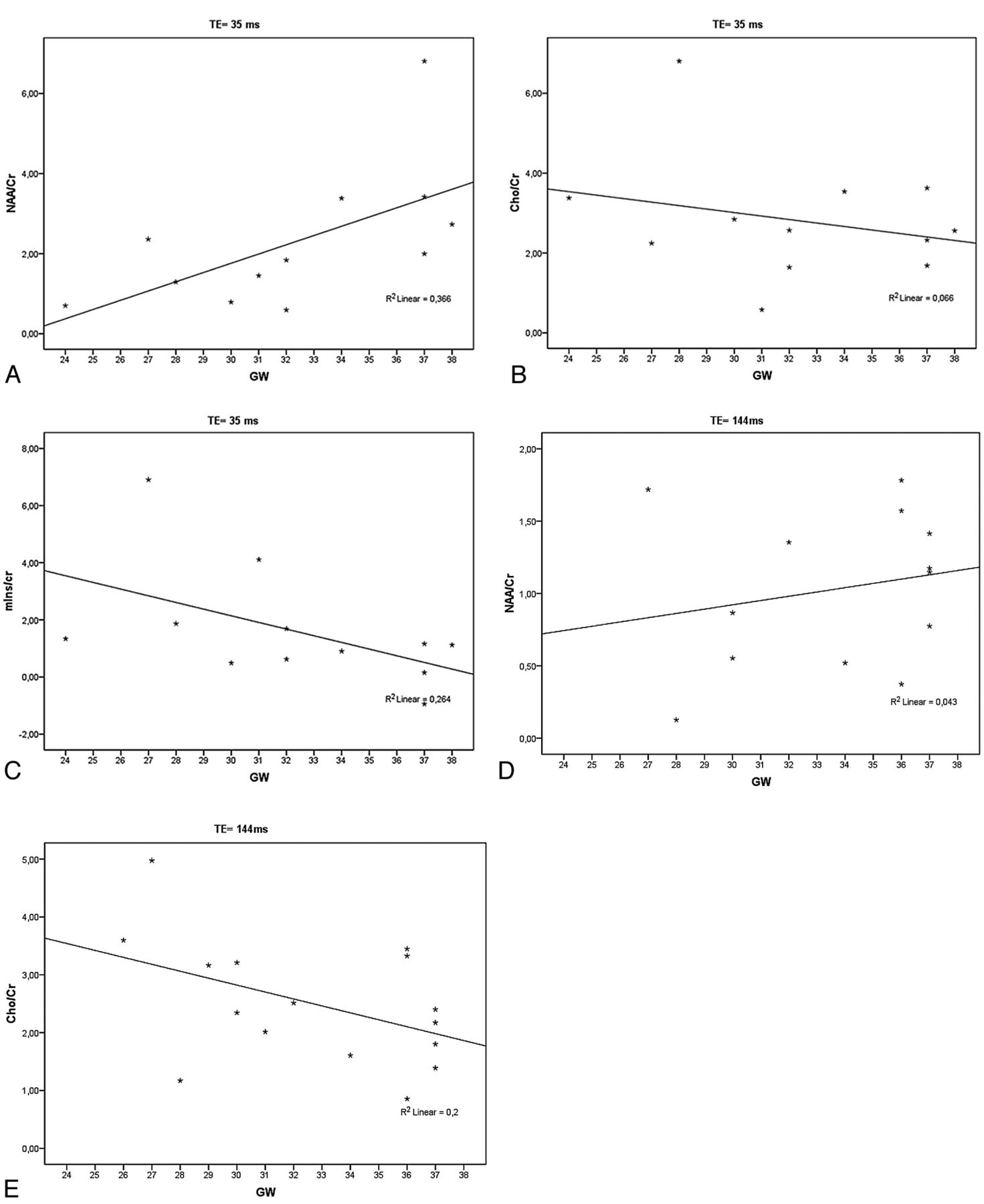
  • Fig 5.
    • Download figure
    • Open in new tab
    • Download powerpoint
    Fig 5.

    A–C, The linear correlation analysis confirmed the significant increase of NAA/Cr ratio and shows a not significant decrease of Cho/Cr and mIns/Cr ratios with GA in the readable short TE spectra of normally developed fetuses. D and E, Note the increase of the NAA/Cr ratio and the decrease of the Cho/Cr ratio with GA in the readable long TE spectra of normally developed fetuses.
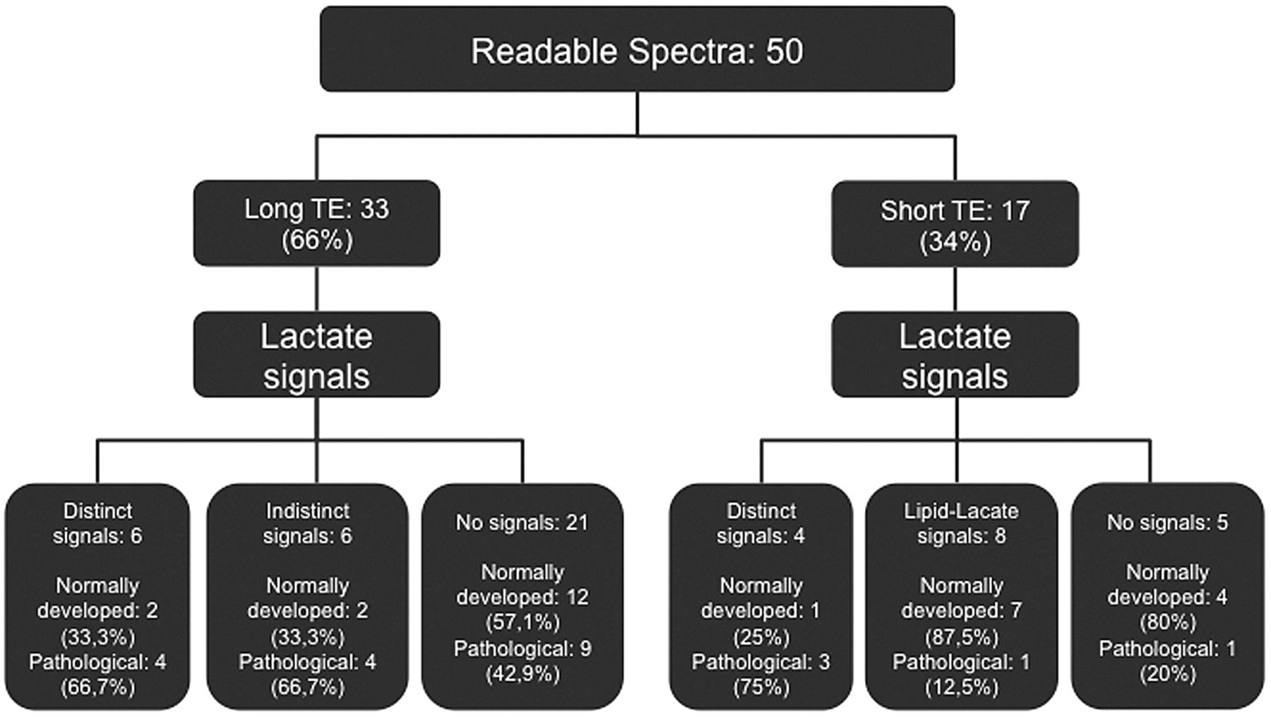
  • Fig 6.
    • Download figure
    • Open in new tab
    • Download powerpoint
    Fig 6.

    Lactate signals found in readable spectra with long and short TEs of normally and pathologically developed fetuses.

Tables

  • Figures
    • View popup
    Table 1:

    Fetal pathologies of readable and nonreadable spectra with short TEs

    Short TEs (35 ms)
    Readable
        Zellweger syndrome
        Solitary median maxillary central incisor
        Infratentorial neoplasm
        IUGR
        Abdominal mass
        Cerebral edema
        Congenital diaphragmatic hernia
        Microphthalmos
        Gastroschisis
    Nonreadable
        Ovarian cyst
        Congenital cystic adenomatoid malformation
        Abdominal mass
    • View popup
    Table 2:

    Fetal pathologies of readable and nonreadable spectra with long TEs

    Long TEs (144 ms)
    Readable
        Corpus callosum agenesia
        Infratentorial mass
        IUGR
        Chiari II malformation
        Asymmetric ventricles
        Cerebral ischemia
        Bilateral cheilognathopalatoschisis
        Cerebral arteriovenous fistula
        Lung hypoplasia
        Kidney agenesia
        Intracranial teratoma
    Nonreadable
        Vitium cordis
        Congenital diaphragmatic hernia
PreviousNext
Back to top

In this issue

American Journal of Neuroradiology: 34 (2)
American Journal of Neuroradiology
Vol. 34, Issue 2
1 Feb 2013
  • Table of Contents
  • Index by author
Advertisement
Print
Download PDF
Email Article

Thank you for your interest in spreading the word on American Journal of Neuroradiology.

NOTE: We only request your email address so that the person you are recommending the page to knows that you wanted them to see it, and that it is not junk mail. We do not capture any email address.

Enter multiple addresses on separate lines or separate them with commas.
MR Spectroscopy of the Fetal Brain: Is It Possible without Sedation?
(Your Name) has sent you a message from American Journal of Neuroradiology
(Your Name) thought you would like to see the American Journal of Neuroradiology web site.
CAPTCHA
This question is for testing whether or not you are a human visitor and to prevent automated spam submissions.
Cite this article
V. Berger-Kulemann, P.C. Brugger, D. Pugash, M. Krssak, M. Weber, A. Wielandner, D. Prayer
MR Spectroscopy of the Fetal Brain: Is It Possible without Sedation?
American Journal of Neuroradiology Feb 2013, 34 (2) 424-431; DOI: 10.3174/ajnr.A3196

Citation Manager Formats

  • BibTeX
  • Bookends
  • EasyBib
  • EndNote (tagged)
  • EndNote 8 (xml)
  • Medlars
  • Mendeley
  • Papers
  • RefWorks Tagged
  • Ref Manager
  • RIS
  • Zotero
0 Responses
Respond to this article
Share
Bookmark this article
MR Spectroscopy of the Fetal Brain: Is It Possible without Sedation?
V. Berger-Kulemann, P.C. Brugger, D. Pugash, M. Krssak, M. Weber, A. Wielandner, D. Prayer
American Journal of Neuroradiology Feb 2013, 34 (2) 424-431; DOI: 10.3174/ajnr.A3196
del.icio.us logo Twitter logo Facebook logo Mendeley logo
  • Tweet Widget
  • Facebook Like
  • Google Plus One
Purchase

Jump to section

  • Article
    • Abstract
    • ABBREVIATIONS:
    • Materials and Methods
    • Results
    • Discussion
    • Conclusions
    • Footnotes
    • References
  • Figures & Data
  • Info & Metrics
  • Responses
  • References
  • PDF

Related Articles

  • No related articles found.
  • PubMed
  • Google Scholar

Cited By...

  • Elucidating Metabolic Maturation in the Healthy Fetal Brain Using 1H-MR Spectroscopy
  • Crossref (18)
  • Google Scholar

This article has been cited by the following articles in journals that are participating in Crossref Cited-by Linking.

  • From fetus to older age: A review of brain metabolic changes across the lifespan
    Monika Cichocka, Anna Bereś
    Ageing Research Reviews 2018 46
  • Elucidating Metabolic Maturation in the Healthy Fetal Brain Using1H-MR Spectroscopy
    I.E. Evangelou, A.J. du Plessis, G. Vezina, R. Noeske, C. Limperopoulos
    American Journal of Neuroradiology 2016 37 2
  • Fetal Cerebral Magnetic Resonance Imaging Beyond Morphology
    András Jakab, Ivana Pogledic, Ernst Schwartz, Gerlinde Gruber, Christian Mitter, Peter C. Brugger, Georg Langs, Veronika Schöpf, Gregor Kasprian, Daniela Prayer
    Seminars in Ultrasound, CT and MRI 2015 36 6
  • Fetal MRI for dummies: what the fetal medicine specialist should know about acquisitions and sequences
    Michael Aertsen, Mariana C. Diogo, Steven Dymarkowski, Jan Deprest, Daniela Prayer
    Prenatal Diagnosis 2020 40 1
  • Current state of MRI of the fetal brain in utero
    Deborah A. Jarvis, Paul D. Griffiths
    Journal of Magnetic Resonance Imaging 2019 49 3
  • The role of magnetic resonance imaging in the diagnosis and prognostic evaluation of fetuses with congenital diaphragmatic hernia
    Ilaria Amodeo, Irene Borzani, Genny Raffaeli, Nicola Persico, Giacomo Simeone Amelio, Silvia Gulden, Mariarosa Colnaghi, Eduardo Villamor, Fabio Mosca, Giacomo Cavallaro
    European Journal of Pediatrics 2022 181 9
  • Harnessing the Power of Advanced Fetal Neuroimaging to Understand In Utero Footprints for Later Neuropsychiatric Disorders
    Josepheen De Asis-Cruz, Catherine Limperopoulos
    Biological Psychiatry 2023 93 10
  • Indikationen und Technik der fetalen Magnetresonanztomographie
    U. Asenbaum, P.C. Brugger, R. Woitek, J. Furtner, D. Prayer
    Der Radiologe 2013 53 2
  • Fetal neuroimaging: an update on technical advances and clinical findings
    Ashley J. Robinson, M. Ashraf Ederies
    Pediatric Radiology 2018 48 4
  • Evaluation of changes in biochemical composition of fetal brain between 18th and 40th gestational week in proton magnetic resonance spectroscopy
    Andrzej Urbanik, Monika Cichocka, Justyna Kozub, Paulina Karcz, Izabela Herman-Sucharska
    The Journal of Maternal-Fetal & Neonatal Medicine 2019 32 15

More in this TOC Section

  • Comparison of Image Quality and Radiation Dose in Pediatric Temporal Bone CT Using Photon-Counting Detector CT and Energy-Integrating Detector CT
  • SyMRI & MR Fingerprinting in Brainstem Myelination
  • Gad Contrast in PEDS Seizure Evaluation
Show more Pediatrics

Similar Articles

Advertisement

Indexed Content

  • Current Issue
  • Accepted Manuscripts
  • Article Preview
  • Past Issues
  • Editorials
  • Editors Choice
  • Fellow Journal Club
  • Letters to the Editor

Cases

  • Case Collection
  • Archive - Case of the Week
  • Archive - Case of the Month
  • Archive - Classic Case

Special Collections

  • Special Collections

Resources

  • News and Updates
  • Turn around Times
  • Submit a Manuscript
  • Author Policies
  • Manuscript Submission Guidelines
  • Evidence-Based Medicine Level Guide
  • Publishing Checklists
  • Graphical Abstract Preparation
  • Imaging Protocol Submission
  • Submit a Case
  • Become a Reviewer/Academy of Reviewers
  • Get Peer Review Credit from Publons

Multimedia

  • AJNR Podcast
  • AJNR SCANtastic
  • Video Articles

About Us

  • About AJNR
  • Editorial Board
  • Not an AJNR Subscriber? Join Now
  • Alerts
  • Feedback
  • Advertise with us
  • Librarian Resources
  • Permissions
  • Terms and Conditions

American Society of Neuroradiology

  • Not an ASNR Member? Join Now

© 2025 by the American Society of Neuroradiology All rights, including for text and data mining, AI training, and similar technologies, are reserved.
Print ISSN: 0195-6108 Online ISSN: 1936-959X

Powered by HighWire