Advertisement

Main menu

  • Home
  • Content
    • Current Issue
    • Accepted Manuscripts
    • Article Preview
    • Past Issue Archive
    • AJNR Case Collection
    • Case of the Week Archive
    • Classic Case Archive
    • Case of the Month Archive
  • Special Collections
    • Spinal CSF Leak Articles (Jan 2020-June 2024)
    • 2024 AJNR Journal Awards
    • Most Impactful AJNR Articles
  • Multimedia
    • AJNR Podcast
    • AJNR Scantastics
    • Video Articles
  • For Authors
    • Submit a Manuscript
    • Author Policies
    • Fast publishing of Accepted Manuscripts
    • Graphical Abstract Preparation
    • Manuscript Submission Guidelines
    • Imaging Protocol Submission
    • Submit a Case for the Case Collection
  • About Us
    • About AJNR
    • Editorial Board
  • More
    • Become a Reviewer/Academy of Reviewers
    • Subscribers
    • Permissions
    • Alerts
    • Feedback
    • Advertisers
    • ASNR Home
  • Other Publications
    • ajnr

User menu

  • Alerts
  • Log in

Search

  • Advanced search
American Journal of Neuroradiology

American Journal of Neuroradiology

ASHNR American Society of Functional Neuroradiology ASHNR American Society of Pediatric Neuroradiology ASSR
  • Alerts
  • Log in

Advanced Search

  • Home
  • Content
    • Current Issue
    • Accepted Manuscripts
    • Article Preview
    • Past Issue Archive
    • AJNR Case Collection
    • Case of the Week Archive
    • Classic Case Archive
    • Case of the Month Archive
  • Special Collections
    • Spinal CSF Leak Articles (Jan 2020-June 2024)
    • 2024 AJNR Journal Awards
    • Most Impactful AJNR Articles
  • Multimedia
    • AJNR Podcast
    • AJNR Scantastics
    • Video Articles
  • For Authors
    • Submit a Manuscript
    • Author Policies
    • Fast publishing of Accepted Manuscripts
    • Graphical Abstract Preparation
    • Manuscript Submission Guidelines
    • Imaging Protocol Submission
    • Submit a Case for the Case Collection
  • About Us
    • About AJNR
    • Editorial Board
  • More
    • Become a Reviewer/Academy of Reviewers
    • Subscribers
    • Permissions
    • Alerts
    • Feedback
    • Advertisers
    • ASNR Home
  • Follow AJNR on Twitter
  • Visit AJNR on Facebook
  • Follow AJNR on Instagram
  • Join AJNR on LinkedIn
  • RSS Feeds

Welcome to the new AJNR, Updated Hall of Fame, and more. Read the full announcements.


AJNR is seeking candidates for the position of Associate Section Editor, AJNR Case Collection. Read the full announcement.


Research ArticleFunctional
Open Access

Functional Homotopic Changes in Multiple Sclerosis with Resting-State Functional MR Imaging

Y. Zhou, M. Milham, X.-N. Zuo, C. Kelly, H. Jaggi, J. Herbert, R.I. Grossman and Y. Ge
American Journal of Neuroradiology June 2013, 34 (6) 1180-1187; DOI: https://doi.org/10.3174/ajnr.A3386
Y. Zhou
aFrom the Radiology/Center for Biomedical Imaging (Y.Z., H.J., J.H., R.I.G., Y.G.)
  • Find this author on Google Scholar
  • Find this author on PubMed
  • Search for this author on this site
M. Milham
bNew York University Child Study Center (M.M., X.-N.Z., C.K.), New York University School of Medicine, New York, New York
  • Find this author on Google Scholar
  • Find this author on PubMed
  • Search for this author on this site
X.-N. Zuo
bNew York University Child Study Center (M.M., X.-N.Z., C.K.), New York University School of Medicine, New York, New York
cLaboratory for Functional Connectome and Development (X.-N.Z.), Key Laboratory of Behavioral Science, Magnetic Resonance Imaging Research Center, Institute of Psychology, Chinese Academy of Sciences, Beijing, China.
  • Find this author on Google Scholar
  • Find this author on PubMed
  • Search for this author on this site
C. Kelly
bNew York University Child Study Center (M.M., X.-N.Z., C.K.), New York University School of Medicine, New York, New York
  • Find this author on Google Scholar
  • Find this author on PubMed
  • Search for this author on this site
H. Jaggi
aFrom the Radiology/Center for Biomedical Imaging (Y.Z., H.J., J.H., R.I.G., Y.G.)
  • Find this author on Google Scholar
  • Find this author on PubMed
  • Search for this author on this site
J. Herbert
aFrom the Radiology/Center for Biomedical Imaging (Y.Z., H.J., J.H., R.I.G., Y.G.)
  • Find this author on Google Scholar
  • Find this author on PubMed
  • Search for this author on this site
R.I. Grossman
aFrom the Radiology/Center for Biomedical Imaging (Y.Z., H.J., J.H., R.I.G., Y.G.)
  • Find this author on Google Scholar
  • Find this author on PubMed
  • Search for this author on this site
Y. Ge
aFrom the Radiology/Center for Biomedical Imaging (Y.Z., H.J., J.H., R.I.G., Y.G.)
  • Find this author on Google Scholar
  • Find this author on PubMed
  • Search for this author on this site
  • Article
  • Figures & Data
  • Info & Metrics
  • Responses
  • References
  • PDF
Loading

Article Figures & Data

Figures

  • Tables
  • Fig 1.
    • Download figure
    • Open in new tab
    • Download powerpoint
    Fig 1.

    Whole-brain voxelwise homotopic RSFC pattern by use of multiple linear regression models in control (A) and MS (B) groups. Homotopic RSFC was computed within 1 hemisphere (left side) for each pair of homotopic voxels and corrected by the Gaussian random field theory (minimum Z > 2.3; cluster level, P < .05, corrected). The final statistical maps are visualized as 6 hemispheric surfaces (cortical regions) with 6-mm full width at half maximum and multiple axial images (subcortical regions). Compared with control participants, patients demonstrated decreased VMHC in several cortical regions (long arrows) including the frontal, temporal, and occipital lobes and increased VMHC mainly in the subcortical regions (short arrows).

  • Fig 2.
    • Download figure
    • Open in new tab
    • Download powerpoint
    Fig 2.

    Comparison results of whole-brain voxelwise VMHC maps between patients with MS and healthy control participants (NC) showed regions with significantly decreased (blue) and increased VMHC (red and yellow) in patients compared with control participants corrected with Gaussian random field theory (minimum Z > 2.3; cluster level; P < .05, corrected).
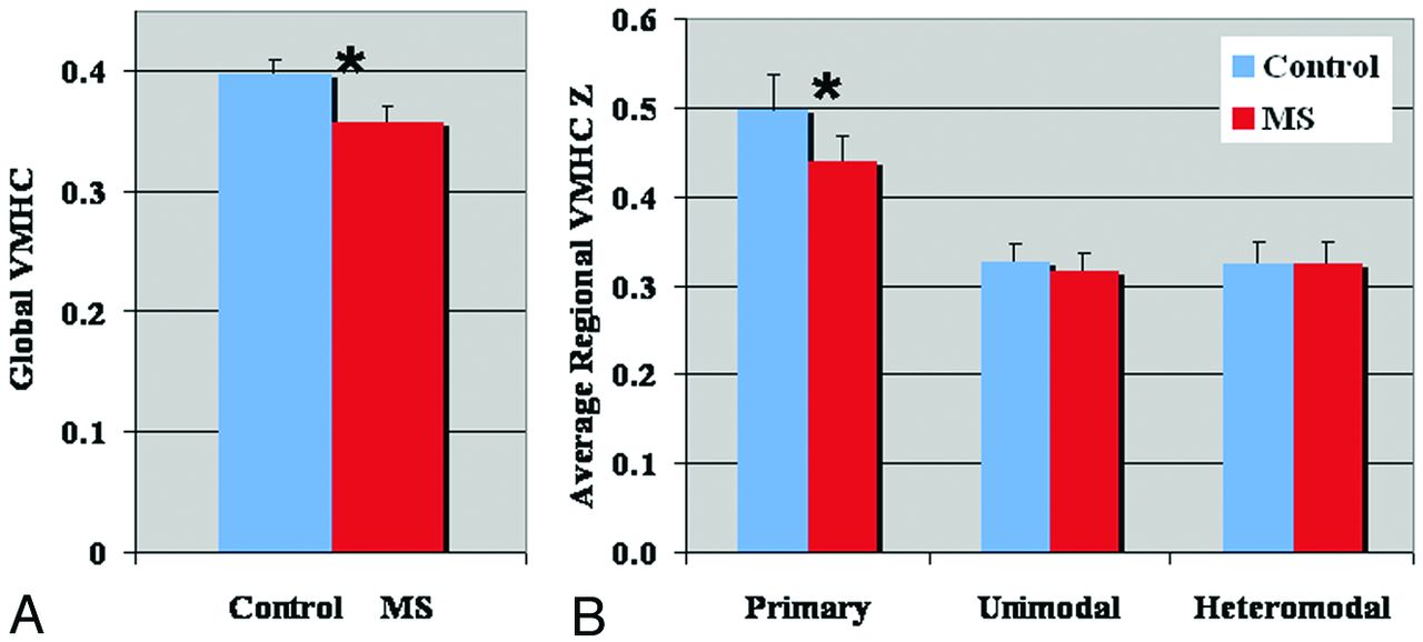
  • Fig 3.
    • Download figure
    • Open in new tab
    • Download powerpoint
    Fig 3.

    Comparison results of global (A) and regional (B) VMHC differences between patients with MS and healthy control participants. There was significantly reduced global VMHC in patients compared with control participants (P = .04) (A). Regional analysis showed significantly higher interhemispheric correlation in the primary cortical regions (ie, visual, somatosensory, motor, and auditory cortices) compared with unimodal and heteromodal regions (B) in both groups. Significantly lower VMHC was found only in the primary cortex in patients compared with control participants (P < .05). Error bars denote standard error in each group.

  • Fig 4.
    • Download figure
    • Open in new tab
    • Download powerpoint
    Fig 4.

    Structural differences measured with midsagittal CC area (A) and the FA of the entire CC (B) between patients with MS and control participants showed a significantly reduced midsagittal CC area (P = .026) and FA (P = .0018) in patients compared with control participants. The boxes have lines at the lower quartile (horizontal blue lines), median (horizontal red lines), and upper quartile values. The whiskers are lines extending from each end of the boxes to show the extent of the rest of the data. The “notch” marks the 95% confidence interval for the medians, which can be used to differentiate 2 groups. Namely 2 medians will differ significantly with P < .05 if the 2 notch intervals do not overlap.
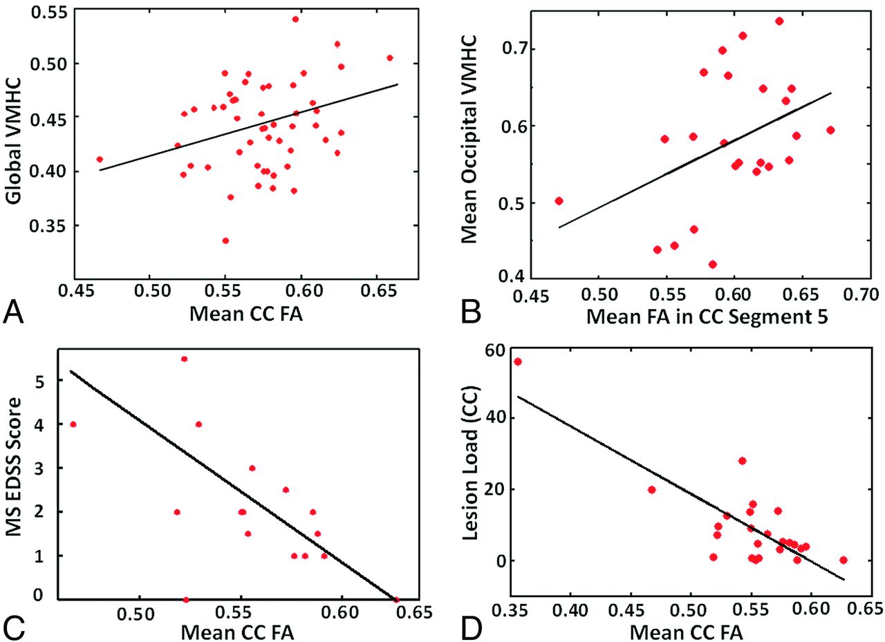
  • Fig 5.
    • Download figure
    • Open in new tab
    • Download powerpoint
    Fig 5.

    Mean FA of the entire CC and global VMHC in all participants (A) showed significant positive correlation (r = 0.3; P = .03). Mean FA of CC segment 5 (occipital projections) and mean VMHC in the occipital regions showed significant positive correlation (r = 0.43; P = .037) in patients with MS (B). There was a significant negative correlation between the Expanded Disability Status Scale score and the mean FA of CC (r = −0.61; P = .013) in the patients (C). The mean FA of CC also significantly correlates with the lesion load (r = −0.92; P < .0001) (D).
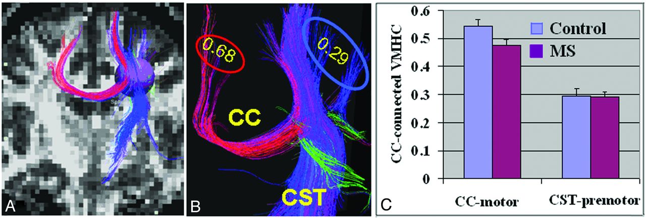
  • Fig 6.
    • Download figure
    • Open in new tab
    • Download powerpoint
    Fig 6.

    Interhemispheric and intrahemispheric structural and functional connectivity of cortical motor regions. A, On the basis of whole-brain fiber tractography from FA images, a sphere with a radius of 4.5 mm was placed in the posterior motor triangle area as a seed to select both transcallosal (interhemispheric fibers indicated in red) and nontranscallosal CST (intrahemispheric fibers indicated in blue) motor fibers to define the connecting terminal cortices for VMHC evaluation. B, A representative example of different VMHC values in 2 separate terminal regions showed higher VMHC (0.68) in motor regions connected with transcallosal fibers compared with premotor regions (0.29) projected by nontranscallosal CST fibers. C, Group comparison of transcallosal VMHC and nontranscallosal VMHCs showed much higher value in transcallosal regions. There was significantly reduced VMHC in transcallosal motor regions in patients compared with control participants (P = .03) but not in nontranscallosal CST regions (P = .09).

Tables

  • Figures
    • View popup
    Table 1:

    Brain regions showing decreased VMHC in patients with MS vs control participants

    Brain RegionBAX,Y,Z, MNI (mm)Cluster SizeMaximal Z Score
    Visual (calcarine)17−10, −94, −29053.83
    Primary somatosensory1−50, −16, 462863.8
    Lateral occipital19−34, −74, −41633.58
    Middle temporal22−66, −46, 81294.07
    Frontal pole11−36, 64, −61263.52
    Superior temporal48−54, −4, −2933.88
    Inferior occipital18−24, −70, −16653.41
    Middle occipital37−38, −42, −28643.06
    Fusiform37−22, −54, −14593.35
    Cuneus19−12, −82, 34573.68
    • Note:—Only the right side is listed because of symmetric computation between the hemispheres, P-values were obtained with cluster-level multiple comparison correction (corrected P < .05; cluster size >20); BA indicates Brodmann area; MNI, Montreal Neurological Institute.

    • View popup
    Table 2:

    Brain regions showing increased VMHC in patients with MS vs control participants

    Brain RegionBAX,Y,Z, MNI (mm)Cluster SizeMaximal Z Score
    Orbital frontal/insular48−26, −12, −141603.45
    Thalamus−−2, −24, 12403.16
    Cerebellum−−10, −82, −26403.44
    Pallidum−−10, 4, −4233.19
    Inferior temporal20−48, −34, −20223.71
    • Note:—Only the right side is listed because of symmetric computation between the hemispheres. P-values were obtained with cluster-level multiple comparison correction (corrected, P < .05; cluster size >20); BA indicates Brodmann area; MNI, Montreal Neurological Institute.

PreviousNext
Back to top

In this issue

American Journal of Neuroradiology: 34 (6)
American Journal of Neuroradiology
Vol. 34, Issue 6
1 Jun 2013
  • Table of Contents
  • Index by author
Advertisement
Print
Download PDF
Email Article

Thank you for your interest in spreading the word on American Journal of Neuroradiology.

NOTE: We only request your email address so that the person you are recommending the page to knows that you wanted them to see it, and that it is not junk mail. We do not capture any email address.

Enter multiple addresses on separate lines or separate them with commas.
Functional Homotopic Changes in Multiple Sclerosis with Resting-State Functional MR Imaging
(Your Name) has sent you a message from American Journal of Neuroradiology
(Your Name) thought you would like to see the American Journal of Neuroradiology web site.
CAPTCHA
This question is for testing whether or not you are a human visitor and to prevent automated spam submissions.
Cite this article
Y. Zhou, M. Milham, X.-N. Zuo, C. Kelly, H. Jaggi, J. Herbert, R.I. Grossman, Y. Ge
Functional Homotopic Changes in Multiple Sclerosis with Resting-State Functional MR Imaging
American Journal of Neuroradiology Jun 2013, 34 (6) 1180-1187; DOI: 10.3174/ajnr.A3386

Citation Manager Formats

  • BibTeX
  • Bookends
  • EasyBib
  • EndNote (tagged)
  • EndNote 8 (xml)
  • Medlars
  • Mendeley
  • Papers
  • RefWorks Tagged
  • Ref Manager
  • RIS
  • Zotero
0 Responses
Respond to this article
Share
Bookmark this article
Functional Homotopic Changes in Multiple Sclerosis with Resting-State Functional MR Imaging
Y. Zhou, M. Milham, X.-N. Zuo, C. Kelly, H. Jaggi, J. Herbert, R.I. Grossman, Y. Ge
American Journal of Neuroradiology Jun 2013, 34 (6) 1180-1187; DOI: 10.3174/ajnr.A3386
del.icio.us logo Twitter logo Facebook logo Mendeley logo
  • Tweet Widget
  • Facebook Like
  • Google Plus One
Purchase

Jump to section

  • Article
    • Abstract
    • ABBREVIATIONS:
    • Materials and Methods
    • Results
    • Discussion
    • Conclusions
    • Acknowledgments
    • Footnotes
    • REFERENCES
  • Figures & Data
  • Info & Metrics
  • Responses
  • References
  • PDF

Related Articles

  • No related articles found.
  • PubMed
  • Google Scholar

Cited By...

  • Acupuncture for poststroke hemiplegia focusing on cerebral bilateral connections: study protocol for a randomised controlled neuroimaging trial
  • Time-Shift Homotopic Connectivity in Mesial Temporal Lobe Epilepsy
  • Crossref (36)
  • Google Scholar

This article has been cited by the following articles in journals that are participating in Crossref Cited-by Linking.

  • Multiparametric MRI Characterization and Prediction in Autism Spectrum Disorder Using Graph Theory and Machine Learning
    Yongxia Zhou, Fang Yu, Timothy Duong, Huafu Chen
    PLoS ONE 2014 9 6
  • Interhemispheric Functional and Structural Disconnection in Alzheimer’s Disease: A Combined Resting-State fMRI and DTI Study
    Zhiqun Wang, Jianli Wang, Han Zhang, Robert Mchugh, Xiaoyu Sun, Kuncheng Li, Qing X. Yang, Yong Liu
    PLOS ONE 2015 10 5
  • Disrupted topological organization of structural and functional brain connectomes in clinically isolated syndrome and multiple sclerosis
    Ni Shu, Yunyun Duan, Mingrui Xia, Menno M. Schoonheim, Jing Huang, Zhuoqiong Ren, Zheng Sun, Jing Ye, Huiqing Dong, Fu-Dong Shi, Frederik Barkhof, Kuncheng Li, Yaou Liu
    Scientific Reports 2016 6 1
  • Decreased Prefrontal Lobe Interhemispheric Functional Connectivity in Adolescents with Internet Gaming Disorder: A Primary Study Using Resting-State fMRI
    Yao Wang, Yan Yin, Ya-wen Sun, Yan Zhou, Xue Chen, Wei-na Ding, Wei Wang, Wei Li, Jian-rong Xu, Ya-song Du, Martin Walter
    PLOS ONE 2015 10 3
  • Major depressive disorder: Findings of reduced homotopic connectivity and investigation of underlying structural mechanisms
    Marco Hermesdorf, Benedikt Sundermann, Stephan Feder, Wolfram Schwindt, Jens Minnerup, Volker Arolt, Klaus Berger, Bettina Pfleiderer, Heike Wersching
    Human Brain Mapping 2016 37 3
  • Modulation of interhemispheric functional coordination in electroconvulsive therapy for depression
    Q Wei, Y Tian, Y Yu, F Zhang, X Hu, Y Dong, Y Chen, P Hu, X Hu, K Wang
    Translational Psychiatry 2014 4 9
  • Intelligence‐related differences in the asymmetry of spontaneous cerebral activity
    Emiliano Santarnecchi, Elisa Tatti, Simone Rossi, Vinicio Serino, Alessandro Rossi
    Human Brain Mapping 2015 36 9
  • Analysis of voxel-mirrored homotopic connectivity in medication-free, current major depressive disorder
    Huanhuan Fan, Xiao Yang, Jian Zhang, Yayun Chen, Tao Li, Xiaohong Ma
    Journal of Affective Disorders 2018 240
  • Resting-state functional connectivity in multiple sclerosis: An examination of group differences and individual differences
    Alisha L. Janssen, Aaron Boster, Beth A. Patterson, Amir Abduljalil, Ruchika Shaurya Prakash
    Neuropsychologia 2013 51 13
  • Altered Functional Connectivity in Patients with Subcortical Vascular Cognitive Impairment—A Resting-State Functional Magnetic Resonance Imaging Study
    Weina Ding, Wenwei Cao, Yao Wang, Yawen Sun, Xue Chen, Yan Zhou, Qun Xu, Jianrong Xu, Xi-Nian Zuo
    PLOS ONE 2015 10 9

More in this TOC Section

  • Glutaric Aciduria Type 1: DK vs. Conventional MRI
  • Kurtosis and Epileptogenic Tubers: A Pilot Study
  • WM Neuroaxonal Loss in Type 1 Diabetes
Show more Functional

Similar Articles

Advertisement

Indexed Content

  • Current Issue
  • Accepted Manuscripts
  • Article Preview
  • Past Issues
  • Editorials
  • Editors Choice
  • Fellow Journal Club
  • Letters to the Editor

Cases

  • Case Collection
  • Archive - Case of the Week
  • Archive - Case of the Month
  • Archive - Classic Case

Special Collections

  • Special Collections

Resources

  • News and Updates
  • Turn around Times
  • Submit a Manuscript
  • Author Policies
  • Manuscript Submission Guidelines
  • Evidence-Based Medicine Level Guide
  • Publishing Checklists
  • Graphical Abstract Preparation
  • Imaging Protocol Submission
  • Submit a Case
  • Become a Reviewer/Academy of Reviewers
  • Get Peer Review Credit from Publons

Multimedia

  • AJNR Podcast
  • AJNR SCANtastic
  • Video Articles

About Us

  • About AJNR
  • Editorial Board
  • Not an AJNR Subscriber? Join Now
  • Alerts
  • Feedback
  • Advertise with us
  • Librarian Resources
  • Permissions
  • Terms and Conditions

American Society of Neuroradiology

  • Not an ASNR Member? Join Now

© 2025 by the American Society of Neuroradiology All rights, including for text and data mining, AI training, and similar technologies, are reserved.
Print ISSN: 0195-6108 Online ISSN: 1936-959X

Powered by HighWire