Advertisement

Main menu

  • Home
  • Content
    • Current Issue
    • Accepted Manuscripts
    • Article Preview
    • Past Issue Archive
    • Video Articles
    • AJNR Case Collection
    • Case of the Week Archive
    • Case of the Month Archive
    • Classic Case Archive
  • Special Collections
    • AJNR Awards
    • Low-Field MRI
    • Alzheimer Disease
    • ASNR Foundation Special Collection
    • Photon-Counting CT
    • View All
  • Multimedia
    • AJNR Podcasts
    • AJNR SCANtastic
    • Trainee Corner
    • MRI Safety Corner
    • Imaging Protocols
  • For Authors
    • Submit a Manuscript
    • Submit a Video Article
    • Submit an eLetter to the Editor/Response
    • Manuscript Submission Guidelines
    • Statistical Tips
    • Fast Publishing of Accepted Manuscripts
    • Graphical Abstract Preparation
    • Imaging Protocol Submission
    • Author Policies
  • About Us
    • About AJNR
    • Editorial Board
    • Editorial Board Alumni
  • More
    • Become a Reviewer/Academy of Reviewers
    • Subscribers
    • Permissions
    • Alerts
    • Feedback
    • Advertisers
    • ASNR Home

User menu

  • Alerts
  • Log in

Search

  • Advanced search
American Journal of Neuroradiology

American Journal of Neuroradiology

ASHNR American Society of Functional Neuroradiology ASHNR American Society of Pediatric Neuroradiology ASSR
  • Alerts
  • Log in

Advanced Search

  • Home
  • Content
    • Current Issue
    • Accepted Manuscripts
    • Article Preview
    • Past Issue Archive
    • Video Articles
    • AJNR Case Collection
    • Case of the Week Archive
    • Case of the Month Archive
    • Classic Case Archive
  • Special Collections
    • AJNR Awards
    • Low-Field MRI
    • Alzheimer Disease
    • ASNR Foundation Special Collection
    • Photon-Counting CT
    • View All
  • Multimedia
    • AJNR Podcasts
    • AJNR SCANtastic
    • Trainee Corner
    • MRI Safety Corner
    • Imaging Protocols
  • For Authors
    • Submit a Manuscript
    • Submit a Video Article
    • Submit an eLetter to the Editor/Response
    • Manuscript Submission Guidelines
    • Statistical Tips
    • Fast Publishing of Accepted Manuscripts
    • Graphical Abstract Preparation
    • Imaging Protocol Submission
    • Author Policies
  • About Us
    • About AJNR
    • Editorial Board
    • Editorial Board Alumni
  • More
    • Become a Reviewer/Academy of Reviewers
    • Subscribers
    • Permissions
    • Alerts
    • Feedback
    • Advertisers
    • ASNR Home
  • Follow AJNR on Twitter
  • Visit AJNR on Facebook
  • Follow AJNR on Instagram
  • Join AJNR on LinkedIn
  • RSS Feeds

AJNR Awards, New Junior Editors, and more. Read the latest AJNR updates

Research ArticleSpine Imaging and Spine Image-Guided Interventions

Facet Joint Signal Change on MRI at Levels of Acute/Subacute Lumbar Compression Fractures

V.T. Lehman, C.P. Wood, C.H. Hunt, R.E. Carter, J.B. Allred, F.E. Diehn, J.M. Morris, J.T. Wald and K.R. Thielen
American Journal of Neuroradiology July 2013, 34 (7) 1468-1473; DOI: https://doi.org/10.3174/ajnr.A3449
V.T. Lehman
aFrom the Division of Neuroradiology (V.T.L., C.P.W., C.H.H., F.E.D., J.M.M., J.T.W., K.R.T.), Department of Radiology, Mayo Clinic College of Graduate Medical Education, Rochester, Minnesota
  • Find this author on Google Scholar
  • Find this author on PubMed
  • Search for this author on this site
C.P. Wood
aFrom the Division of Neuroradiology (V.T.L., C.P.W., C.H.H., F.E.D., J.M.M., J.T.W., K.R.T.), Department of Radiology, Mayo Clinic College of Graduate Medical Education, Rochester, Minnesota
  • Find this author on Google Scholar
  • Find this author on PubMed
  • Search for this author on this site
C.H. Hunt
aFrom the Division of Neuroradiology (V.T.L., C.P.W., C.H.H., F.E.D., J.M.M., J.T.W., K.R.T.), Department of Radiology, Mayo Clinic College of Graduate Medical Education, Rochester, Minnesota
  • Find this author on Google Scholar
  • Find this author on PubMed
  • Search for this author on this site
R.E. Carter
bDivision of Biomedical Statistics and Informatics (R.E.C., J.B.A.), Mayo Clinic Department of Health Sciences Research, Rochester, Minnesota.
  • Find this author on Google Scholar
  • Find this author on PubMed
  • Search for this author on this site
J.B. Allred
bDivision of Biomedical Statistics and Informatics (R.E.C., J.B.A.), Mayo Clinic Department of Health Sciences Research, Rochester, Minnesota.
  • Find this author on Google Scholar
  • Find this author on PubMed
  • Search for this author on this site
F.E. Diehn
aFrom the Division of Neuroradiology (V.T.L., C.P.W., C.H.H., F.E.D., J.M.M., J.T.W., K.R.T.), Department of Radiology, Mayo Clinic College of Graduate Medical Education, Rochester, Minnesota
  • Find this author on Google Scholar
  • Find this author on PubMed
  • Search for this author on this site
J.M. Morris
aFrom the Division of Neuroradiology (V.T.L., C.P.W., C.H.H., F.E.D., J.M.M., J.T.W., K.R.T.), Department of Radiology, Mayo Clinic College of Graduate Medical Education, Rochester, Minnesota
  • Find this author on Google Scholar
  • Find this author on PubMed
  • Search for this author on this site
J.T. Wald
aFrom the Division of Neuroradiology (V.T.L., C.P.W., C.H.H., F.E.D., J.M.M., J.T.W., K.R.T.), Department of Radiology, Mayo Clinic College of Graduate Medical Education, Rochester, Minnesota
  • Find this author on Google Scholar
  • Find this author on PubMed
  • Search for this author on this site
K.R. Thielen
aFrom the Division of Neuroradiology (V.T.L., C.P.W., C.H.H., F.E.D., J.M.M., J.T.W., K.R.T.), Department of Radiology, Mayo Clinic College of Graduate Medical Education, Rochester, Minnesota
  • Find this author on Google Scholar
  • Find this author on PubMed
  • Search for this author on this site
  • Article
  • Figures & Data
  • Info & Metrics
  • Responses
  • References
  • PDF
Loading

Article Figures & Data

Figures

  • Tables
  • Fig 1.
    • Download figure
    • Open in new tab
    • Download powerpoint
    Fig 1.

    For the L2 vertebral body, the facet joint signal-change score (0–16) is the sum of the facet joint signal-change grades of each of the 4 associated facet joints (0–4). Anatomically, the L2 vertebral body–associated facet joints are shared with the adjacent vertebral bodies. Therefore, the L2-associated facet joints could be subject to stress from compression fractures of the L2 or adjacent L1 and L3 vertebral bodies. For the L2 level, the above facet joint signal-change score for the L1/L2 and L2/L3 facet joints of patients with an acute/subacute compression fracture of L1, L2, and/or L3 was compared with the facet joint signal-change score for the L1/L2 and L2/L3 facet joints of patients without an acute/subacute compression fracture at L1, L2, or L3. This same method was then applied to the remaining lumbar vertebrae.

  • Fig 2.
    • Download figure
    • Open in new tab
    • Download powerpoint
    Fig 2.

    Level-by-level distribution of acute/subacute vertebral body compression fractures, chronic compression fractures, and prior vertebral augmentation.
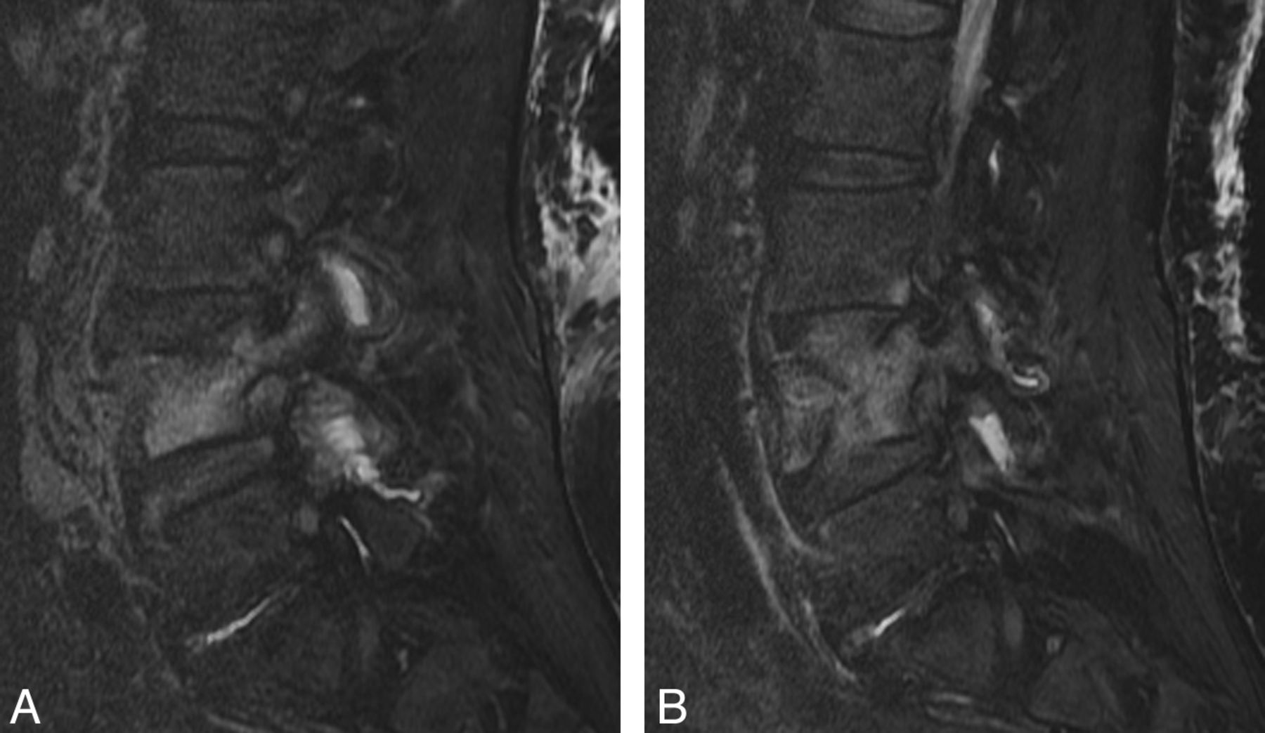
  • Fig 3.
    • Download figure
    • Open in new tab
    • Download powerpoint
    Fig 3.

    Facet joint inflammatory changes are more prevalent at levels of acute/subacute compression fracture than in levels without such fracture. A 66-year-old woman presented with an acute/subacute L4 compression fracture. Sagittal fat-saturated T2-weighted images demonstrate facet edema of the left (A) and right (B) L3/L4 and L4/5 facet joints. There was no facet joint inflammatory change associated with the remaining lumbar facet joints (images not shown).

  • Fig 4.
    • Download figure
    • Open in new tab
    • Download powerpoint
    Fig 4.

    Level-by-level distribution of the degree of vertebral body height loss.

Tables

  • Figures
    • View popup
    Table 1:

    Grading of facet joint inflammation using fat-saturated MRI signal characteristicsa

    Grade
    0No signal abnormality
    1Signal abnormality limited to joint capsule
    2Periarticular signal abnormality involving <50% of the facet joint perimeterb
    3Periarticular signal abnormality involving >50% of the facet joint perimeter
    4Grade 3 with additional signal abnormality within the neural foramen, pedicle, ligamentum flavum, transverse process, or vertebral bodyc
    • ↵a Grading scale adapted from Czervionke and Fenton.6

    • ↵b Periarticular signal abnormality includes T2 hyperintensity or gadolinium enhancement on T1-weighted images.

    • ↵c Signal abnormality within the pedicle must be continuous with other perifacet signal abnormalities and cannot represent posterior extension of vertebral body edema.

    • View popup
    Table 2:

    Facet joint signal-change scores of acute/subacute lumbar levels compared with other levels

    Facet Joint Signal-ChangeScorea
    Acute/subacute (mean) (SD)2.2 (3.3)2.6 (3.3)3.7 (3.7)4.6 (5.2)4.8 (5.3)
    Other (mean) (SD)b0.0 (0.0)0.3 (0.9)1.0 (2.3)1.2 (2.5)0.4 (0.9)
    P valuec<.01.01<.01<.01<.01
    • ↵a A vertebral body was considered influenced by an acute or subacute fracture if either the body of interest or an adjacent vertebral body was fractured.

    • ↵b “Other” is used to denote the complement to this case, namely vertebral bodies with no fracture or nearby fracture. Vertebral bodies with a chronic compression fracture could be considered “other” if there were no adjacent acute/subacute fractures.

    • ↵c Wilcoxon rank sum test.

    • View popup
    Table 3:

    Facet joint signal-change score versus degree of height loss for patients with acute/subacute fractures

    L1 (n = 75)L2 (n = 75)L3 (n = 75)L4 (n = 75)L5 (n = 75)
    Spearman rank correlation (ρ)0.410.150.020.220.14
    P value<.01.21.84.05.22
PreviousNext
Back to top

In this issue

American Journal of Neuroradiology: 34 (7)
American Journal of Neuroradiology
Vol. 34, Issue 7
1 Jul 2013
  • Table of Contents
  • Index by author
Advertisement
Print
Download PDF
Email Article

Thank you for your interest in spreading the word on American Journal of Neuroradiology.

NOTE: We only request your email address so that the person you are recommending the page to knows that you wanted them to see it, and that it is not junk mail. We do not capture any email address.

Enter multiple addresses on separate lines or separate them with commas.
Facet Joint Signal Change on MRI at Levels of Acute/Subacute Lumbar Compression Fractures
(Your Name) has sent you a message from American Journal of Neuroradiology
(Your Name) thought you would like to see the American Journal of Neuroradiology web site.
CAPTCHA
This question is for testing whether or not you are a human visitor and to prevent automated spam submissions.
Cite this article
V.T. Lehman, C.P. Wood, C.H. Hunt, R.E. Carter, J.B. Allred, F.E. Diehn, J.M. Morris, J.T. Wald, K.R. Thielen
Facet Joint Signal Change on MRI at Levels of Acute/Subacute Lumbar Compression Fractures
American Journal of Neuroradiology Jul 2013, 34 (7) 1468-1473; DOI: 10.3174/ajnr.A3449

Citation Manager Formats

  • BibTeX
  • Bookends
  • EasyBib
  • EndNote (tagged)
  • EndNote 8 (xml)
  • Medlars
  • Mendeley
  • Papers
  • RefWorks Tagged
  • Ref Manager
  • RIS
  • Zotero
0 Responses
Respond to this article
Share
Bookmark this article
Facet Joint Signal Change on MRI at Levels of Acute/Subacute Lumbar Compression Fractures
V.T. Lehman, C.P. Wood, C.H. Hunt, R.E. Carter, J.B. Allred, F.E. Diehn, J.M. Morris, J.T. Wald, K.R. Thielen
American Journal of Neuroradiology Jul 2013, 34 (7) 1468-1473; DOI: 10.3174/ajnr.A3449
del.icio.us logo Twitter logo Facebook logo Mendeley logo
  • Tweet Widget
  • Facebook Like
  • Google Plus One
Purchase

Jump to section

  • Article
    • Abstract
    • ABBREVIATIONS:
    • Materials and Methods
    • Results
    • Discussion
    • Conclusions
    • Acknowledgments
    • Footnotes
    • REFERENCES
  • Figures & Data
  • Info & Metrics
  • Responses
  • References
  • PDF

Related Articles

  • No related articles found.
  • PubMed
  • Google Scholar

Cited By...

  • Review of the Imaging Features of Benign Osteoporotic and Malignant Vertebral Compression Fractures
  • Crossref
  • Google Scholar

This article has not yet been cited by articles in journals that are participating in Crossref Cited-by Linking.

More in this TOC Section

  • Management Outcomes For VO Spine Biopsy
  • Characteristics of SIH Type I Culprit Lesions
  • Advanced Imaging of Type 2 Spinal CSF Leaks
Show more Spine Imaging and Spine Image-Guided Interventions

Similar Articles

Advertisement

Indexed Content

  • Current Issue
  • Accepted Manuscripts
  • Article Preview
  • Past Issues
  • Editorials
  • Editor's Choice
  • Fellows' Journal Club
  • Letters to the Editor
  • Video Articles

Cases

  • Case Collection
  • Archive - Case of the Week
  • Archive - Case of the Month
  • Archive - Classic Case

More from AJNR

  • Trainee Corner
  • Imaging Protocols
  • MRI Safety Corner
  • Book Reviews

Multimedia

  • AJNR Podcasts
  • AJNR Scantastics

Resources

  • Turnaround Time
  • Submit a Manuscript
  • Submit a Video Article
  • Submit an eLetter to the Editor/Response
  • Manuscript Submission Guidelines
  • Statistical Tips
  • Fast Publishing of Accepted Manuscripts
  • Graphical Abstract Preparation
  • Imaging Protocol Submission
  • Evidence-Based Medicine Level Guide
  • Publishing Checklists
  • Author Policies
  • Become a Reviewer/Academy of Reviewers
  • News and Updates

About Us

  • About AJNR
  • Editorial Board
  • Editorial Board Alumni
  • Alerts
  • Permissions
  • Not an AJNR Subscriber? Join Now
  • Advertise with Us
  • Librarian Resources
  • Feedback
  • Terms and Conditions
  • AJNR Editorial Board Alumni

American Society of Neuroradiology

  • Not an ASNR Member? Join Now

© 2025 by the American Society of Neuroradiology All rights, including for text and data mining, AI training, and similar technologies, are reserved.
Print ISSN: 0195-6108 Online ISSN: 1936-959X

Powered by HighWire