
Article Figures & Data
Figures
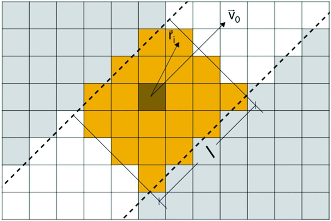
- Fig 1.
2D representation of the segmentation procedure. Dashed lines represent the vessel lumen; yellow pixels are the segmentation result. White pixels represent pixel intensity of ≥18%. Dark pixel is the user-defined reference pixel whose velocity direction was used to label a segment of length l. Segmentation was not updated between cardiac frames. Typical segmentations of the ICA, basilar artery, MCA, and ACA contained 230, 110, 80, and 60 pixels, respectively.
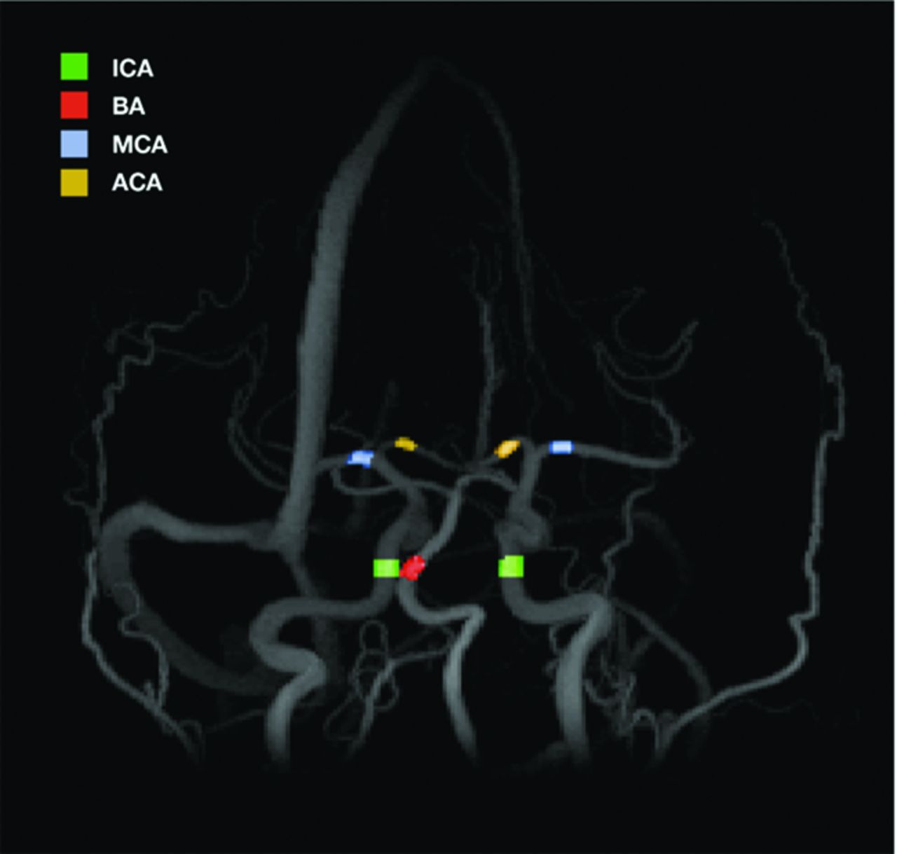
- Fig 2.
Maximum intensity projection image. Colored sections show segmentations produced for comparison with the 2D PCMRI data.
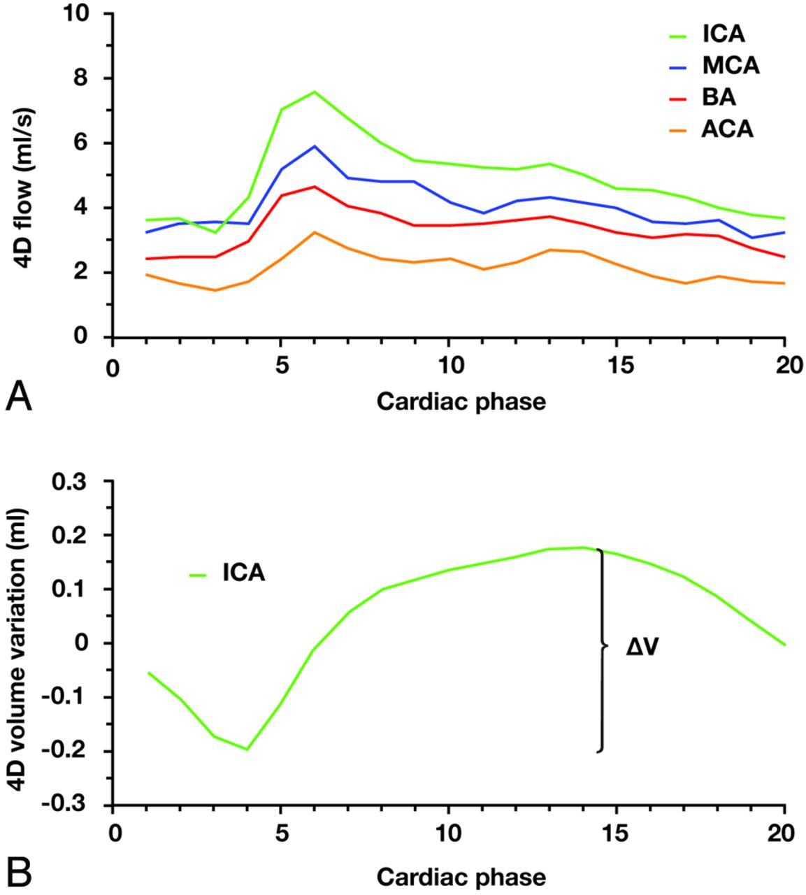
- Fig 3.
Arterial hemodynamics provided from a 4D PCMRI quantification. A, Flow curves for vessels evaluated in this study. The right ICA, MCA, and ACA are shown. Cardiac phase 1–20 represents the time positions in the cardiac cycle of the reconstructed frames. B, ICA ΔV calculation. Subtraction of net flow and calculation of the cumulative integral on the remaining waveform yield the cyclic volume variation of downstream arteries required to convert the pulsatile right ICA waveform to a nonpulsatile capillary flow. ICA ΔV was defined as the maximum minus minimum of this volume variation.
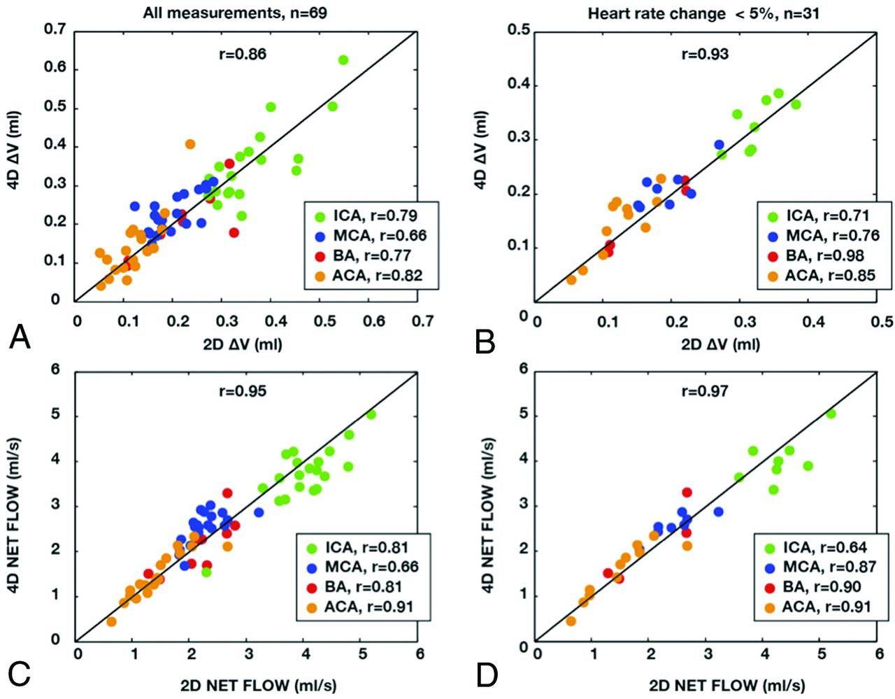
- Fig 4.
Correlation between 4D and 2D PCMRI quantifications of pulsatility and net flow. A, ΔV quantifications for all vessels. B, ΔV quantifications, excluding measurements with large differences in heart rate. C, Net flow quantifications for all vessels. D, Net flow quantifications, excluding measurements with large differences in heart rate.
- Fig 5.
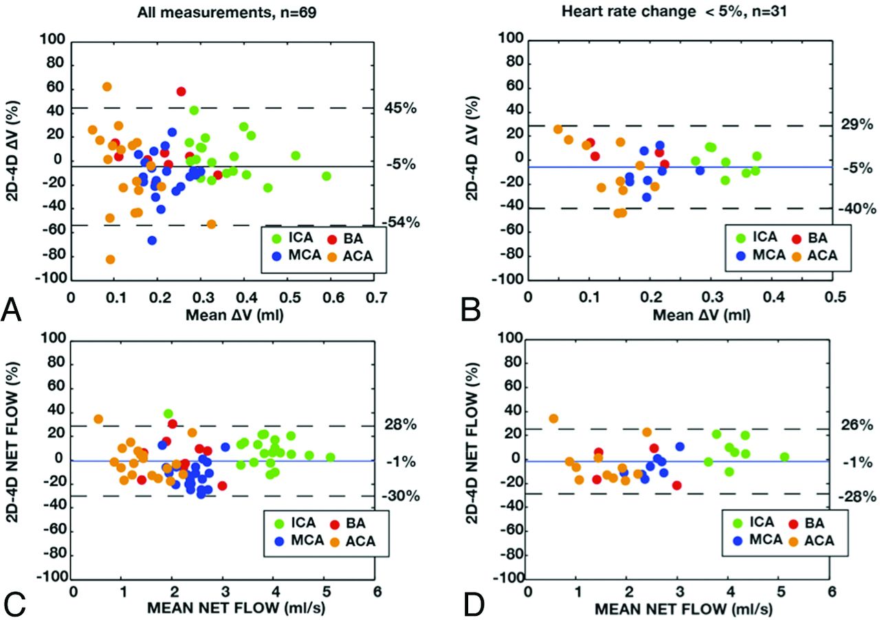
Agreement between 4D and 2D PCMRI quantifications of pulsatility and net flow. Solid lines represent average difference; dashed lines represent 95% limits of agreements. The relative difference was calculated as (2D − 4D)/([2D + 4D]/2). A, ΔV quantifications for all vessels. B, ΔV quantifications, excluding measurements with large differences in heart rate. C, Net flow quantifications for all vessels. D, Net flow quantifications, excluding measurements with large differences in heart rate.


Tables
Vessel Type n ΔV 4D Mean ± SD (mL) ΔV 2D Mean ± SD (mL) Difference Mean ± SD (mL) P Value All BA 10 0.20 ± 0.08 0.21 ± 0.08 0.01 ± 0.05 .504 ICA 20 0.35 ± 0.10 0.36 ± 0.10 0.01 ± 0.06 .656 ICAa 20 0.35 ± 0.10 MCA 20 0.23 ± 0.05 0.20 ± 0.05 −0.03 ± 0.04 .006 ACA 19 0.14 ± 0.08 0.12 ± 0.08 −0.02 ± 0.05 .102 ΔHR BA 4 0.16 ± 0.07 0.17 ± 0.07 0.01 ± 0.01 .268 <5% ICA 8 0.33 ± 0.05 0.33 ± 0.05 −0.01 ± 0.03 .668 MCA 8 0.21 ± 0.04 0.20 ± 0.04 −0.02 ± 0.03 .116 ACA 11 0.14 ± 0.06 0.13 ± 0.06 −0.02 ± 0.03 .074 Note:—BA indicates basilar artery; ICA, internal carotid artery; MCA, middle cerebral artery; ACA, anterior cerebral artery; ΔHR, difference in heart rate; ΔV, volume of arterial pulsatility.
↵a Indicates proximal ICA measurement for internal consistency analyses.
Vessel Type n Net Flow 4D Mean ± SD (mL/s) Net Flow 2D Mean ± SD (mL/s) Difference Mean ± SD (mL/s) P Value All BA 10 2.15 ± 0.58 2.18 ± 0.49 0.03 ± 0.34 .787 ICA 20 3.72 ± 0.70 4.02 ± 0.62 0.29 ± 0.40 .004 ICAa 20 3.81 ± 0.71 MCA 20 2.54 ± 0.36 2.26 ± 0.34 −0.28 ± 0.29 <.001 ACA 19 1.47 ± 0.52 1.44 ± 0.49 −0.02 ± 0.21 .627 ΔHR BA 4 2.16 ± 0.89 2.02 ± 0.74 −0.13 ± 0.39 .538 <5% ICA 8 4.04 ± 0.51 4.32 ± 0.51 0.28 ± 0.43 .108 MCA 8 2.58 ± 0.27 2.46 ± 0.42 −0.12 ± 0.23 .173 ACA 11 1.55 ± 0.61 1.49 ± 0.61 −0.06 ± 0.26 .427 Note:—BA indicates basilar artery; ICA, internal carotid artery; MCA, middle cerebral artery; ACA, anterior cerebral artery; ΔHR, difference in heart rate; ΔV, volume of arterial pulsatility.
↵a Indicates proximal ICA measurement for internal consistency analyses.


Jump to section
Related Articles
Cited By...
- Microvascular pulsatility of the ageing brain and confounding effects of anaesthesia
- Macrovascular blood flow and microvascular cerebrovascular reactivity are regionally coupled in adolescence
- Assessing Cerebral Microvascular Volumetric Pulsatility with High-Resolution 4D CBV MRI at 7T
- Intracranial aneurysm stiffness assessment using 4D flow MRI
- Flow control in the middle cerebral artery during thrombectomy: the effect of anatomy, catheter size and tip location
- Cerebral blood flow pattern in patients with carotid artery stenosis with low trans-stenotic blood flow
- Flow control in the middle cerebral artery during thrombectomy: the effect of anatomy, catheter size and tip location
- A Novel Method for Improving the Accuracy of MR-derived Patient-specific Vascular Models using X-ray Angiography
- Toward noninvasive assessment of stroke risk in pediatric cerebrovascular disease
- Aneurysmal Parent Artery-Specific Inflow Conditions for Complete and Incomplete Circle of Willis Configurations
- Risk of distal embolization with stent retriever thrombectomy and ADAPT
- Age-Related Changes of Normal Cerebral and Cardiac Blood Flow in Children and Adults Aged 7 Months to 61 Years
- Time window for recanalization in basilar artery occlusion: Speculative synthesis
- Attenuation of Blood Flow Pulsatility along the Atlas Slope: A Physiologic Property of the Distal Vertebral Artery?
- Reproducibility of Cerebrospinal Venous Blood Flow and Vessel Anatomy with the Use of Phase Contrast-Vastly Undersampled Isotropic Projection Reconstruction and Contrast-Enhanced MRA