
Article Figures & Data
Figures
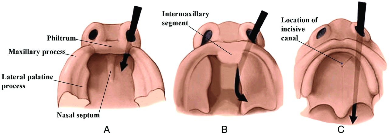
- Fig 1.
Drawings from below show the development of the palate from 6 to 7 weeks (A), 7 to 8 weeks (B), and 8 to 10 weeks (C). The lateral palatine processes grow medially and eventually merge in the midline and with the intermaxillary segment (primitive palate). The incisive canal marks the junction of the primitive and secondary palates. (Modified with permission from Levine HL, Clemente MP, eds. Chapter 1, Surgical Anatomy of the Paranasal Sinus. China: 2005. Sinus Surgery Endoscopic and Microscopic Approaches. Figures 1–3. Thieme Medical Publishers Inc., Georg Thieme Verlag Stuttgart).
- Fig 2.
Serial frontal diagrams (A–D) from approximately 6–10 fetal weeks shown just posterior to the intermaxillary segment illustrate the progressive development of the secondary palate and its fusion with the nasal septum.
- Fig 3.
Drawing from above and in front (A) of the developing lips and gums. The labiogingival lamina develops in the common mesenchymal tissue of this region. When it dissolves, the labiogingival sulcus that remains separates the lips and gums. The dental lamina develops just behind this region and will give rise to the dental buds, which will form the deciduous and permanent teeth. B, Frontal photograph shows the frenulum of the upper lip, the only remaining vestige of the labiogingival lamina.
- Fig 4.
Lateral drawing of the developing teeth. Note that the permanent teeth (blue) develop medial to the deciduous teeth. (Modified with permission from Frank H. Netter, Atlas of Human Anatomy, 5th Edition, Saunders Elsevier, Philadelphia, 2011, Figure 56. Netter Illustrations from www.netterimages.com, ©Elsevier Inc, All rights reserved).
- Fig 5.
Drawings of the progressive development of the teeth from the tooth bud stage that comes from the dental lamina to the adult tooth. (Modified from http://embryology.ch/anglais/sdigestive/gesicht05.htm and www.embryo.chronolab.com/teeth.html).
- Fig 6.
Sagittal drawings of the progressive development of the facial muscles from the dense mesenchyme that arises near the first branchial cleft. (Modified with permission from Gasser R. The Development of the Facial Muscles in Man. Am J Anat 1967;120:357–376).
- Fig 7.
Lateral drawings of a 7- to 8-week embryo (A) and an 8- to 10-week fetus (B) show that the opening of the external auditory canal remains stationary but appears to rise because the progressive elongation of the jaw creates this impression. (Modified with permission from Netter's Atlas of Human Embryology. Edited by Cochard, L.R., PhD. 2002. Icon Learning Systems, Teterboro, New Jersey, Figure 9.27. Netter Illustrations from www.netterimages.com, ©Elsevier Inc, All rights reserved) .
- Fig 8.
Drawing in an anterior oblique view of the late fetal face showing the contributions of the various facial processes. Green indicates the frontonasal process; yellow, the lateral nasal processes; purple, the medial nasal processes; orange, the maxillary processes; and blue, the mandibular processes.
- Fig 9.
Lateral oblique drawings of the 6 hillocks that develop about the first branchial cleft and how they eventually form the pinna of the ear.
- Fig 10.
Frontal drawing of a neonate skull (A) shows the sagittal suture system that divides the cranium and face into 2 halves. This system is made up of the metopic suture, the internasal suture, the intermaxillary suture, and the mandibular symphysis (outlined with a black line). (Modified with permission from Sobotta: Atlas der Anatomie des Menschen. ©Elsevier GmbH, Urban & Fischer Verlag Munchen. Volume 1, Edited by Putz, R. and Pabst, R. Lippincott Williams and Wilkins, Philadelphia 2001, Figure 82). B, Frontal drawing of the body of the sphenoid bone, the greater sphenoid wings, and the cartilage between them (black). The sagittal suture system divides to run on either side of the body of the sphenoid bone because it is separated from the greater sphenoid wings by cartilage. C, Frontal drawing of the midfacial structures at approximately 3 years of age. The union of the ethmoid bodies (pink) with the perpendicular plate (orange) as a result of ossification of the cribriform plates (green) makes the ethmoid bone a single bone and stabilizes the interocular and upper nasal regions. The maxilla is beige; vomer, yellow; septal cartilage, light blue.
- Fig 11.
Lateral (A) and frontal (B) drawings of neonate facial bones and skull and adult facial bones and skull in the lateral (C) and frontal (D) views. In general, the facial structures grow proportionally more and for a longer time the further they are from the neurocranium. Thus, growth of the mandible begins later and continues longer than midfacial and orbital development. The forehead grows in an anterior and slightly upward direction. The forward and upward growth of the forehead contributes to elevation and widening of the nasal bridge. (Modified with permission from Sobotta: Atlas der Anatomie des Menschen. ©Elsevier GmbH, Urban & Fischer Verlag Munchen. Volume 1, Edited by Putz, R. and Pabst, R. Lippincott Williams and Wilkins, Philadelphia 2001, Figures 66, 68, 82 and 83).
- Fig 12.
Lateral diagram of the fetal skull (A) (darker areas) and the adult skull (B) (lighter areas) shows that the inferior orbital rim and the superior orbital rim are in the same plane in the young face (dark line in A), but in the older face, the supraorbital region protrudes forward of the cheek (dark line in B). Frontal diagrams (C and D) show that as the maxillary bones and the ethmoid bodies separate from one another, this movement increases the lateral growth of the interocular distance. As a result, the orbits enlarge and shift laterally (C). The movement of the nasal area combined with the malar movement results in an increase in the vertical size and the width of the upper part of each nasal cavity (D). (Modified with permission from Enlow D. A Morphogenetic Analysis of Facial Growth. Am J Orthodontics 1966;52:283–299. Figures 1 and 4)


