Advertisement

Main menu

  • Home
  • Content
    • Current Issue
    • Accepted Manuscripts
    • Article Preview
    • Past Issue Archive
    • Video Articles
    • AJNR Case Collection
    • Case of the Week Archive
    • Case of the Month Archive
    • Classic Case Archive
  • Special Collections
    • AJNR Awards
    • Low-Field MRI
    • Alzheimer Disease
    • ASNR Foundation Special Collection
    • Photon-Counting CT
    • View All
  • Multimedia
    • AJNR Podcasts
    • AJNR SCANtastic
    • Trainee Corner
    • MRI Safety Corner
    • Imaging Protocols
  • For Authors
    • Submit a Manuscript
    • Submit a Video Article
    • Submit an eLetter to the Editor/Response
    • Manuscript Submission Guidelines
    • Statistical Tips
    • Fast Publishing of Accepted Manuscripts
    • Graphical Abstract Preparation
    • Imaging Protocol Submission
    • Author Policies
  • About Us
    • About AJNR
    • Editorial Board
    • Editorial Board Alumni
  • More
    • Become a Reviewer/Academy of Reviewers
    • Subscribers
    • Permissions
    • Alerts
    • Feedback
    • Advertisers
    • ASNR Home

User menu

  • Alerts
  • Log in

Search

  • Advanced search
American Journal of Neuroradiology

American Journal of Neuroradiology

ASHNR American Society of Functional Neuroradiology ASHNR American Society of Pediatric Neuroradiology ASSR
  • Alerts
  • Log in

Advanced Search

  • Home
  • Content
    • Current Issue
    • Accepted Manuscripts
    • Article Preview
    • Past Issue Archive
    • Video Articles
    • AJNR Case Collection
    • Case of the Week Archive
    • Case of the Month Archive
    • Classic Case Archive
  • Special Collections
    • AJNR Awards
    • Low-Field MRI
    • Alzheimer Disease
    • ASNR Foundation Special Collection
    • Photon-Counting CT
    • View All
  • Multimedia
    • AJNR Podcasts
    • AJNR SCANtastic
    • Trainee Corner
    • MRI Safety Corner
    • Imaging Protocols
  • For Authors
    • Submit a Manuscript
    • Submit a Video Article
    • Submit an eLetter to the Editor/Response
    • Manuscript Submission Guidelines
    • Statistical Tips
    • Fast Publishing of Accepted Manuscripts
    • Graphical Abstract Preparation
    • Imaging Protocol Submission
    • Author Policies
  • About Us
    • About AJNR
    • Editorial Board
    • Editorial Board Alumni
  • More
    • Become a Reviewer/Academy of Reviewers
    • Subscribers
    • Permissions
    • Alerts
    • Feedback
    • Advertisers
    • ASNR Home
  • Follow AJNR on Twitter
  • Visit AJNR on Facebook
  • Follow AJNR on Instagram
  • Join AJNR on LinkedIn
  • RSS Feeds

AJNR Awards, New Junior Editors, and more. Read the latest AJNR updates

Research ArticleBrain

Decreased Infarct Volume and Intracranial Hemorrhage Associated with Intra-Arterial Nonionic Iso-Osmolar Contrast Material in an MCA Occlusion/Reperfusion Model

H. Morales, A. Lu, Y. Kurosawa, J.F. Clark, J. Leach, K. Weiss and T. Tomsick
American Journal of Neuroradiology October 2014, 35 (10) 1885-1891; DOI: https://doi.org/10.3174/ajnr.A3953
H. Morales
aFrom the Departments of Radiology (H.M., J.L., T.T.)
  • Find this author on Google Scholar
  • Find this author on PubMed
  • Search for this author on this site
A. Lu
bNeurology (A.L., Y.K., J.F.C.), University of Cincinnati, Cincinnati, Ohio
  • Find this author on Google Scholar
  • Find this author on PubMed
  • Search for this author on this site
Y. Kurosawa
bNeurology (A.L., Y.K., J.F.C.), University of Cincinnati, Cincinnati, Ohio
  • Find this author on Google Scholar
  • Find this author on PubMed
  • Search for this author on this site
J.F. Clark
bNeurology (A.L., Y.K., J.F.C.), University of Cincinnati, Cincinnati, Ohio
  • Find this author on Google Scholar
  • Find this author on PubMed
  • Search for this author on this site
J. Leach
aFrom the Departments of Radiology (H.M., J.L., T.T.)
  • Find this author on Google Scholar
  • Find this author on PubMed
  • Search for this author on this site
K. Weiss
cDepartment of Radiology (K.W.), University of Mississippi, Oxford, Mississippi.
  • Find this author on Google Scholar
  • Find this author on PubMed
  • Search for this author on this site
T. Tomsick
aFrom the Departments of Radiology (H.M., J.L., T.T.)
  • Find this author on Google Scholar
  • Find this author on PubMed
  • Search for this author on this site
  • Article
  • Figures & Data
  • Info & Metrics
  • Responses
  • References
  • PDF
Loading

Article Figures & Data

Figures

  • Tables
  • Fig 1.
    • Download figure
    • Open in new tab
    • Download powerpoint
    Fig 1.

    T2WI (A1, B1, C1) and DWI (A2, B2, C2) of 3 different rats at 24 hours after ischemia, corresponding to the iodixanol (A1, A2), iopamidol (B1, B2), and saline (C1, C2) groups, show decreased infarcted volume in the iodixanol group compared with iopamidol and saline groups.
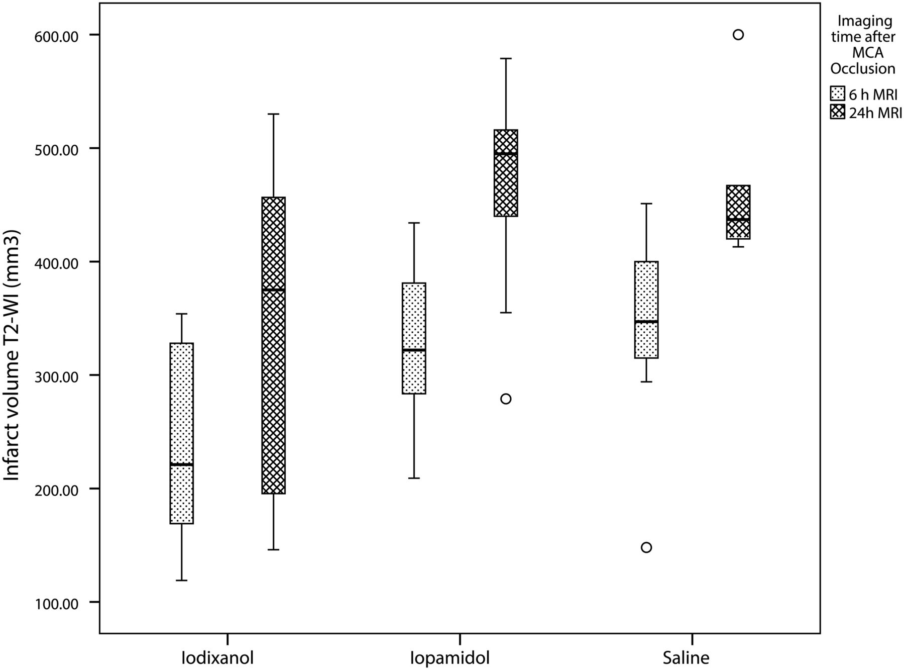
  • Fig 2.
    • Download figure
    • Open in new tab
    • Download powerpoint
    Fig 2.

    Boxplot analysis of absolute infarct volume per group and at 2 different times after ischemia (6 hours and 24 hours). There is increased volume of infarct in the iopamidol and saline groups compared with the iodixanol group.

  • Fig 3.
    • Download figure
    • Open in new tab
    • Download powerpoint
    Fig 3.

    Hemorrhagic transformation on sample animals. A, Iodixanol group. B, Iopamidol group. C, Saline group. Note the small amount of hemorrhage in the iodixanol rat (arrows in A) compared with larger areas in the other 2 animals (arrows in B and C).
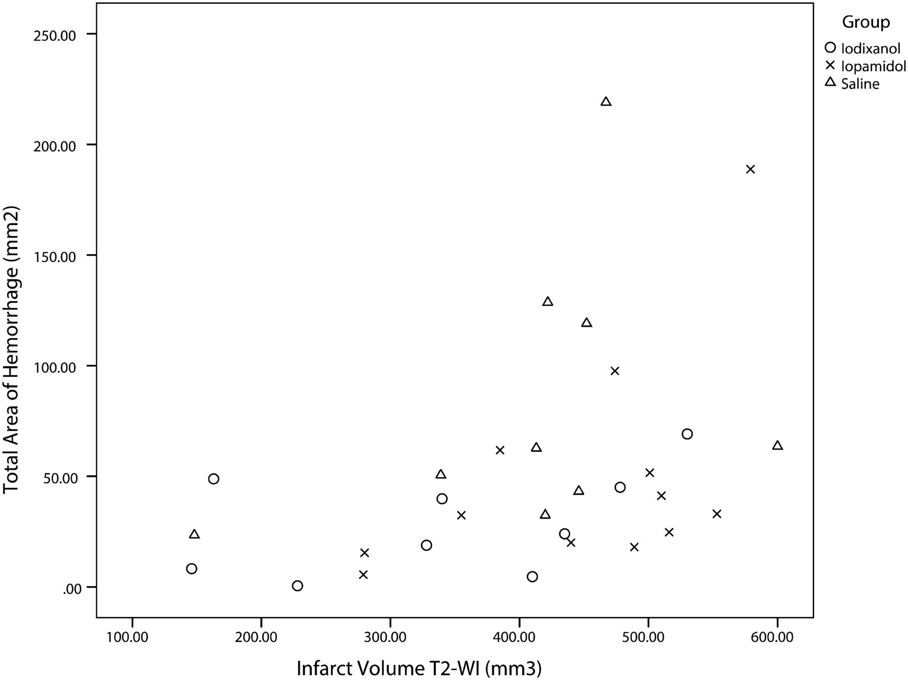
  • Fig 4.
    • Download figure
    • Open in new tab
    • Download powerpoint
    Fig 4.

    Scatterplot shows positive linear correlation between infarct volume and hemorrhagic transformation in all groups. The correlation is mild (r = 0.415), and in some cases, large infarct volumes were not associated with large hemorrhagic areas.

Tables

  • Figures
    • View popup
    Table 1:

    Absolute and relative infarct volumes at 6 and 24 hours after ischemia

    GroupRelative Infarct Volume (% Total Brain)Absolute Infarct Volume: T2WI (mm3)Absolute Infarct Volume: DWI (mm3)
    6 hours24 hours6 hours24 hours6 hours24 hours
    Iodixanol16.4 ± 6 (n = 9)22.7 ± 10 (n = 8)242 ± 89 (n = 9)341 ± 147 (n = 8)208 ± 90 (n = 7)328 ± 144 (n = 7)
    Iopamidol22.8 ± 5.5 (n = 12)32 ± 6.3 (n = 10)324 ± 70 (n = 12)470 ± 91 (n = 10)309 ± 49 (n = 10)438 ± 90 (n = 9)
    Saline24.4 ± 6.2 (n = 9)32.4 ± 5.5 (n = 6)345 ± 92 (n = 9)462 ± 71 (n = 6)331 ± 103 (n = 8)468 ± 63 (n = 5)
    Statistics (ANOVA)P < .018P < .028P < .03P < .047P < .018n.s.
    • Note:—n.s. indicates not significant.

    • View popup
    Table 2:

    Incidence and area of cortical and deep intracranial hemorrhage on cut-section inspection

    GroupCortexDeep (Basal Ganglia)Total
    IncidencebArea (mm2)aIncidenceArea (mm2)bIncidenceArea (mm2)b
    Iodixanol (n = 9)5 (56%)0.8 ± 1.59 (100%)28 ± 239 (100%)28.8 ± 23.3
    Iopamidol (n = 12)9 (75%)18.2 ± 3112 (100%)31 ± 24.612 (100%)49.2 ± 50.3
    Saline (n = 9)8 (89%)25.7 ± 39.79 (100%)56.7 ± 29.39 (100%)82.5 ± 62.7
    • ↵a P < .01.

    • ↵b Not significant.

PreviousNext
Back to top

In this issue

American Journal of Neuroradiology: 35 (10)
American Journal of Neuroradiology
Vol. 35, Issue 10
1 Oct 2014
  • Table of Contents
  • Index by author
  • Complete Issue (PDF)
Advertisement
Print
Download PDF
Email Article

Thank you for your interest in spreading the word on American Journal of Neuroradiology.

NOTE: We only request your email address so that the person you are recommending the page to knows that you wanted them to see it, and that it is not junk mail. We do not capture any email address.

Enter multiple addresses on separate lines or separate them with commas.
Decreased Infarct Volume and Intracranial Hemorrhage Associated with Intra-Arterial Nonionic Iso-Osmolar Contrast Material in an MCA Occlusion/Reperfusion Model
(Your Name) has sent you a message from American Journal of Neuroradiology
(Your Name) thought you would like to see the American Journal of Neuroradiology web site.
CAPTCHA
This question is for testing whether or not you are a human visitor and to prevent automated spam submissions.
Cite this article
H. Morales, A. Lu, Y. Kurosawa, J.F. Clark, J. Leach, K. Weiss, T. Tomsick
Decreased Infarct Volume and Intracranial Hemorrhage Associated with Intra-Arterial Nonionic Iso-Osmolar Contrast Material in an MCA Occlusion/Reperfusion Model
American Journal of Neuroradiology Oct 2014, 35 (10) 1885-1891; DOI: 10.3174/ajnr.A3953

Citation Manager Formats

  • BibTeX
  • Bookends
  • EasyBib
  • EndNote (tagged)
  • EndNote 8 (xml)
  • Medlars
  • Mendeley
  • Papers
  • RefWorks Tagged
  • Ref Manager
  • RIS
  • Zotero
0 Responses
Respond to this article
Share
Bookmark this article
Decreased Infarct Volume and Intracranial Hemorrhage Associated with Intra-Arterial Nonionic Iso-Osmolar Contrast Material in an MCA Occlusion/Reperfusion Model
H. Morales, A. Lu, Y. Kurosawa, J.F. Clark, J. Leach, K. Weiss, T. Tomsick
American Journal of Neuroradiology Oct 2014, 35 (10) 1885-1891; DOI: 10.3174/ajnr.A3953
del.icio.us logo Twitter logo Facebook logo Mendeley logo
  • Tweet Widget
  • Facebook Like
  • Google Plus One
Purchase

Jump to section

  • Article
    • Abstract
    • ABBREVIATIONS:
    • Materials and Methods
    • Results
    • Discussion
    • Conclusions
    • Acknowledgments
    • Footnotes
    • REFERENCES
  • Figures & Data
  • Info & Metrics
  • Responses
  • References
  • PDF

Related Articles

  • No related articles found.
  • PubMed
  • Google Scholar

Cited By...

  • Hemorrhagic Transformation Rates following Contrast Media Administration in Patients Hospitalized with Ischemic Stroke
  • Variable MR and pathologic patterns of hemorrhage after iodinated contrast infusion in MCA occlusion/reperfusion model
  • Outcome Differences between Intra-Arterial Iso- and Low-Osmolality Iodinated Radiographic Contrast Media in the Interventional Management of Stroke III Trial
  • Relevance of Blood-Brain Barrier Disruption After Endovascular Treatment of Ischemic Stroke: Dual-Energy Computed Tomographic Study
  • Crossref
  • Google Scholar

This article has not yet been cited by articles in journals that are participating in Crossref Cited-by Linking.

More in this TOC Section

  • Quiet PROPELLER MRI Techniques Match the Quality of Conventional PROPELLER Brain Imaging Techniques
  • Predictors of Reperfusion in Patients with Acute Ischemic Stroke
  • Enhanced Axonal Metabolism during Early Natalizumab Treatment in Relapsing-Remitting Multiple Sclerosis
Show more Brain

Similar Articles

Advertisement

Indexed Content

  • Current Issue
  • Accepted Manuscripts
  • Article Preview
  • Past Issues
  • Editorials
  • Editor's Choice
  • Fellows' Journal Club
  • Letters to the Editor
  • Video Articles

Cases

  • Case Collection
  • Archive - Case of the Week
  • Archive - Case of the Month
  • Archive - Classic Case

More from AJNR

  • Trainee Corner
  • Imaging Protocols
  • MRI Safety Corner
  • Book Reviews

Multimedia

  • AJNR Podcasts
  • AJNR Scantastics

Resources

  • Turnaround Time
  • Submit a Manuscript
  • Submit a Video Article
  • Submit an eLetter to the Editor/Response
  • Manuscript Submission Guidelines
  • Statistical Tips
  • Fast Publishing of Accepted Manuscripts
  • Graphical Abstract Preparation
  • Imaging Protocol Submission
  • Evidence-Based Medicine Level Guide
  • Publishing Checklists
  • Author Policies
  • Become a Reviewer/Academy of Reviewers
  • News and Updates

About Us

  • About AJNR
  • Editorial Board
  • Editorial Board Alumni
  • Alerts
  • Permissions
  • Not an AJNR Subscriber? Join Now
  • Advertise with Us
  • Librarian Resources
  • Feedback
  • Terms and Conditions
  • AJNR Editorial Board Alumni

American Society of Neuroradiology

  • Not an ASNR Member? Join Now

© 2025 by the American Society of Neuroradiology All rights, including for text and data mining, AI training, and similar technologies, are reserved.
Print ISSN: 0195-6108 Online ISSN: 1936-959X

Powered by HighWire