Advertisement

Main menu

  • Home
  • Content
    • Current Issue
    • Accepted Manuscripts
    • Article Preview
    • Past Issue Archive
    • Video Articles
    • AJNR Case Collection
    • Case of the Week Archive
    • Case of the Month Archive
    • Classic Case Archive
  • Special Collections
    • AJNR Awards
    • Low-Field MRI
    • Alzheimer Disease
    • ASNR Foundation Special Collection
    • Photon-Counting CT
    • View All
  • Multimedia
    • AJNR Podcasts
    • AJNR SCANtastic
    • Trainee Corner
    • MRI Safety Corner
    • Imaging Protocols
  • For Authors
    • Submit a Manuscript
    • Submit a Video Article
    • Submit an eLetter to the Editor/Response
    • Manuscript Submission Guidelines
    • Statistical Tips
    • Fast Publishing of Accepted Manuscripts
    • Graphical Abstract Preparation
    • Imaging Protocol Submission
    • Author Policies
  • About Us
    • About AJNR
    • Editorial Board
    • Editorial Board Alumni
  • More
    • Become a Reviewer/Academy of Reviewers
    • Subscribers
    • Permissions
    • Alerts
    • Feedback
    • Advertisers
    • ASNR Home

User menu

  • Alerts
  • Log in

Search

  • Advanced search
American Journal of Neuroradiology

American Journal of Neuroradiology

ASHNR American Society of Functional Neuroradiology ASHNR American Society of Pediatric Neuroradiology ASSR
  • Alerts
  • Log in

Advanced Search

  • Home
  • Content
    • Current Issue
    • Accepted Manuscripts
    • Article Preview
    • Past Issue Archive
    • Video Articles
    • AJNR Case Collection
    • Case of the Week Archive
    • Case of the Month Archive
    • Classic Case Archive
  • Special Collections
    • AJNR Awards
    • Low-Field MRI
    • Alzheimer Disease
    • ASNR Foundation Special Collection
    • Photon-Counting CT
    • View All
  • Multimedia
    • AJNR Podcasts
    • AJNR SCANtastic
    • Trainee Corner
    • MRI Safety Corner
    • Imaging Protocols
  • For Authors
    • Submit a Manuscript
    • Submit a Video Article
    • Submit an eLetter to the Editor/Response
    • Manuscript Submission Guidelines
    • Statistical Tips
    • Fast Publishing of Accepted Manuscripts
    • Graphical Abstract Preparation
    • Imaging Protocol Submission
    • Author Policies
  • About Us
    • About AJNR
    • Editorial Board
    • Editorial Board Alumni
  • More
    • Become a Reviewer/Academy of Reviewers
    • Subscribers
    • Permissions
    • Alerts
    • Feedback
    • Advertisers
    • ASNR Home
  • Follow AJNR on Twitter
  • Visit AJNR on Facebook
  • Follow AJNR on Instagram
  • Join AJNR on LinkedIn
  • RSS Feeds

AJNR Awards, New Junior Editors, and more. Read the latest AJNR updates

Research ArticleBrain

Calvarial Fracture Patterns on CT Imaging Predict Risk of a Delayed Epidural Hematoma following Decompressive Craniectomy for Traumatic Brain Injury

J.F. Talbott, A. Gean, E.L. Yuh and S.I. Stiver
American Journal of Neuroradiology October 2014, 35 (10) 1930-1935; DOI: https://doi.org/10.3174/ajnr.A4001
J.F. Talbott
aFrom the Department of Radiology and Biomedical Imaging (J.F.T., A.G., E.L.Y.)
bBrain and Spine Injury Center (J.F.T., A.G., E.L.Y.), San Francisco General Hospital, San Francisco, California
  • Find this author on Google Scholar
  • Find this author on PubMed
  • Search for this author on this site
A. Gean
aFrom the Department of Radiology and Biomedical Imaging (J.F.T., A.G., E.L.Y.)
bBrain and Spine Injury Center (J.F.T., A.G., E.L.Y.), San Francisco General Hospital, San Francisco, California
cDepartment of Neurological Surgery (A.G., S.I.S.), University of California, San Francisco, California.
  • Find this author on Google Scholar
  • Find this author on PubMed
  • Search for this author on this site
E.L. Yuh
aFrom the Department of Radiology and Biomedical Imaging (J.F.T., A.G., E.L.Y.)
bBrain and Spine Injury Center (J.F.T., A.G., E.L.Y.), San Francisco General Hospital, San Francisco, California
  • Find this author on Google Scholar
  • Find this author on PubMed
  • Search for this author on this site
S.I. Stiver
cDepartment of Neurological Surgery (A.G., S.I.S.), University of California, San Francisco, California.
  • Find this author on Google Scholar
  • Find this author on PubMed
  • Search for this author on this site
  • Article
  • Figures & Data
  • Supplemental
  • Info & Metrics
  • Responses
  • References
  • PDF
Loading

Article Figures & Data

Figures

  • Tables
  • Fig 1.
    • Download figure
    • Open in new tab
    • Download powerpoint
    Fig 1.

    DEDH following decompressive hemicraniectomy for traumatic brain injury. A, Axial noncontrast CT at the level of the corona radiata before initial decompression demonstrates diffuse effacement of the cerebral sulci and small holohemispheric subdural hematoma on the left. Small bilateral frontal hemorrhagic contusions and scattered subarachnoid hemorrhage were also present. B, Three-dimensional volume-rendered image reconstructed from bone algorithm noncontrast head CT demonstrates complex calvarial fractures on the right involving the right temporal, occipital, and parietal bones. C, Noncontrast head CT performed after left decompressive hemicraniectomy shows interval development of a new, large, heterogeneously attenuated epidural hematoma in the right parietal region with new right to left midline shift and left frontotemporal external herniation.

  • Fig 2.
    • Download figure
    • Open in new tab
    • Download powerpoint
    Fig 2.

    A, Axial noncontrast CT at the level of the internal capsules demonstrates a left frontal extra-axial hematoma (arrow). There is also diffuse sulcal effacement. B, Nondisplaced right-sided parietal calvarial fracture is present on the preoperative CT (arrow). Right frontal and temporal calvarial fractures were also present (not shown). Coronal suture diastasis is seen on the left. C, Following left-sided decompressive craniectomy, a large, predominantly parietal, DEDH developed adjacent to the patient's right parietal bone fracture. D, A decompressive craniectomy was subsequently performed on the right side with evacuation of DEDH and significant improvement in mass effect.
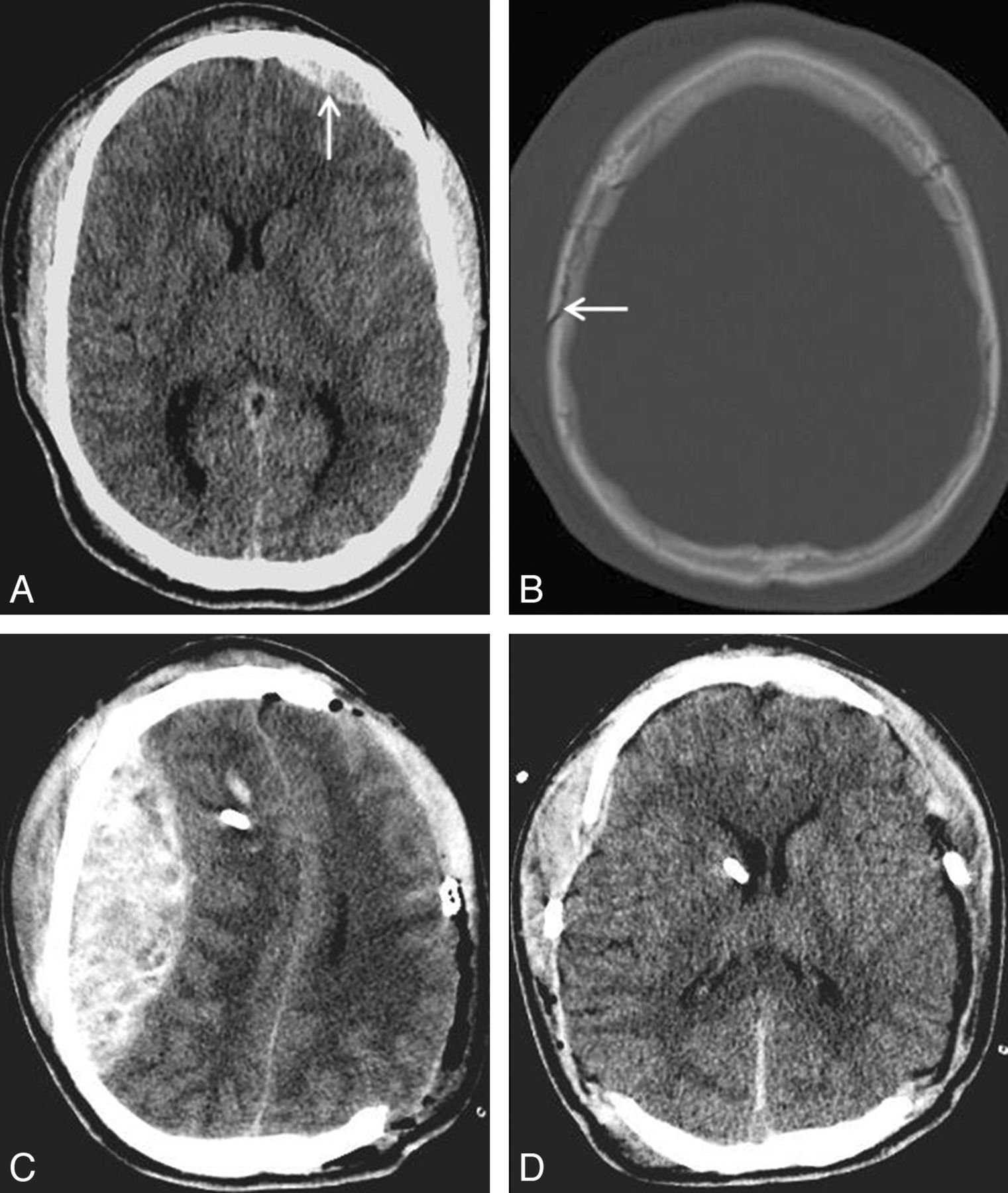
  • Fig 3.
    • Download figure
    • Open in new tab
    • Download powerpoint
    Fig 3.

    A, Axial noncontrast CT at the level of the internal capsules demonstrates a left-sided frontoparietal subdural hematoma (arrow). There is also bilateral subarachnoid hemorrhage, diffuse sulcal effacement, left to right midline shift, and near complete effacement of the left lateral ventricle with early trapping of the right lateral ventricle. B, Nondisplaced right occipital calvarial fracture paralleling the ipsilateral lambdoid suture is also identified on the preoperative head CT. A right sided parietal calvarial fracture was also present (not shown). C, Following left-sided decompressive craniectomy, postoperative head CT reveals a large right-sided occipital DEDH in the posterior fossa, adjacent to right-sided occipital bone fracture, with severe mass effect on the cerebellum and brain stem and effacement of the fourth ventricle.

Tables

  • Figures
  • Sensitivity, specificity, and diagnostic odds ratio analyses of contralateral calvarial skull fractures for DEDHs after DC

    Any Contralateral Calvarial Fracture (n = 55)Fractures Involving a Single Bone Plate (n = 33)Fractures Involving ≥2 Bone Plates (n = 22)
    Sensitivity100%25%75%
    Specificity77%83%94%
    Diagnostic odds ratioUndefineda1.841
    • ↵a The sensitivity of the finding “any calvarial fracture” is 100%, therefore false negatives = 0 and odds ratio is undefined.

PreviousNext
Back to top

In this issue

American Journal of Neuroradiology: 35 (10)
American Journal of Neuroradiology
Vol. 35, Issue 10
1 Oct 2014
  • Table of Contents
  • Index by author
  • Complete Issue (PDF)
Advertisement
Print
Download PDF
Email Article

Thank you for your interest in spreading the word on American Journal of Neuroradiology.

NOTE: We only request your email address so that the person you are recommending the page to knows that you wanted them to see it, and that it is not junk mail. We do not capture any email address.

Enter multiple addresses on separate lines or separate them with commas.
Calvarial Fracture Patterns on CT Imaging Predict Risk of a Delayed Epidural Hematoma following Decompressive Craniectomy for Traumatic Brain Injury
(Your Name) has sent you a message from American Journal of Neuroradiology
(Your Name) thought you would like to see the American Journal of Neuroradiology web site.
CAPTCHA
This question is for testing whether or not you are a human visitor and to prevent automated spam submissions.
Cite this article
J.F. Talbott, A. Gean, E.L. Yuh, S.I. Stiver
Calvarial Fracture Patterns on CT Imaging Predict Risk of a Delayed Epidural Hematoma following Decompressive Craniectomy for Traumatic Brain Injury
American Journal of Neuroradiology Oct 2014, 35 (10) 1930-1935; DOI: 10.3174/ajnr.A4001

Citation Manager Formats

  • BibTeX
  • Bookends
  • EasyBib
  • EndNote (tagged)
  • EndNote 8 (xml)
  • Medlars
  • Mendeley
  • Papers
  • RefWorks Tagged
  • Ref Manager
  • RIS
  • Zotero
0 Responses
Respond to this article
Share
Bookmark this article
Calvarial Fracture Patterns on CT Imaging Predict Risk of a Delayed Epidural Hematoma following Decompressive Craniectomy for Traumatic Brain Injury
J.F. Talbott, A. Gean, E.L. Yuh, S.I. Stiver
American Journal of Neuroradiology Oct 2014, 35 (10) 1930-1935; DOI: 10.3174/ajnr.A4001
del.icio.us logo Twitter logo Facebook logo Mendeley logo
  • Tweet Widget
  • Facebook Like
  • Google Plus One
Purchase

Jump to section

  • Article
    • Abstract
    • ABBREVIATIONS:
    • Materials and Methods
    • Results
    • Discussion
    • Conclusions
    • Footnotes
    • REFERENCES
  • Figures & Data
  • Supplemental
  • Info & Metrics
  • Responses
  • References
  • PDF

Related Articles

  • PubMed
  • Google Scholar

Cited By...

  • New-onset contralateral delayed extradural haematoma in an operated case of extradural haematoma: life-threatening if not diagnosed early
  • Head injury in the elderly - an overview for the physician
  • Predictive Value of Calvarial Fracture for Delayed Epidural Hematoma following Decompressive Craniectomy
  • Reply:
  • Crossref
  • Google Scholar

This article has not yet been cited by articles in journals that are participating in Crossref Cited-by Linking.

More in this TOC Section

  • Fast Contrast-Enhanced 4D MRA and 4D Flow MRI Using Constrained Reconstruction (HYPRFlow): Potential Applications for Brain Arteriovenous Malformations
  • Multimodal CT Provides Improved Performance for Lacunar Infarct Detection
  • Optimal MRI Sequence for Identifying Occlusion Location in Acute Stroke: Which Value of Time-Resolved Contrast-Enhanced MRA?
Show more Brain

Similar Articles

Advertisement

Indexed Content

  • Current Issue
  • Accepted Manuscripts
  • Article Preview
  • Past Issues
  • Editorials
  • Editor's Choice
  • Fellows' Journal Club
  • Letters to the Editor
  • Video Articles

Cases

  • Case Collection
  • Archive - Case of the Week
  • Archive - Case of the Month
  • Archive - Classic Case

More from AJNR

  • Trainee Corner
  • Imaging Protocols
  • MRI Safety Corner
  • Book Reviews

Multimedia

  • AJNR Podcasts
  • AJNR Scantastics

Resources

  • Turnaround Time
  • Submit a Manuscript
  • Submit a Video Article
  • Submit an eLetter to the Editor/Response
  • Manuscript Submission Guidelines
  • Statistical Tips
  • Fast Publishing of Accepted Manuscripts
  • Graphical Abstract Preparation
  • Imaging Protocol Submission
  • Evidence-Based Medicine Level Guide
  • Publishing Checklists
  • Author Policies
  • Become a Reviewer/Academy of Reviewers
  • News and Updates

About Us

  • About AJNR
  • Editorial Board
  • Editorial Board Alumni
  • Alerts
  • Permissions
  • Not an AJNR Subscriber? Join Now
  • Advertise with Us
  • Librarian Resources
  • Feedback
  • Terms and Conditions
  • AJNR Editorial Board Alumni

American Society of Neuroradiology

  • Not an ASNR Member? Join Now

© 2025 by the American Society of Neuroradiology All rights, including for text and data mining, AI training, and similar technologies, are reserved.
Print ISSN: 0195-6108 Online ISSN: 1936-959X

Powered by HighWire