
Article Figures & Data
Figures
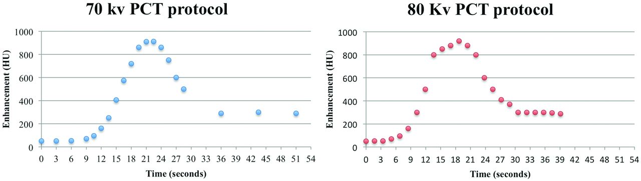
- Fig 1.
Sample middle-brain time-attenuation curves for the 2 CTP protocols.
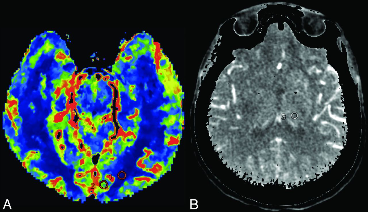
- Fig 2.
A, Parametric map showing placement of regions of interest in the WM and GM of the left medial occipital lobe. B, The region of interest is measured at the peak arterial phase over the left thalamus.
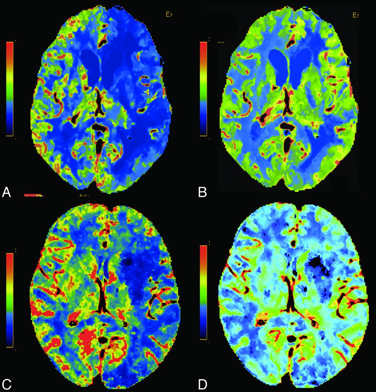
- Fig 3.
Normal-sample CTP datasets from the 70-kVp protocol: CBF (A), CBV (B), time-to-maximum (C), TTP (D). From the 80-kVp protocol: CBF (E), CBV (F), time-to-maximum (G), TTP (H).
- Fig 4.
Two different patients with left MCA ischemia. CBF (A) and CBV (B) at 70-kVp and CBF (C) and CBV (D) at 80-kVp CTP. Note the decreased CBF and preserved CBV.


Tables
70 kVp 80 kVp Tube current 150 mAs 80 mAs Section thickness 5 mm 5 mm Passes 20 passes 24 passes Range 144 mm 120 mm Time per pass 3, 1.5, 7.5 seconds 1.7 seconds Scan window 51 seconds 39 seconds 70 kVp 80 kVp P Value CBF WM Mean 5.47 8.76 .05 SD 2.26 4.47 .35 SNR 2.81 3.20 .66 GM Mean 40.03 40.76 .87 SD 14.84 16.94 .94 SNR 4.38 3.95 .92 CBV WM Mean 1.50 0.95 .44 SD 0.41 0.35 .21 SNR 3.15 4.97 .07 GM Mean 4.88 3.49 .75 SD 1.45 1.08 .21 SNR 5.34 6.53 .18 Tmax WM Mean 7.81 6.52 .10 SD 1.72 1.63 .47 SNR 6.22 7.08 .96 GM Mean 3.94 4.51 .13 SD 0.68 1.03 .31 SNR 6.82 6.72 .82 Arterial max Thalamus Mean 59.71 50.25 .0005 SD 11.96 11.34 .426 SNR 6.43 4.69 .3316 Venous max Thalamus Mean 56.72 48.68 .0032 SD 11.67 10.91 .3811 SNR 5.11 4.62 .4738 Note:—max indicates maximum; Tmax, time-to-maximum.
↵a Data are presented as mean, SD, and SNR.
- Table 3:
Qualitative analysis of overall quality and gray-white matter differentiation in 80-kVp and 70-kVp whole-brain CTPa
70 kVp 80 kVp P Value Overall quality CBF 2.95 ± 0.167 2.81 ± 2.84 .0459 CBV 2.95 ± 0.167 2.76 ± 0.305 .0173 TT 2.89 ± 0.224 2.69 ± 0.276 .0113 GM-WM differentiation CBF 2.91 ± 0.187 2.83 ± 0.314 .5598 CBV 2.93 ± 0.178 2.70 ± 0.347 .0278 TT 2.86 ± 0.231 2.69 ± 0.276 .0630 Note:—TT indicates transit time.
↵a Data are presented as means and SD.
- Table 4:
CTDIvol and DLP/ED values for whole-brain CTP examinations at 80 kVp and 70 kVp with different scanners
70 kVp 80 kVp Percentage Reduction 100% − (70 kVp/80 kVp) × 100 CTDIvol (mGy) 105 192 45.31 DLP (mGy·cm) 1588 1831 13.27 ED (mSv) 3.65 4.21 13.27 Note:—ED indicates effective dose.


