Advertisement

Main menu

  • Home
  • Content
    • Current Issue
    • Accepted Manuscripts
    • Article Preview
    • Past Issue Archive
    • AJNR Case Collection
    • Case of the Week Archive
    • Classic Case Archive
    • Case of the Month Archive
  • Special Collections
    • Spinal CSF Leak Articles (Jan 2020-June 2024)
    • 2024 AJNR Journal Awards
    • Most Impactful AJNR Articles
  • Multimedia
    • AJNR Podcast
    • AJNR Scantastics
    • Video Articles
  • For Authors
    • Submit a Manuscript
    • Author Policies
    • Fast publishing of Accepted Manuscripts
    • Graphical Abstract Preparation
    • Manuscript Submission Guidelines
    • Imaging Protocol Submission
    • Submit a Case for the Case Collection
  • About Us
    • About AJNR
    • Editorial Board
  • More
    • Become a Reviewer/Academy of Reviewers
    • Subscribers
    • Permissions
    • Alerts
    • Feedback
    • Advertisers
    • ASNR Home
  • Other Publications
    • ajnr

User menu

  • Alerts
  • Log in

Search

  • Advanced search
American Journal of Neuroradiology

American Journal of Neuroradiology

ASHNR American Society of Functional Neuroradiology ASHNR American Society of Pediatric Neuroradiology ASSR
  • Alerts
  • Log in

Advanced Search

  • Home
  • Content
    • Current Issue
    • Accepted Manuscripts
    • Article Preview
    • Past Issue Archive
    • AJNR Case Collection
    • Case of the Week Archive
    • Classic Case Archive
    • Case of the Month Archive
  • Special Collections
    • Spinal CSF Leak Articles (Jan 2020-June 2024)
    • 2024 AJNR Journal Awards
    • Most Impactful AJNR Articles
  • Multimedia
    • AJNR Podcast
    • AJNR Scantastics
    • Video Articles
  • For Authors
    • Submit a Manuscript
    • Author Policies
    • Fast publishing of Accepted Manuscripts
    • Graphical Abstract Preparation
    • Manuscript Submission Guidelines
    • Imaging Protocol Submission
    • Submit a Case for the Case Collection
  • About Us
    • About AJNR
    • Editorial Board
  • More
    • Become a Reviewer/Academy of Reviewers
    • Subscribers
    • Permissions
    • Alerts
    • Feedback
    • Advertisers
    • ASNR Home
  • Follow AJNR on Twitter
  • Visit AJNR on Facebook
  • Follow AJNR on Instagram
  • Join AJNR on LinkedIn
  • RSS Feeds

Welcome to the new AJNR, Updated Hall of Fame, and more. Read the full announcements.


AJNR is seeking candidates for the position of Associate Section Editor, AJNR Case Collection. Read the full announcement.


Research ArticleBrain

Evaluation of Normal Changes in Pons Metabolites due to Aging Using Turbo Spectroscopic Imaging

A. Yang, X.h. Xiao and Z.l. Wang
American Journal of Neuroradiology November 2014, 35 (11) 2099-2105; DOI: https://doi.org/10.3174/ajnr.A4031
A. Yang
aFrom the Medical Imaging Center, Affiliated Zhongshan City Hospital of Sun Yat-sen University, Zhongshan City, Guangdong Province, China.
  • Find this author on Google Scholar
  • Find this author on PubMed
  • Search for this author on this site
X.h. Xiao
aFrom the Medical Imaging Center, Affiliated Zhongshan City Hospital of Sun Yat-sen University, Zhongshan City, Guangdong Province, China.
  • Find this author on Google Scholar
  • Find this author on PubMed
  • Search for this author on this site
Z.l. Wang
aFrom the Medical Imaging Center, Affiliated Zhongshan City Hospital of Sun Yat-sen University, Zhongshan City, Guangdong Province, China.
  • Find this author on Google Scholar
  • Find this author on PubMed
  • Search for this author on this site
  • Article
  • Figures & Data
  • Info & Metrics
  • Responses
  • References
  • PDF
Loading

Article Figures & Data

Figures

  • Tables
  • Fig 1.
    • Download figure
    • Open in new tab
    • Download powerpoint
    Fig 1.

    Positioning of the pons TSI scan and spectrum data processing. A, T2-weighted images show the placement of volumes of interest in the pons. The left side is sagittal, and the right side is transversal. The TSI was positioned parallel to the frontal cranial fossa. Note that the box is the signal-generating area. B, The upper row of images shows the region-of-interest selection area, and the lower image shows the reconstructed average metabolite ratios of the region of interest.

  • Fig 2.
    • Download figure
    • Open in new tab
    • Download powerpoint
    Fig 2.

    Proton MR spectrum of the pons in subjects of different ages. A, Spectrum of a 1-month-old baby's pons. B, Spectrum of a 5-year-old boy's pons. C, Spectrum of a 63-year-old man's pons. Note the relatively high Cho and low NAA levels in the baby. There is a higher NAA level and a lower Cho level in the 5-year-old child compared with the baby. The Cho level is lower in the 63-year-old man compared with the 5-year-old child.
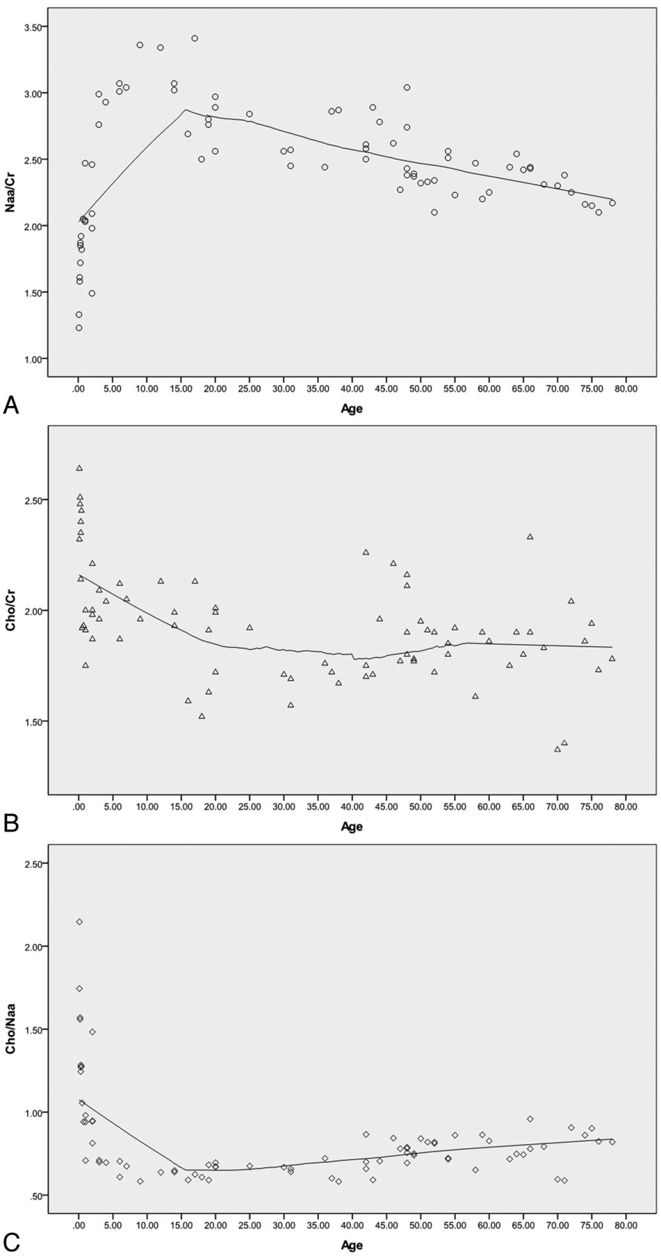
  • Fig 3.
    • Download figure
    • Open in new tab
    • Download powerpoint
    Fig 3.

    Scatterplot showing the metabolite changes in the pons with age. A, The scatterplot shows that NAA/Cr was low at birth, rose rapidly at 0–5 years, reaching a peak at approximately 10–20 years, and then gradually decreased. B, The Cho/Cr decreased rapidly at 0–5 years, then continued to decline, and was stable after 20 years of age. C, The Cho/NAA decreased rapidly at 0–5 years, then continued to decline, and finally rose after 20 years of age. Note that the curve-fitting model used is locally weighted scatterplot smoothing (LOESS), and the percentage of fitting points is 50%.

Tables

  • Figures
    • View popup
    Table 1:

    Comparison of NAA/Cr among different age groups

    Age Groups (yr)MeanNo.P Value
    0–52.14 ± 0.2520.001,a <.001b
    6–201.90 ± 0.2015.468,c .261d
    21–501.85 ± 0.1921.665e
    Older than 501.82 ± 0.2022<.001f
    • ↵a Comparison between 0 to 5- and 6 to 20-year-old subjects.

    • ↵b Comparison between 0 to 5- and 21 to 50-year-old subjects.

    • ↵c Comparison between 6 to 20- and 21 to 50-year-old subjects.

    • ↵d Comparison between 6 to 20- and older than 50-year-old subjects.

    • ↵e Comparison between 21 to 50- and older than 50-year-old subjects.

    • ↵f Comparison between 0 to 5- and older than 50-year-old subjects.

    • View popup
    Table 2:

    Comparison of Cho/Cr among different age groups

    Age Groups (yr)MeanNo.P Value
    0–52.14 ± 0.25200.001,a <.001b
    6–201.90 ± 0.20150.468,c .261d
    21–501.85 ± 0.1921.665e
    Older than 501.82 ± 0.2022<.001f
    • ↵a Comparison between 0 to 5- and 6 to 20-year-old subjects.

    • ↵b Comparison between 0 to 5- and 21 to 50-year-old subjects.

    • ↵c Comparison between 6 to 20- and 21 to 50-year-old subjects.

    • ↵d Comparison between 6 to 20- and older than 50-year-old subjects.

    • ↵e Comparison between 21 to 50- and older than 50-year-old subjects.

    • ↵f Comparison between 0 to 5- and older than 50-year-old subjects.

    • View popup
    Table 3:

    Comparison of Cho/NAA among different age groups

    Age Groups (yr)Median/QuartileNo.P Value
    0–51.01/0.84–1.4320<.001,a <.001b
    6–200.63/0.60–0.6715.005,c <.001d
    21–500.70/0.65–0.7821.015e
    Older than 500.81/0.72–0.8622.001f
    • ↵a Comparison between 0 to 5- and 6 to 20-year-old subjects.

    • ↵b Comparison between 0 to 5- and 21 to 50-year-old subjects.

    • ↵c Comparison between 6 to 20- and 21 to 50-year-old subjects.

    • ↵d Comparison between 6 to 20- and older than 50-year-old subjects.

    • ↵e Comparison between 21 to 50- and older than 50-year-old subjects.

    • ↵f Comparison between 0 to 5- and older than 50-year-old subjects.

PreviousNext
Back to top

In this issue

American Journal of Neuroradiology: 35 (11)
American Journal of Neuroradiology
Vol. 35, Issue 11
1 Nov 2014
  • Table of Contents
  • Index by author
  • Complete Issue (PDF)
Advertisement
Print
Download PDF
Email Article

Thank you for your interest in spreading the word on American Journal of Neuroradiology.

NOTE: We only request your email address so that the person you are recommending the page to knows that you wanted them to see it, and that it is not junk mail. We do not capture any email address.

Enter multiple addresses on separate lines or separate them with commas.
Evaluation of Normal Changes in Pons Metabolites due to Aging Using Turbo Spectroscopic Imaging
(Your Name) has sent you a message from American Journal of Neuroradiology
(Your Name) thought you would like to see the American Journal of Neuroradiology web site.
CAPTCHA
This question is for testing whether or not you are a human visitor and to prevent automated spam submissions.
Cite this article
A. Yang, X.h. Xiao, Z.l. Wang
Evaluation of Normal Changes in Pons Metabolites due to Aging Using Turbo Spectroscopic Imaging
American Journal of Neuroradiology Nov 2014, 35 (11) 2099-2105; DOI: 10.3174/ajnr.A4031

Citation Manager Formats

  • BibTeX
  • Bookends
  • EasyBib
  • EndNote (tagged)
  • EndNote 8 (xml)
  • Medlars
  • Mendeley
  • Papers
  • RefWorks Tagged
  • Ref Manager
  • RIS
  • Zotero
0 Responses
Respond to this article
Share
Bookmark this article
Evaluation of Normal Changes in Pons Metabolites due to Aging Using Turbo Spectroscopic Imaging
A. Yang, X.h. Xiao, Z.l. Wang
American Journal of Neuroradiology Nov 2014, 35 (11) 2099-2105; DOI: 10.3174/ajnr.A4031
del.icio.us logo Twitter logo Facebook logo Mendeley logo
  • Tweet Widget
  • Facebook Like
  • Google Plus One
Purchase

Jump to section

  • Article
    • Abstract
    • ABBREVIATION:
    • Materials and Methods
    • Results
    • Discussion
    • Conclusions
    • References
  • Figures & Data
  • Info & Metrics
  • Responses
  • References
  • PDF

Related Articles

  • No related articles found.
  • PubMed
  • Google Scholar

Cited By...

  • No citing articles found.
  • Crossref (6)
  • Google Scholar

This article has been cited by the following articles in journals that are participating in Crossref Cited-by Linking.

  • Neurochemical changes in the aging brain: A systematic review
    Carlee Cleeland, Andrew Pipingas, Andrew Scholey, David White
    Neuroscience & Biobehavioral Reviews 2019 98
  • From fetus to older age: A review of brain metabolic changes across the lifespan
    Monika Cichocka, Anna Bereś
    Ageing Research Reviews 2018 46
  • Fast magnetic resonance spectroscopic imaging techniques in human brain- applications in multiple sclerosis
    Oun Al-iedani, Jeannette Lechner-Scott, Karen Ribbons, Saadallah Ramadan
    Journal of Biomedical Science 2017 24 1
  • Metabolic Imaging in Humans
    Taylor L. Fuss, Leo L. Cheng
    Topics in Magnetic Resonance Imaging 2016 25 5
  • Organization of neurochemical interactions in young and older brains as revealed with a network approach: Evidence from proton magnetic resonance spectroscopy (1H-MRS)
    Geraldine Rodríguez-Nieto, Oron Levin, Lize Hermans, Akila Weerasekera, Anca Croitor Sava, Astrid Haghebaert, Astrid Huybrechts, Koen Cuypers, Dante Mantini, Uwe Himmelreich, Stephan P. Swinnen
    NeuroImage 2023 266
  • Metabolomic Biomarkers Are Associated With Area of the Pons in Fragile X Premutation Carriers at Risk for Developing FXTAS
    Marwa Zafarullah, Blythe Durbin-Johnson, Emily S. Fourie, David R. Hessl, Susan M. Rivera, Flora Tassone
    Frontiers in Psychiatry 2021 12

More in this TOC Section

  • Evaluating the Effects of White Matter Multiple Sclerosis Lesions on the Volume Estimation of 6 Brain Tissue Segmentation Methods
  • White Matter Alterations in the Brains of Patients with Active, Remitted, and Cured Cushing Syndrome: A DTI Study
  • Enhanced Axonal Metabolism during Early Natalizumab Treatment in Relapsing-Remitting Multiple Sclerosis
Show more Brain

Similar Articles

Advertisement

Indexed Content

  • Current Issue
  • Accepted Manuscripts
  • Article Preview
  • Past Issues
  • Editorials
  • Editors Choice
  • Fellow Journal Club
  • Letters to the Editor

Cases

  • Case Collection
  • Archive - Case of the Week
  • Archive - Case of the Month
  • Archive - Classic Case

Special Collections

  • Special Collections

Resources

  • News and Updates
  • Turn around Times
  • Submit a Manuscript
  • Author Policies
  • Manuscript Submission Guidelines
  • Evidence-Based Medicine Level Guide
  • Publishing Checklists
  • Graphical Abstract Preparation
  • Imaging Protocol Submission
  • Submit a Case
  • Become a Reviewer/Academy of Reviewers
  • Get Peer Review Credit from Publons

Multimedia

  • AJNR Podcast
  • AJNR SCANtastic
  • Video Articles

About Us

  • About AJNR
  • Editorial Board
  • Not an AJNR Subscriber? Join Now
  • Alerts
  • Feedback
  • Advertise with us
  • Librarian Resources
  • Permissions
  • Terms and Conditions

American Society of Neuroradiology

  • Not an ASNR Member? Join Now

© 2025 by the American Society of Neuroradiology All rights, including for text and data mining, AI training, and similar technologies, are reserved.
Print ISSN: 0195-6108 Online ISSN: 1936-959X

Powered by HighWire