
Article Figures & Data
Figures
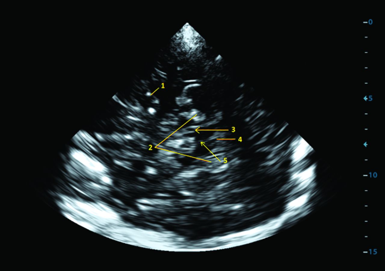
- Fig 1.
Transcranial sonography: brain stem with the substantia nigra imaged from a transtemporal approach, axial mesencephalic plane. 1) middle cerebral artery, 2) perimesencephalic cisterns, 3) substantia nigra, 4) fourth ventricle, and 5) brain raphe.
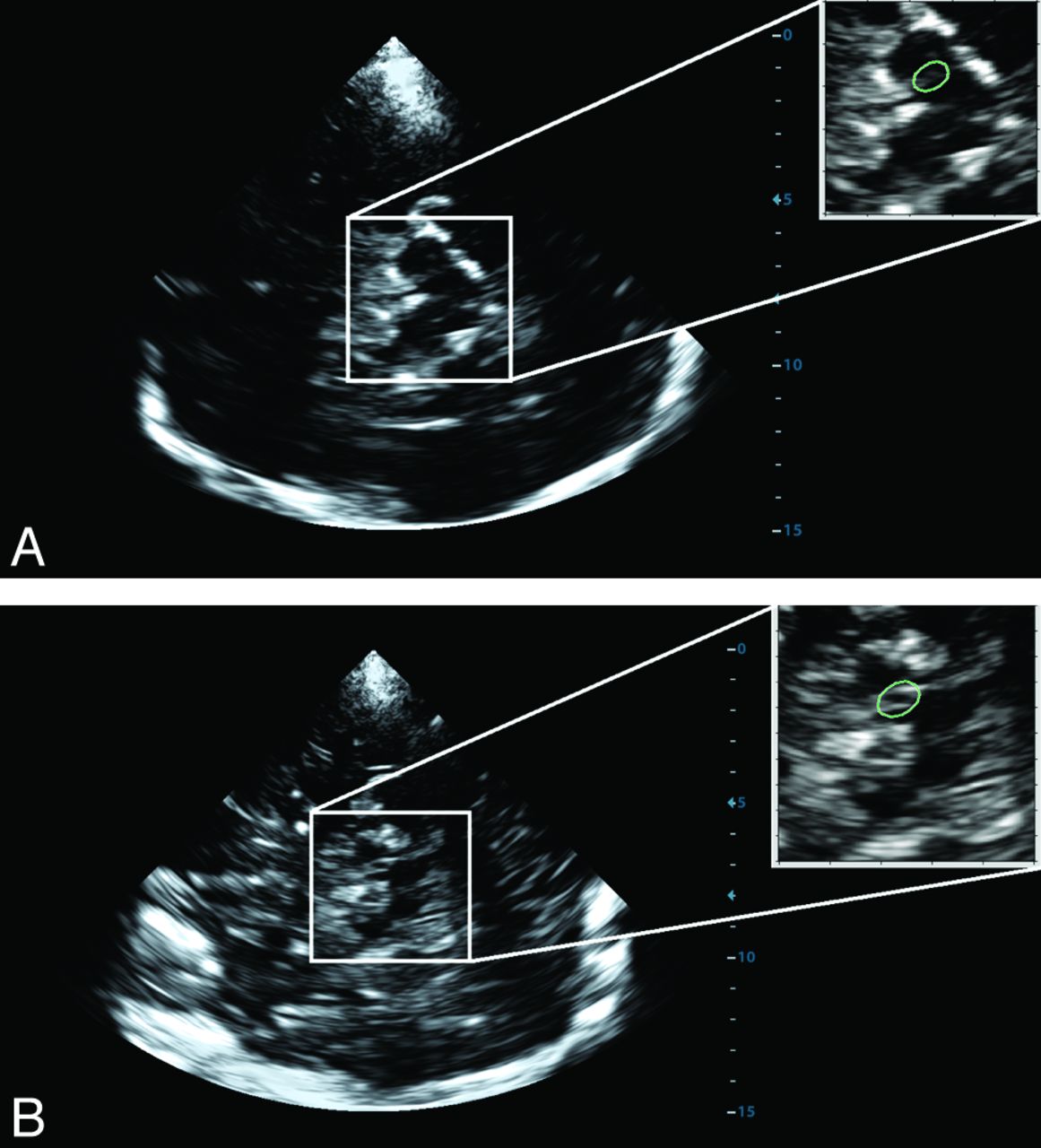
- Fig 2.
Placement of the region of interest (green) for digital analysis on the TCS brain stem images obtained from a healthy volunteer (A) and a patient with Parkinson disease (B).
- Fig 3.
The difference between the 90th percentile of the derivation cohort (red line; echogenicity index, 18.536) and the counted value in the validation cohort (black) measured by using machine 1. A, Normal echogenicity of the substantia nigra (echogenicity index, 11.760). B, Hyperechogenic substantia nigra (echogenicity index, 31.810).
- Fig 4.
The 90th percentile of the derivation cohort for the Esaote My Lab Twice (machine 1) and GE Healthcare Vivid 7 Pro (machine 2) machines.
- Fig 5.
Receiver operating characteristic curve for Parkinson disease diagnosis using manual measurement (blue) and digital analysis (red) of the substantia nigra for machine 1.


Tables
Derivation Cohort Healthy Volunteers Validation Cohort Healthy Volunteers Patients with PD No. of subjects 113 50 30 Mean age (yr) 52.3 ± 13.1 54.1 ± 12.2 56.9 ± 10.5 Male sex (No.) (%) 58 (51.3) 25 (50.0) 19 (63.3) Median UPDRS-III (IQR, range) NA NA 29.5 (20.5–38.5; 12–47) Median Hoehn and Yahr stage (IQR, range) NA NA 2 (1–3; 1–3) L-DOPA therapy (No.) (%) NA NA 19 (63.3%) DA therapy (No.) (%) NA NA 15 (50.0) Mean of disease duration (mo) (range) NA NA 28.4 ± 12.0 (6–48) Note:—DA indicates dopamine agonist; IQR, interquartile range; NA, not applicable; UPDRS, Unified Parkinson's Rating Scale.
- Table 2:
Correlations between manual measurement and digital analysis of substantia nigra echogenicity for both machines, between measurements performed using different machines, and between the right and left substantia nigra using both manual measurement and digital analysis
Correlations Spearman Coefficient P Value Between manual measurement and digital analysis for My Lab Twice 0.630 <.0001 Between manual measurement and digital analysis for Vivid Pro 7 0.553 <.0001 Between digital analysis using different machines 0.686 <.0001 Between visual measurements using different machines 0.721 <.0001 Between the right and left substantia nigra using digital analysis and My Lab Twice 0.575 <.0001 Between the right and left substantia nigra using digital analysis and Vivid Pro 7 0.512 .0001 Between the right and left substantia nigra using manual measurement and My Lab Twice 0.494 .0003 Between the right and left substantia nigra using manual measurement and Vivid Pro 7 0.631 <.0001


Jump to section
Related Articles
Cited By...
- No citing articles found.