Advertisement

Main menu

  • Home
  • Content
    • Current Issue
    • Accepted Manuscripts
    • Article Preview
    • Past Issue Archive
    • Video Articles
    • AJNR Case Collection
    • Case of the Week Archive
    • Case of the Month Archive
    • Classic Case Archive
  • Special Collections
    • AJNR Awards
    • Low-Field MRI
    • Alzheimer Disease
    • ASNR Foundation Special Collection
    • Photon-Counting CT
    • View All
  • Multimedia
    • AJNR Podcasts
    • AJNR SCANtastic
    • Trainee Corner
    • MRI Safety Corner
    • Imaging Protocols
  • For Authors
    • Submit a Manuscript
    • Submit a Video Article
    • Submit an eLetter to the Editor/Response
    • Manuscript Submission Guidelines
    • Statistical Tips
    • Fast Publishing of Accepted Manuscripts
    • Graphical Abstract Preparation
    • Imaging Protocol Submission
    • Author Policies
  • About Us
    • About AJNR
    • Editorial Board
    • Editorial Board Alumni
  • More
    • Become a Reviewer/Academy of Reviewers
    • Subscribers
    • Permissions
    • Alerts
    • Feedback
    • Advertisers
    • ASNR Home

User menu

  • Alerts
  • Log in

Search

  • Advanced search
American Journal of Neuroradiology

American Journal of Neuroradiology

ASHNR American Society of Functional Neuroradiology ASHNR American Society of Pediatric Neuroradiology ASSR
  • Alerts
  • Log in

Advanced Search

  • Home
  • Content
    • Current Issue
    • Accepted Manuscripts
    • Article Preview
    • Past Issue Archive
    • Video Articles
    • AJNR Case Collection
    • Case of the Week Archive
    • Case of the Month Archive
    • Classic Case Archive
  • Special Collections
    • AJNR Awards
    • Low-Field MRI
    • Alzheimer Disease
    • ASNR Foundation Special Collection
    • Photon-Counting CT
    • View All
  • Multimedia
    • AJNR Podcasts
    • AJNR SCANtastic
    • Trainee Corner
    • MRI Safety Corner
    • Imaging Protocols
  • For Authors
    • Submit a Manuscript
    • Submit a Video Article
    • Submit an eLetter to the Editor/Response
    • Manuscript Submission Guidelines
    • Statistical Tips
    • Fast Publishing of Accepted Manuscripts
    • Graphical Abstract Preparation
    • Imaging Protocol Submission
    • Author Policies
  • About Us
    • About AJNR
    • Editorial Board
    • Editorial Board Alumni
  • More
    • Become a Reviewer/Academy of Reviewers
    • Subscribers
    • Permissions
    • Alerts
    • Feedback
    • Advertisers
    • ASNR Home
  • Follow AJNR on Twitter
  • Visit AJNR on Facebook
  • Follow AJNR on Instagram
  • Join AJNR on LinkedIn
  • RSS Feeds

AJNR Awards, New Junior Editors, and more. Read the latest AJNR updates

Research ArticleBrain

Automatic Quantification of Subarachnoid Hemorrhage on Noncontrast CT

A.M. Boers, I.A. Zijlstra, C.S. Gathier, R. van den Berg, C.H. Slump, H.A. Marquering and C.B. Majoie
American Journal of Neuroradiology December 2014, 35 (12) 2279-2286; DOI: https://doi.org/10.3174/ajnr.A4042
A.M. Boers
aFrom the Departments of Radiology (A.M.B., I.A.Z., R.v.d.B., H.A.M., C.B.M.)
bBiomedical Engineering and Physics (A.M.B., H.A.M.), Academic Medical Center, Amsterdam, the Netherlands
cInstitute of Technical Medicine (A.M.B.)
  • Find this author on Google Scholar
  • Find this author on PubMed
  • Search for this author on this site
I.A. Zijlstra
aFrom the Departments of Radiology (A.M.B., I.A.Z., R.v.d.B., H.A.M., C.B.M.)
  • Find this author on Google Scholar
  • Find this author on PubMed
  • Search for this author on this site
C.S. Gathier
eDepartment of Neurology (C.S.G.), University Medical Centre Utrecht, Utrecht, the Netherlands.
  • Find this author on Google Scholar
  • Find this author on PubMed
  • Search for this author on this site
R. van den Berg
aFrom the Departments of Radiology (A.M.B., I.A.Z., R.v.d.B., H.A.M., C.B.M.)
  • Find this author on Google Scholar
  • Find this author on PubMed
  • Search for this author on this site
C.H. Slump
dMIRA Institute for Biomedical Technology and Technical Medicine (C.H.S.), University of Twente, Enschede, the Netherlands
  • Find this author on Google Scholar
  • Find this author on PubMed
  • Search for this author on this site
H.A. Marquering
aFrom the Departments of Radiology (A.M.B., I.A.Z., R.v.d.B., H.A.M., C.B.M.)
bBiomedical Engineering and Physics (A.M.B., H.A.M.), Academic Medical Center, Amsterdam, the Netherlands
  • Find this author on Google Scholar
  • Find this author on PubMed
  • Search for this author on this site
C.B. Majoie
aFrom the Departments of Radiology (A.M.B., I.A.Z., R.v.d.B., H.A.M., C.B.M.)
  • Find this author on Google Scholar
  • Find this author on PubMed
  • Search for this author on this site
  • Article
  • Figures & Data
  • Info & Metrics
  • Responses
  • References
  • PDF
Loading

Article Figures & Data

Figures

  • Tables
  • Fig 1.
    • Download figure
    • Open in new tab
    • Download powerpoint
    Fig 1.

    Illustration of differentiation between partial volume effect and hemorrhage in the vicinity of the skull. A, Calculation of the density gradient of an NCCT image with high hypodensities near the skull. High gradients are expected to be caused by partial volume effects in contrast to hemorrhages, which result in low gradients as seen in B and C. The pixels corresponding to low gradients (blue) are excluded from further segmentation. The white arrows mark the areas with high gradients present in the CSF image.
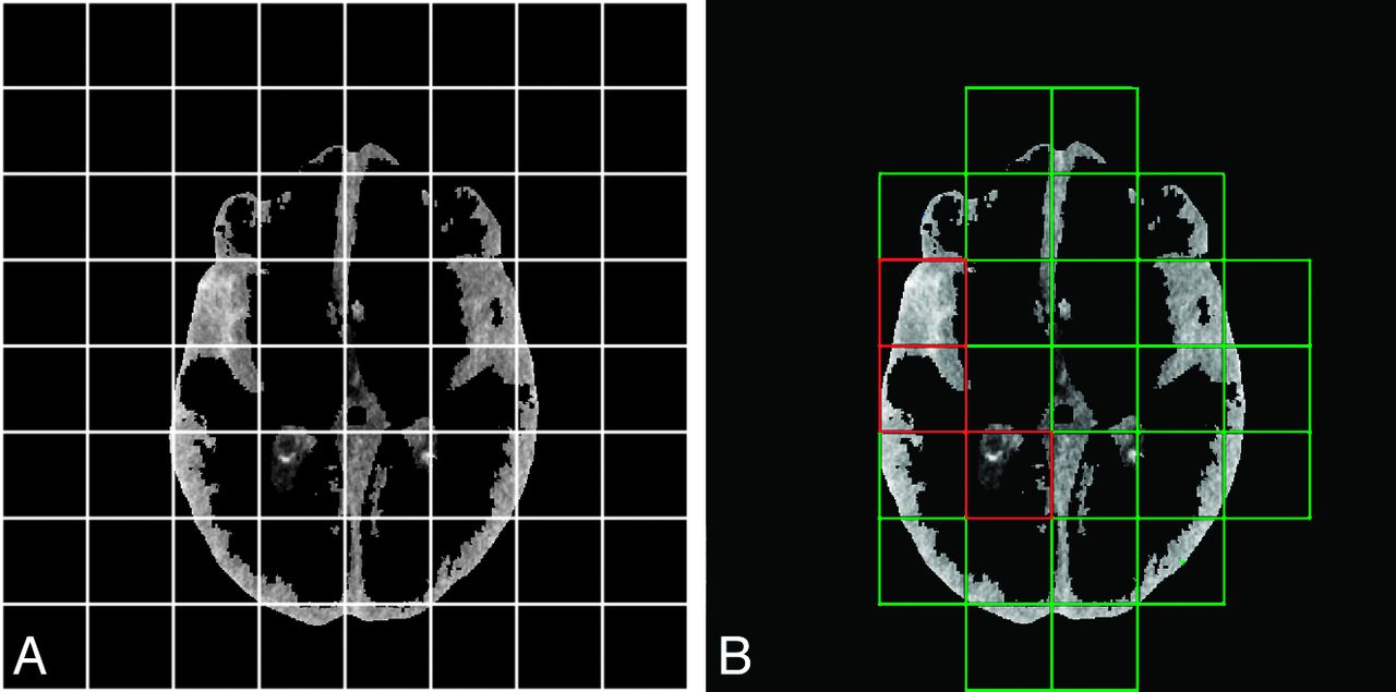
  • Fig 2.
    • Download figure
    • Open in new tab
    • Download powerpoint
    Fig 2.

    Illustration of the correction for patient-specific density differences. A, Each section of a specific tissue type (here CSF) is divided into 64 tiles, and the SDs of the density were calculated. B, Green tiles represent those with a low SD of the density and are expected to be free of a substantial amount of extravasated blood and therefore mainly consist of healthy brain tissue, whereas tiles with a high SD (red tiles) are more likely to contain hemorrhage. The densities in the green tiles were included in the calculation of the mean density of that tissue type. Comparison with the mean density of that tissue type in the reference image resulted in a density offset, which was corrected.
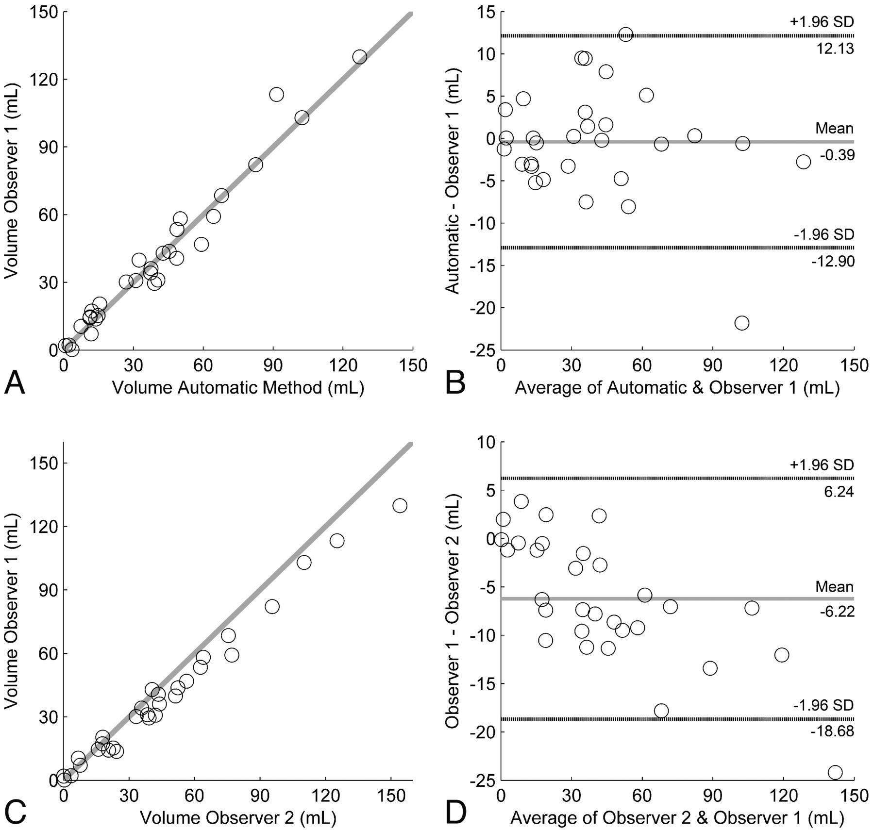
  • Fig 3.
    • Download figure
    • Open in new tab
    • Download powerpoint
    Fig 3.

    The accuracy of the volume measurement of the automatic method compared with that in observer 1. A, Accuracy depicted as a scatterplot. B, Accuracy shown by the Bland-Altman plot. Interobserver variability of the manual hemorrhage volume measurement depicted as C, scatterplot, and D, Bland-Altman analysis.

  • Fig 4.
    • Download figure
    • Open in new tab
    • Download powerpoint
    Fig 4.

    Correlation of the Hijdra score and Fisher score with hemorrhage volume after SAH assessed with scatterplots. A, The Fisher score with automatic volume measurement (blue) and manual volume measurement (red). B, The Hijdra score with automatic volume measurement (blue) and manual volume measurement (red).

  • Fig 5.
    • Download figure
    • Open in new tab
    • Download powerpoint
    Fig 5.

    Example of the results of the automatic segmentation of extravasated blood after SAH and manual measurement. A, NCCT image, shown in red, with results of the automatic method of a relatively small hemorrhage. B, The same NCCT image and hemorrhage as delineated by observer 1 (blue). C, NCCT image with beam-hardening in an extreme degree. The automatic method, in red, shows deviations of the hemorrhage volume as delineated by observer 1 (D) in blue.

Tables

  • Figures
    • View popup
    Table 1:

    Clinical and radiographic characteristics of the study population

    CharacteristicsTest Set (No.) (%)Training Set (No.) (%)
    Sex
        Male12 (40)5 (75)
        Female18 (60)15 (25)
    Age (yr)
        45 or younger7 (23)5 (25)
        46–6016 (53)11 (55)
        Older than 607 (23)4 (20)
    IVH
        Yes16 (53)4 (20)
        No14 (47)16 (80)
    ICH
        Yes19 (63)12 (60)
        No11 (37)8 (40)
    MCA location
        Left14 (47)18 (10)
        Right16 (53)2 (90)
    WFNS at admission
        Grade I11 (37)9 (45)
        Grade II2 (7)2 (10)
        Grade III2 (7)4 (20)
        Grade IV8 (27)3 (15)
        Grade V7 (23)2 (10)
    History of hypertension
        Yes3 (10)7 (35)
        No22 (73)13 (65)
    Aneurysm size
        ≤5 mm9 (30)6 (30)
        6–10 mm14 (47)12 (60)
        11–15 mm3 (10)2 (10)
        >15 mm3 (10)–
    Signs of DCI
        No21 (70)19 (95)
        Yes9 (30)1 (5)
            Paresis1 (3)1 (5)
            Decreased consciousness4 (13)–
            Hemiparesis and aphasia3 (10)–
            Vasospasm1 (3)–
    • Note:—IVH indicates intraventricular hemorrhage; ICH, intracerebral hemorrhage; DCI, delayed cerebral ischemia; WFNS, World Federation of Neurosurgical Societies.

    • View popup
    Table 2:

    Interobserver variability of manual SAH volume measurement and comparison of the manual and automatic methods

    ICC Volumea (95% CI)Bland-Altman Volume Limits of Agreement (mL)ICC Densitya (95% CI)Bland-Altman Density Limits of Agreement (HU)Dice Coefficient (Mean and Range)No.b
    Automatic interobserver0.98 (0.96–0.99)−12.90–12.130.80 (0.62–0.90)−7.58–9.180.55 (0.00–0.83)30
    Manual interobserver0.97 (0.77–0.99)−18.68–6.240.98 (0.89–0.99)c−1.52–3.44c0.64 (0.00–0.86)30
    • ↵a Case 3 intraclass correlation coefficients using an absolute agreement definition.

    • ↵b Number of NCCT scans included in the calculation.

    • ↵c Number of NCCT scans included for calculation = 29.

PreviousNext
Back to top

In this issue

American Journal of Neuroradiology: 35 (12)
American Journal of Neuroradiology
Vol. 35, Issue 12
1 Dec 2014
  • Table of Contents
  • Index by author
  • Complete Issue (PDF)
Advertisement
Print
Download PDF
Email Article

Thank you for your interest in spreading the word on American Journal of Neuroradiology.

NOTE: We only request your email address so that the person you are recommending the page to knows that you wanted them to see it, and that it is not junk mail. We do not capture any email address.

Enter multiple addresses on separate lines or separate them with commas.
Automatic Quantification of Subarachnoid Hemorrhage on Noncontrast CT
(Your Name) has sent you a message from American Journal of Neuroradiology
(Your Name) thought you would like to see the American Journal of Neuroradiology web site.
CAPTCHA
This question is for testing whether or not you are a human visitor and to prevent automated spam submissions.
Cite this article
A.M. Boers, I.A. Zijlstra, C.S. Gathier, R. van den Berg, C.H. Slump, H.A. Marquering, C.B. Majoie
Automatic Quantification of Subarachnoid Hemorrhage on Noncontrast CT
American Journal of Neuroradiology Dec 2014, 35 (12) 2279-2286; DOI: 10.3174/ajnr.A4042

Citation Manager Formats

  • BibTeX
  • Bookends
  • EasyBib
  • EndNote (tagged)
  • EndNote 8 (xml)
  • Medlars
  • Mendeley
  • Papers
  • RefWorks Tagged
  • Ref Manager
  • RIS
  • Zotero
0 Responses
Respond to this article
Share
Bookmark this article
Automatic Quantification of Subarachnoid Hemorrhage on Noncontrast CT
A.M. Boers, I.A. Zijlstra, C.S. Gathier, R. van den Berg, C.H. Slump, H.A. Marquering, C.B. Majoie
American Journal of Neuroradiology Dec 2014, 35 (12) 2279-2286; DOI: 10.3174/ajnr.A4042
del.icio.us logo Twitter logo Facebook logo Mendeley logo
  • Tweet Widget
  • Facebook Like
  • Google Plus One
Purchase

Jump to section

  • Article
    • Abstract
    • ABBREVIATIONS:
    • Materials and Methods
    • Results
    • Discussion
    • Conclusions
    • Footnotes
    • References
  • Figures & Data
  • Info & Metrics
  • Responses
  • References
  • PDF

Related Articles

  • No related articles found.
  • PubMed
  • Google Scholar

Cited By...

  • Optimizing Photon-Counting Detector CT for Imaging Intracranial Aneurysms
  • Integrating Clinical Data and Radiomics and Deep Learning Features for End-to-End Delayed Cerebral Ischemia Prediction on Noncontrast CT
  • Deep Learning-based Multiclass Segmentation in Aneurysmal Subarachnoid Hemorrhage
  • Predicting vasospasm risk using first presentation aneurysmal subarachnoid haemorrhage volume: a semi-automated CT image segmentation analysis in ITK-SNAP
  • Prediction of Outcome Using Quantified Blood Volume in Aneurysmal SAH
  • Association of Quantified Location-Specific Blood Volumes with Delayed Cerebral Ischemia after Aneurysmal Subarachnoid Hemorrhage
  • Association of Computed Tomography Ischemic Lesion Location With Functional Outcome in Acute Large Vessel Occlusion Ischemic Stroke
  • Topographic distribution of cerebral infarct probability in patients with acute ischemic stroke: mapping of intra-arterial treatment effect
  • Association of Automatically Quantified Total Blood Volume after Aneurysmal Subarachnoid Hemorrhage with Delayed Cerebral Ischemia
  • Crossref
  • Google Scholar

This article has not yet been cited by articles in journals that are participating in Crossref Cited-by Linking.

More in this TOC Section

  • Progression of Microstructural Damage in Spinocerebellar Ataxia Type 2: A Longitudinal DTI Study
  • SWI or T2*: Which MRI Sequence to Use in the Detection of Cerebral Microbleeds? The Karolinska Imaging Dementia Study
  • Statin Therapy Does Not Affect the Radiographic and Clinical Profile of Patients with TIA and Minor Stroke
Show more Brain

Similar Articles

Advertisement

Indexed Content

  • Current Issue
  • Accepted Manuscripts
  • Article Preview
  • Past Issues
  • Editorials
  • Editor's Choice
  • Fellows' Journal Club
  • Letters to the Editor
  • Video Articles

Cases

  • Case Collection
  • Archive - Case of the Week
  • Archive - Case of the Month
  • Archive - Classic Case

More from AJNR

  • Trainee Corner
  • Imaging Protocols
  • MRI Safety Corner
  • Book Reviews

Multimedia

  • AJNR Podcasts
  • AJNR Scantastics

Resources

  • Turnaround Time
  • Submit a Manuscript
  • Submit a Video Article
  • Submit an eLetter to the Editor/Response
  • Manuscript Submission Guidelines
  • Statistical Tips
  • Fast Publishing of Accepted Manuscripts
  • Graphical Abstract Preparation
  • Imaging Protocol Submission
  • Evidence-Based Medicine Level Guide
  • Publishing Checklists
  • Author Policies
  • Become a Reviewer/Academy of Reviewers
  • News and Updates

About Us

  • About AJNR
  • Editorial Board
  • Editorial Board Alumni
  • Alerts
  • Permissions
  • Not an AJNR Subscriber? Join Now
  • Advertise with Us
  • Librarian Resources
  • Feedback
  • Terms and Conditions
  • AJNR Editorial Board Alumni

American Society of Neuroradiology

  • Not an ASNR Member? Join Now

© 2025 by the American Society of Neuroradiology All rights, including for text and data mining, AI training, and similar technologies, are reserved.
Print ISSN: 0195-6108 Online ISSN: 1936-959X

Powered by HighWire