
Article Figures & Data
Figures
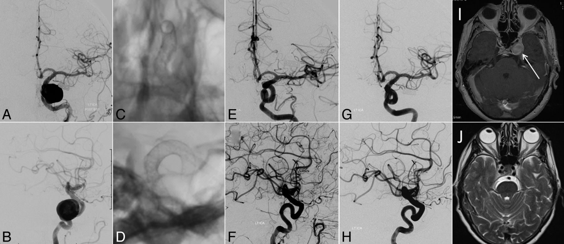
- Fig 1.
A 65-year-old woman who presented with progressive left-sided ophthalmoparesis due to third and fourth cranial nerve palsy. Digital subtraction angiography in frontal (A) and lateral (B) views demonstrates a large (19-mm-diameter) aneurysm arising from the cavernous segment of the left internal carotid artery. The patient was treated by endoluminal reconstruction of the LICA with 3 overlapping PEDs (frontal, C, and lateral, D). One-year follow-up digital subtraction angiography in frontal (E) and lateral (F) views and 5-year follow-up digital subtraction angiography in frontal (G) and lateral (H) views confirm stable angiographic cure. Regression of symptoms was correlated with the resolution of aneurysm mass effect as illustrated by comparison of the pretreatment gadolinium-enhanced axial T1-weighted MR image (I, white arrow) with the 5-year follow-up axial T2-weighted MR image (J).
- Fig 2.
Radiographic outcomes at 6, 12, and 36 months.


Tables
Indications Symptomatic CCAs Symptomatic mass effect (ophthalmoplegia or intractable retro-orbital pain) Symptomatic with acute thrombotic changes Symptomatic or asymptomatic CCAs Ruptured aneurysms Bony erosion Radiographic evidence of projection into subarachnoid space Underlying coagulopathy Large aneurysms (>10 mm) Evidence of growth of aneurysms No. or Mean Age (yr) 57 ± 14.2 Female 83.7% (36) Presenting symptom Visual 65.1% (28) Headaches 16.3% (7) Thromboembolic event 4.7% (2) Memory 2.3% (1) Facial pain/numbness 2.3% (1) Aneurysm maximum diameter (mm) 24.3 ± 9.7 Small, <10 mm 0 Large, 10–25 mm 23 Giant, ≥25 mm 20 Aneurysm neck (mm) 13.6 ± 11.6 Dome-to-neck ratio 2.2 ± .9 Study Total Aneurysms in Study Silk or PED CCAs (No.) Major Morbidity (No.) Mortality (No.) Berge et al29 77 Silk 29 3 0 Becske et al7,a 108 PED 28 1 0 Chan et al30 13 PED 5 0 0 Chitale et al31 42 PED 16 2 0 Cinar et al32 55 PED 5 0 0 Fischer et al33,b 101 PED 15 1 0 Lubicz et al34 34 Silk 5 1 0 Lylyk et al5 63 PED 11 0 0 McAuliffe et al35,c 57 PED 11 0 0 Nelson et al6 31 PED 5 0 0 O'Kelly et al8 94 PED 28 0 0 Piano et al36 104 Silk 16 1 0 PED Saatci et al37 251 PED 28 1 0 Velioglu et al38,d 87 Silk 19 0 1 Yu et al27,e 178 PED 32 1 1 Current study 43 PED 43 1 0 Total 1338 296 12 (4.1%) 2 (0.7%) PED only 227 7 (3.1%) 1 (0.44%) 0.68 Silk only 53 4 (7.5%) 1 (1.9%)


Jump to section
Related Articles
Cited By...
- Reconstruction of the sphenoid sinus erosion or dehiscence after treatment of unruptured intracavernous aneurysms with flow diverter stents
- Long-term outcomes of flow diversion for unruptured intracranial aneurysms: a systematic review and meta-analysis
- Flow diversion for compressive unruptured internal carotid artery aneurysms with neuro-ophthalmological symptoms: a systematic review and meta-analysis
- Onset-to-treatment time and aneurysmal regression predict improvement of cranial neuropathy after flow diversion treatment in patients with symptomatic internal carotid artery aneurysms
- Flow diversion for internal carotid artery aneurysms with compressive neuro-ophthalmologic symptoms: clinical and anatomical results in an international multicenter study
- Use of flow diverter stent for treatment of a cervical carotid artery dissection and pseudoaneurysm causing Horners syndrome
- Flow Diversion in Ruptured Intracranial Aneurysms: A Meta-Analysis
- Carotid cavernous fistula after Pipeline placement: a single-center experience and review of the literature
- Treatment of direct carotid-cavernous fistulas with a double lumen balloon
- Therapeutic Internal Carotid Artery Occlusion for Large and Giant Aneurysms: A Single Center Cohort of 146 Patients