Advertisement

Main menu

  • Home
  • Content
    • Current Issue
    • Accepted Manuscripts
    • Article Preview
    • Past Issue Archive
    • Video Articles
    • AJNR Case Collection
    • Case of the Week Archive
    • Case of the Month Archive
    • Classic Case Archive
  • Special Collections
    • AJNR Awards
    • Low-Field MRI
    • Alzheimer Disease
    • ASNR Foundation Special Collection
    • Photon-Counting CT
    • View All
  • Multimedia
    • AJNR Podcasts
    • AJNR SCANtastic
    • Trainee Corner
    • MRI Safety Corner
    • Imaging Protocols
  • For Authors
    • Submit a Manuscript
    • Submit a Video Article
    • Submit an eLetter to the Editor/Response
    • Manuscript Submission Guidelines
    • Statistical Tips
    • Fast Publishing of Accepted Manuscripts
    • Graphical Abstract Preparation
    • Imaging Protocol Submission
    • Author Policies
  • About Us
    • About AJNR
    • Editorial Board
    • Editorial Board Alumni
  • More
    • Become a Reviewer/Academy of Reviewers
    • Subscribers
    • Permissions
    • Alerts
    • Feedback
    • Advertisers
    • ASNR Home

User menu

  • Alerts
  • Log in

Search

  • Advanced search
American Journal of Neuroradiology

American Journal of Neuroradiology

ASHNR American Society of Functional Neuroradiology ASHNR American Society of Pediatric Neuroradiology ASSR
  • Alerts
  • Log in

Advanced Search

  • Home
  • Content
    • Current Issue
    • Accepted Manuscripts
    • Article Preview
    • Past Issue Archive
    • Video Articles
    • AJNR Case Collection
    • Case of the Week Archive
    • Case of the Month Archive
    • Classic Case Archive
  • Special Collections
    • AJNR Awards
    • Low-Field MRI
    • Alzheimer Disease
    • ASNR Foundation Special Collection
    • Photon-Counting CT
    • View All
  • Multimedia
    • AJNR Podcasts
    • AJNR SCANtastic
    • Trainee Corner
    • MRI Safety Corner
    • Imaging Protocols
  • For Authors
    • Submit a Manuscript
    • Submit a Video Article
    • Submit an eLetter to the Editor/Response
    • Manuscript Submission Guidelines
    • Statistical Tips
    • Fast Publishing of Accepted Manuscripts
    • Graphical Abstract Preparation
    • Imaging Protocol Submission
    • Author Policies
  • About Us
    • About AJNR
    • Editorial Board
    • Editorial Board Alumni
  • More
    • Become a Reviewer/Academy of Reviewers
    • Subscribers
    • Permissions
    • Alerts
    • Feedback
    • Advertisers
    • ASNR Home
  • Follow AJNR on Twitter
  • Visit AJNR on Facebook
  • Follow AJNR on Instagram
  • Join AJNR on LinkedIn
  • RSS Feeds

AJNR Awards, New Junior Editors, and more. Read the latest AJNR updates

Research ArticleNeurointervention

Optimized Angiographic CT Using Intravenous Contrast Injection: A Noninvasive Imaging Option for the Follow-Up of Coiled Aneurysms?

P. Gölitz, T. Struffert, I. Kaschka, K. Roessler, F. Knossalla and A. Doerfler
American Journal of Neuroradiology December 2014, 35 (12) 2341-2347; DOI: https://doi.org/10.3174/ajnr.A4039
P. Gölitz
aFrom the Departments of Neuroradiology (P.G., T.S., I.K., A.D.)
  • Find this author on Google Scholar
  • Find this author on PubMed
  • Search for this author on this site
T. Struffert
aFrom the Departments of Neuroradiology (P.G., T.S., I.K., A.D.)
  • Find this author on Google Scholar
  • Find this author on PubMed
  • Search for this author on this site
I. Kaschka
aFrom the Departments of Neuroradiology (P.G., T.S., I.K., A.D.)
  • Find this author on Google Scholar
  • Find this author on PubMed
  • Search for this author on this site
K. Roessler
bNeurosurgery (K.R.)
  • Find this author on Google Scholar
  • Find this author on PubMed
  • Search for this author on this site
F. Knossalla
cNeurology (F.K.), University of Erlangen, Nuremberg, Germany.
  • Find this author on Google Scholar
  • Find this author on PubMed
  • Search for this author on this site
A. Doerfler
aFrom the Departments of Neuroradiology (P.G., T.S., I.K., A.D.)
  • Find this author on Google Scholar
  • Find this author on PubMed
  • Search for this author on this site
  • Article
  • Figures & Data
  • Info & Metrics
  • Responses
  • References
  • PDF
Loading

Article Figures & Data

Figures

  • Tables
  • Fig 1.
    • Download figure
    • Open in new tab
    • Download powerpoint
    Fig 1.

    ivACT (A) and intra-arterial 3D-DSA (B) show, in comparable image quality, complete occlusion after coiling of a posterior communicating artery aneurysm. 2D-DSA (C) confirms complete aneurysm occlusion. TOF-MRA (D) demonstrates complete occlusion.
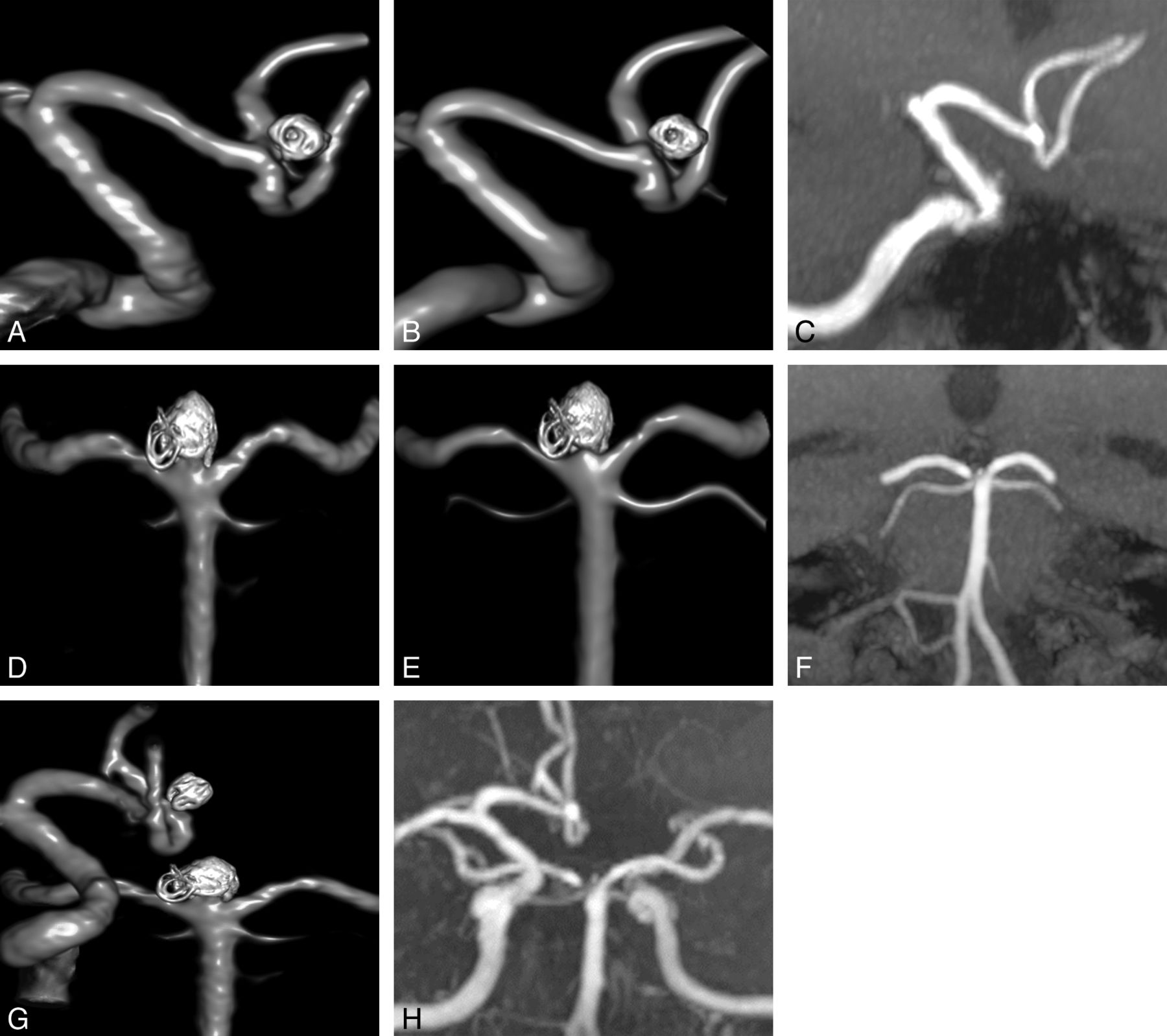
  • Fig 2.
    • Download figure
    • Open in new tab
    • Download powerpoint
    Fig 2.

    ivACT (A) and intra-arterial 3D-DSA (B) show, in a comparable way, complete occlusion of an anterior communicating artery aneurysm after coiling. MRA reveals no remnant (C). In addition, a second aneurysm located at the tip of the basilar artery was treated by coil embolization. ivACT (D) and 3D-DSA (E) demonstrate, in nearly identical quality, complete aneurysm occlusion with only mild irregularity at the aneurysm base. In addition, MRA reveals no remnant (F). As an advantage of ivACT (G) and MRA (H), aneurysm evaluation can be performed with only 1 examination step, in contrast to DSA, requiring several vessel catheterizations and contrast material injections.

  • Fig 3.
    • Download figure
    • Open in new tab
    • Download powerpoint
    Fig 3.

    ivACT (A) and 3D-DSA (B) reveal a small remnant after coiling of a posterior inferior cerebellar artery aneurysm in nearly identical quality. DSA confirms this finding (C). TOF-MRA also detects the small remnant (D).
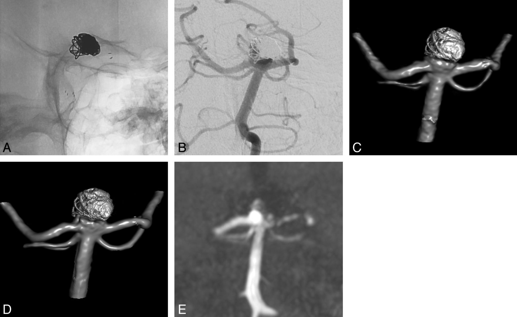
  • Fig 4.
    • Download figure
    • Open in new tab
    • Download powerpoint
    Fig 4.

    Native (A) and DSA images (B) after stent-assisted coil embolization of a basilar artery tip aneurysm show a small, laterally located remnant. In ivACT (C) and intra-arterial rotational 3D-DSA (D), the remnant can be revealed reliably in nearly identical image quality. In TOF-MRA (E), the remnant can be delineated clearly, but in contrast to ivACT and 3D-DSA, artifacts are induced by the implanted stent, making stent assessment impossible. In contrast, ivACT allows precise assessment of the stent lumen.

  • Fig 5.
    • Download figure
    • Open in new tab
    • Download powerpoint
    Fig 5.

    Native (A) and contrast-enhanced 2D-DSA (B) images show complete occlusion of a posterior communicating artery aneurysm, treated initially by subtotal clipping, followed by stent-assisted coil embolization. ivACT (C) and 3D-DSA (D) illustrate complete aneurysm occlusion in a comparable quality. In ivACT, no compromising artifacts by the metal implants are observed. MRA fails in delineating the coiled aneurysm because of severe susceptibility artifacts (E).

Tables

  • Figures
  • Mean size and SD of aneurysm remnants measured by ivACT and TOF-MRA in correlation with DSA

    RemnantDSA (Mean) (SD)ivACT (Mean) (SD)Pearson Correlation CoefficientTOF-MRA (Mean) (SD)Pearson Correlation Coefficient
    Height3.1 mm (±1.7)2.9 mm (±1.8)0.963 (P < .001)2.8 mm (±1.9)0.925 (P < .001)
    Width3.1 mm (±2.0)3.1 mm (±2.6)0.930 (P < 0.001)2.8 mm (±2.1)0.941 (P < .001)
PreviousNext
Back to top

In this issue

American Journal of Neuroradiology: 35 (12)
American Journal of Neuroradiology
Vol. 35, Issue 12
1 Dec 2014
  • Table of Contents
  • Index by author
  • Complete Issue (PDF)
Advertisement
Print
Download PDF
Email Article

Thank you for your interest in spreading the word on American Journal of Neuroradiology.

NOTE: We only request your email address so that the person you are recommending the page to knows that you wanted them to see it, and that it is not junk mail. We do not capture any email address.

Enter multiple addresses on separate lines or separate them with commas.
Optimized Angiographic CT Using Intravenous Contrast Injection: A Noninvasive Imaging Option for the Follow-Up of Coiled Aneurysms?
(Your Name) has sent you a message from American Journal of Neuroradiology
(Your Name) thought you would like to see the American Journal of Neuroradiology web site.
CAPTCHA
This question is for testing whether or not you are a human visitor and to prevent automated spam submissions.
Cite this article
P. Gölitz, T. Struffert, I. Kaschka, K. Roessler, F. Knossalla, A. Doerfler
Optimized Angiographic CT Using Intravenous Contrast Injection: A Noninvasive Imaging Option for the Follow-Up of Coiled Aneurysms?
American Journal of Neuroradiology Dec 2014, 35 (12) 2341-2347; DOI: 10.3174/ajnr.A4039

Citation Manager Formats

  • BibTeX
  • Bookends
  • EasyBib
  • EndNote (tagged)
  • EndNote 8 (xml)
  • Medlars
  • Mendeley
  • Papers
  • RefWorks Tagged
  • Ref Manager
  • RIS
  • Zotero
0 Responses
Respond to this article
Share
Bookmark this article
Optimized Angiographic CT Using Intravenous Contrast Injection: A Noninvasive Imaging Option for the Follow-Up of Coiled Aneurysms?
P. Gölitz, T. Struffert, I. Kaschka, K. Roessler, F. Knossalla, A. Doerfler
American Journal of Neuroradiology Dec 2014, 35 (12) 2341-2347; DOI: 10.3174/ajnr.A4039
del.icio.us logo Twitter logo Facebook logo Mendeley logo
  • Tweet Widget
  • Facebook Like
  • Google Plus One
Purchase

Jump to section

  • Article
    • Abstract
    • ABBREVIATION:
    • Materials and Methods
    • Results
    • Discussion
    • Conclusions
    • Footnotes
    • References
  • Figures & Data
  • Info & Metrics
  • Responses
  • References
  • PDF

Related Articles

  • No related articles found.
  • PubMed
  • Google Scholar

Cited By...

  • MRA versus DSA for the follow-up imaging of intracranial aneurysms treated using endovascular techniques: a meta-analysis
  • Metal artifact reduction for flat panel detector intravenous CT angiography in patients with intracranial metallic implants after endovascular and surgical treatment
  • Crossref
  • Google Scholar

This article has not yet been cited by articles in journals that are participating in Crossref Cited-by Linking.

More in this TOC Section

  • A Retrospective Study in Tentorial DAVFs
  • Proximal Protection Devices for Carotid Stenting
  • Hydrocoils: an Updated Meta-Analysis
Show more NEUROINTERVENTION

Similar Articles

Advertisement

Indexed Content

  • Current Issue
  • Accepted Manuscripts
  • Article Preview
  • Past Issues
  • Editorials
  • Editor's Choice
  • Fellows' Journal Club
  • Letters to the Editor
  • Video Articles

Cases

  • Case Collection
  • Archive - Case of the Week
  • Archive - Case of the Month
  • Archive - Classic Case

More from AJNR

  • Trainee Corner
  • Imaging Protocols
  • MRI Safety Corner
  • Book Reviews

Multimedia

  • AJNR Podcasts
  • AJNR Scantastics

Resources

  • Turnaround Time
  • Submit a Manuscript
  • Submit a Video Article
  • Submit an eLetter to the Editor/Response
  • Manuscript Submission Guidelines
  • Statistical Tips
  • Fast Publishing of Accepted Manuscripts
  • Graphical Abstract Preparation
  • Imaging Protocol Submission
  • Evidence-Based Medicine Level Guide
  • Publishing Checklists
  • Author Policies
  • Become a Reviewer/Academy of Reviewers
  • News and Updates

About Us

  • About AJNR
  • Editorial Board
  • Editorial Board Alumni
  • Alerts
  • Permissions
  • Not an AJNR Subscriber? Join Now
  • Advertise with Us
  • Librarian Resources
  • Feedback
  • Terms and Conditions
  • AJNR Editorial Board Alumni

American Society of Neuroradiology

  • Not an ASNR Member? Join Now

© 2025 by the American Society of Neuroradiology All rights, including for text and data mining, AI training, and similar technologies, are reserved.
Print ISSN: 0195-6108 Online ISSN: 1936-959X

Powered by HighWire