Advertisement

Main menu

  • Home
  • Content
    • Current Issue
    • Accepted Manuscripts
    • Article Preview
    • Past Issue Archive
    • AJNR Case Collection
    • Case of the Week Archive
    • Classic Case Archive
    • Case of the Month Archive
  • Special Collections
    • Spinal CSF Leak Articles (Jan 2020-June 2024)
    • 2024 AJNR Journal Awards
    • Most Impactful AJNR Articles
  • Multimedia
    • AJNR Podcast
    • AJNR Scantastics
    • Video Articles
  • For Authors
    • Submit a Manuscript
    • Author Policies
    • Fast publishing of Accepted Manuscripts
    • Graphical Abstract Preparation
    • Manuscript Submission Guidelines
    • Imaging Protocol Submission
    • Submit a Case for the Case Collection
  • About Us
    • About AJNR
    • Editorial Board
  • More
    • Become a Reviewer/Academy of Reviewers
    • Subscribers
    • Permissions
    • Alerts
    • Feedback
    • Advertisers
    • ASNR Home
  • Other Publications
    • ajnr

User menu

  • Alerts
  • Log in

Search

  • Advanced search
American Journal of Neuroradiology

American Journal of Neuroradiology

ASHNR American Society of Functional Neuroradiology ASHNR American Society of Pediatric Neuroradiology ASSR
  • Alerts
  • Log in

Advanced Search

  • Home
  • Content
    • Current Issue
    • Accepted Manuscripts
    • Article Preview
    • Past Issue Archive
    • AJNR Case Collection
    • Case of the Week Archive
    • Classic Case Archive
    • Case of the Month Archive
  • Special Collections
    • Spinal CSF Leak Articles (Jan 2020-June 2024)
    • 2024 AJNR Journal Awards
    • Most Impactful AJNR Articles
  • Multimedia
    • AJNR Podcast
    • AJNR Scantastics
    • Video Articles
  • For Authors
    • Submit a Manuscript
    • Author Policies
    • Fast publishing of Accepted Manuscripts
    • Graphical Abstract Preparation
    • Manuscript Submission Guidelines
    • Imaging Protocol Submission
    • Submit a Case for the Case Collection
  • About Us
    • About AJNR
    • Editorial Board
  • More
    • Become a Reviewer/Academy of Reviewers
    • Subscribers
    • Permissions
    • Alerts
    • Feedback
    • Advertisers
    • ASNR Home
  • Follow AJNR on Twitter
  • Visit AJNR on Facebook
  • Follow AJNR on Instagram
  • Join AJNR on LinkedIn
  • RSS Feeds

Welcome to the new AJNR, Updated Hall of Fame, and more. Read the full announcements.


AJNR is seeking candidates for the position of Associate Section Editor, AJNR Case Collection. Read the full announcement.


Research ArticleBrain
Open Access

Perfusion Measurement in Brain Gliomas with Intravoxel Incoherent Motion MRI

C. Federau, R. Meuli, K. O'Brien, P. Maeder and P. Hagmann
American Journal of Neuroradiology February 2014, 35 (2) 256-262; DOI: https://doi.org/10.3174/ajnr.A3686
C. Federau
aFrom the Department of Diagnostic and Interventional Radiology (C.F., R.M., P.M., P.H.), Centre Hospitalier Universitaire Vaudois and University of Lausanne, Lausanne, Switzerland
  • Find this author on Google Scholar
  • Find this author on PubMed
  • Search for this author on this site
R. Meuli
aFrom the Department of Diagnostic and Interventional Radiology (C.F., R.M., P.M., P.H.), Centre Hospitalier Universitaire Vaudois and University of Lausanne, Lausanne, Switzerland
  • Find this author on Google Scholar
  • Find this author on PubMed
  • Search for this author on this site
K. O'Brien
bCenter for Biomedical Imaging (K.O.), University of Geneva, Geneva, Switzerland.
  • Find this author on Google Scholar
  • Find this author on PubMed
  • Search for this author on this site
P. Maeder
aFrom the Department of Diagnostic and Interventional Radiology (C.F., R.M., P.M., P.H.), Centre Hospitalier Universitaire Vaudois and University of Lausanne, Lausanne, Switzerland
  • Find this author on Google Scholar
  • Find this author on PubMed
  • Search for this author on this site
P. Hagmann
aFrom the Department of Diagnostic and Interventional Radiology (C.F., R.M., P.M., P.H.), Centre Hospitalier Universitaire Vaudois and University of Lausanne, Lausanne, Switzerland
  • Find this author on Google Scholar
  • Find this author on PubMed
  • Search for this author on this site
  • Article
  • Figures & Data
  • Supplemental
  • Info & Metrics
  • Responses
  • References
  • PDF
Loading

Article Figures & Data

Figures

  • Tables
  • Fig 1.
    • Download figure
    • Open in new tab
    • Download powerpoint
    Fig 1.

    A–C, Glioblastoma multiforme involving the paraventricular region of the right frontal lobe seen on axial T2-weighted, T1-weighted postgadolinium, and DSC CBV. D–F, IVIM parametric maps f, D*, and fD* (scale bars: f = unitless; D*, fD* = mm2s−1). G, Logarithmic plot of signal-intensity decay as a function of b of the region of interest of maximal perfusion fraction, with the corresponding biexponential fit.

  • Fig 2.
    • Download figure
    • Open in new tab
    • Download powerpoint
    Fig 2.

    A–C, Oligoastrocytoma, grade 2, centered on the pre-central gyrus of the right frontal lobe, seen on axial T2-weighted, T1-weighted postgadolinium, and DSC CBV. D–F, IVIM parametric maps f, D*, and fD* (scale bars: f = unitless; D*, fD* = mm2s−1). G, Logarithmic plot of signal-intensity decay as a function of b of the region of interest of the maximal perfusion fraction, with the corresponding biexponential fit.
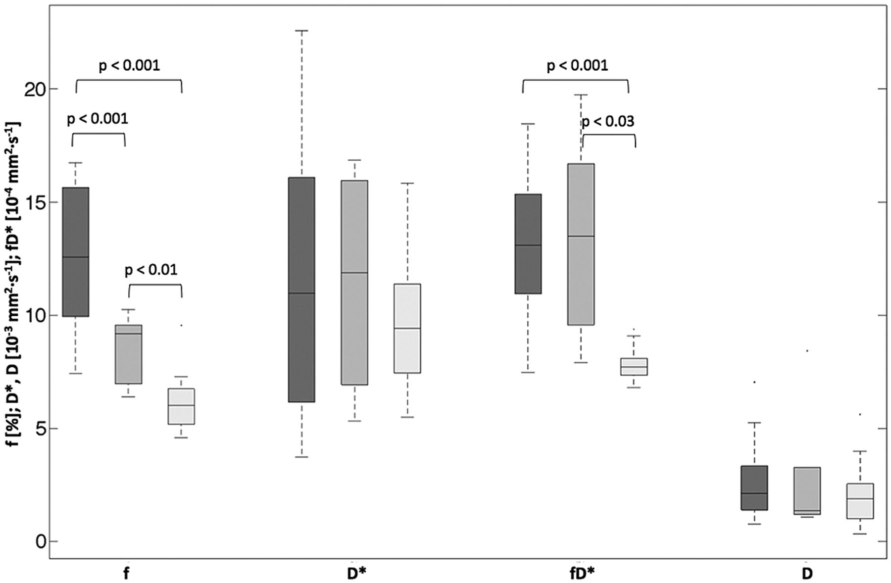
  • Fig 3.
    • Download figure
    • Open in new tab
    • Download powerpoint
    Fig 3.

    Box-and-whisker plot (median, 25th and 75th percentiles, minimum, maximum, and outliers) of f, D*, fD*, and D, as measured in ROIs of the maximum perfusion fraction. Dark gray indicates high-grade tumors; medium gray, low-grade tumors; light gray, contralateral control region of both high- and low- grade. P values are indicated when <.05.

  • Fig 4.
    • Download figure
    • Open in new tab
    • Download powerpoint
    Fig 4.

    Scatterplots comparing relative DSC CBV (y-axis) with absolute IVIM f (x-axis). Pearson r correlation coefficient is given.
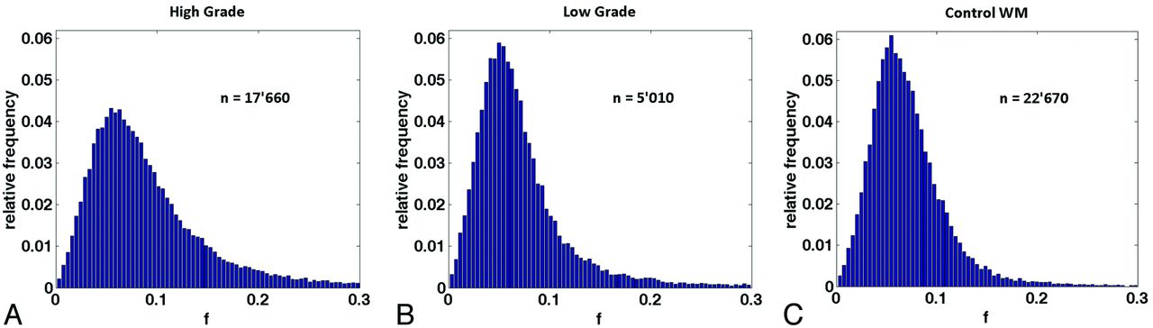
  • Fig 5.
    • Download figure
    • Open in new tab
    • Download powerpoint
    Fig 5.

    Normalized histograms of the perfusion fraction comprising the voxels of all whole tumor volumes for the high-grade (A) and low-grade (B) groups and for the control group (C). The total number of voxels included is indicated by n. An increase in the normalized number of highly perfused voxels can be observed in the high-grade tumor group in comparison with the low-grade and control groups.

Tables

  • Figures
    • View popup
    Table 1:

    Patient demographics, tumor localization, and histologic diagnosis

    GradeAge (yr)SexLocalizationSample ObtainedPathologic DiagnosisWHO Grade
    High
        177MaleLeft parietal lobeSurgical resectionGlioblastoma multiformeIV
        267MaleRight parietal lobeSurgical resectionGlioblastoma multiformeIV
        384FemaleLeft parietal lobeStereotaxic biopsyGlioblastoma multiformeIV
        461FemaleRight frontal lobeSurgical resectionGlioblastoma multiformeIV
        560MaleRight frontal lobeSurgical resectionGlioblastoma multiformeIV
        668MaleRight insulaSurgical resectionGlioblastoma multiformeIV
        773MaleRight frontotemporal lobesSurgical resectionAnaplastic oligoastrocytomaIII
        836MaleLeft insulaStereotaxic biopsyDiffuse gliomaIII
        950MaleLeft temporal lobeStereotaxic biopsyGlioblastoma multiformeIV
        1043MaleLeft temporal lobeSurgical resectionGlioblastoma multiformeIV
        1173MaleLeft temporal lobeSurgical resectionGlioblastoma multiformeIV
        1253MaleLeft parietal lobeSurgical resectionGlioblastoma multiformeIV
        1360FemaleLeft operculumStereotaxic biopsyGlioblastoma multiformeIV
        1424MaleLeft cingulumStereotaxic biopsyAnaplastic oligoastrocytomaIII
        1561MaleLeft temporal lobeSurgical resectionGlioblastoma multiformeIV
        1663FemaleRight frontal lobeSurgical resectionGlioblastoma multiformeIV
    Low
        138MaleRight frontal lobeSurgical resectionOligoastrocytomaII
        258MaleRight frontal lobeSurgical resectionNeuroglial tumorII
        354MaleLeft temporo-occipital lobesStereotaxic biopsyDiffuse astrocytomaII
        438MaleLeft frontal lobeStereotaxic biopsyOligodendrogliomaII
        52MaleCentered on 3rd ventricleStereotaxic biopsyPilomyxoid astrocytomaII
    • Note:—WHO indicates World Health Organization.

PreviousNext
Back to top

In this issue

American Journal of Neuroradiology: 35 (2)
American Journal of Neuroradiology
Vol. 35, Issue 2
1 Feb 2014
  • Table of Contents
  • Index by author
  • Complete Issue (PDF)
Advertisement
Print
Download PDF
Email Article

Thank you for your interest in spreading the word on American Journal of Neuroradiology.

NOTE: We only request your email address so that the person you are recommending the page to knows that you wanted them to see it, and that it is not junk mail. We do not capture any email address.

Enter multiple addresses on separate lines or separate them with commas.
Perfusion Measurement in Brain Gliomas with Intravoxel Incoherent Motion MRI
(Your Name) has sent you a message from American Journal of Neuroradiology
(Your Name) thought you would like to see the American Journal of Neuroradiology web site.
CAPTCHA
This question is for testing whether or not you are a human visitor and to prevent automated spam submissions.
Cite this article
C. Federau, R. Meuli, K. O'Brien, P. Maeder, P. Hagmann
Perfusion Measurement in Brain Gliomas with Intravoxel Incoherent Motion MRI
American Journal of Neuroradiology Feb 2014, 35 (2) 256-262; DOI: 10.3174/ajnr.A3686

Citation Manager Formats

  • BibTeX
  • Bookends
  • EasyBib
  • EndNote (tagged)
  • EndNote 8 (xml)
  • Medlars
  • Mendeley
  • Papers
  • RefWorks Tagged
  • Ref Manager
  • RIS
  • Zotero
0 Responses
Respond to this article
Share
Bookmark this article
Perfusion Measurement in Brain Gliomas with Intravoxel Incoherent Motion MRI
C. Federau, R. Meuli, K. O'Brien, P. Maeder, P. Hagmann
American Journal of Neuroradiology Feb 2014, 35 (2) 256-262; DOI: 10.3174/ajnr.A3686
del.icio.us logo Twitter logo Facebook logo Mendeley logo
  • Tweet Widget
  • Facebook Like
  • Google Plus One
Purchase

Jump to section

  • Article
    • Abstract
    • ABBREVIATIONS:
    • Materials and Methods
    • Results
    • Discussion
    • Conclusions
    • Footnotes
    • References
  • Figures & Data
  • Supplemental
  • Info & Metrics
  • Responses
  • References
  • PDF

Related Articles

  • PubMed
  • Google Scholar

Cited By...

  • Relaxation-selective Intravoxel Incoherent Motion Imaging of Microvascular Perfusion and Fluid Compartments in the Human Choroid Plexus
  • Interobserver Reliability on Intravoxel Incoherent Motion Imaging in Patients with Acute Ischemic Stroke
  • Collateral blood flow measurement with intravoxel incoherent motion perfusion imaging in hyperacute brain stroke
  • A Simplified Model for Intravoxel Incoherent Motion Perfusion Imaging of the Brain
  • Crossref (124)
  • Google Scholar

This article has been cited by the following articles in journals that are participating in Crossref Cited-by Linking.

  • Clinical Intravoxel Incoherent Motion and Diffusion MR Imaging: Past, Present, and Future
    Mami Iima, Denis Le Bihan
    Radiology 2016 278 1
  • What can we see with IVIM MRI?
    Denis Le Bihan
    NeuroImage 2019 187
  • Differentiation of high-grade and low-grade diffuse gliomas by intravoxel incoherent motion MR imaging
    Osamu Togao, Akio Hiwatashi, Koji Yamashita, Kazufumi Kikuchi, Masahiro Mizoguchi, Koji Yoshimoto, Satoshi O. Suzuki, Toru Iwaki, Makoto Obara, Marc Van Cauteren, Hiroshi Honda
    Neuro-Oncology 2016 18 1
  • Intravoxel incoherent motion diffusion-weighted MR imaging of breast cancer at 3.0 tesla: Comparison of different curve-fitting methods
    Shiteng Suo, Naier Lin, He Wang, Liangbin Zhang, Rui Wang, Su Zhang, Jia Hua, Jianrong Xu
    Journal of Magnetic Resonance Imaging 2015 42 2
  • Optimal differentiation of high- and low-grade glioma and metastasis: a meta-analysis of perfusion, diffusion, and spectroscopy metrics
    Jurgita Usinskiene, Agne Ulyte, Atle Bjørnerud, Jonas Venius, Vasileios K. Katsaros, Ryte Rynkeviciene, Simona Letautiene, Darius Norkus, Kestutis Suziedelis, Saulius Rocka, Andrius Usinskas, Eduardas Aleknavicius
    Neuroradiology 2016 58 4
  • Intravoxel incoherent motion MRI as a means to measure in vivo perfusion: A review of the evidence
    Christian Federau
    NMR in Biomedicine 2017 30 11
  • Intravoxel incoherent motion diffusion‐weighted imaging analysis of diffusion and microperfusion in grading gliomas and comparison with arterial spin labeling for evaluation of tumor perfusion
    Nanxi Shen, Lingyun Zhao, Jingjing Jiang, Rifeng Jiang, Changliang Su, Shun Zhang, Xiangyu Tang, Wenzhen Zhu
    Journal of Magnetic Resonance Imaging 2016 44 3
  • Intravoxel incoherent motion diffusion-weighted MR imaging of gliomas: efficacy in preoperative grading
    Yu-Chuan Hu, Lin-Feng Yan, Lang Wu, Pang Du, Bao-Ying Chen, Liang Wang, Shu-Mei Wang, Yu Han, Qiang Tian, Ying Yu, Tian-Yong Xu, Wen Wang, Guang-Bin Cui
    Scientific Reports 2014 4 1
  • Intravoxel incoherent motion diffusion-weighted imaging in head and neck squamous cell carcinoma: Assessment of perfusion-related parameters compared to dynamic contrast-enhanced MRI
    Noriyuki Fujima, Daisuke Yoshida, Tomohiro Sakashita, Akihiro Homma, Akiko Tsukahara, Khin Khin Tha, Kohsuke Kudo, Hiroki Shirato
    Magnetic Resonance Imaging 2014 32 10
  • Imaging brain tumour microstructure
    Markus Nilsson, Elisabet Englund, Filip Szczepankiewicz, Danielle van Westen, Pia C. Sundgren
    NeuroImage 2018 182

More in this TOC Section

  • Optimal MRI Sequence for Identifying Occlusion Location in Acute Stroke: Which Value of Time-Resolved Contrast-Enhanced MRA?
  • SWI or T2*: Which MRI Sequence to Use in the Detection of Cerebral Microbleeds? The Karolinska Imaging Dementia Study
  • Progression of Microstructural Damage in Spinocerebellar Ataxia Type 2: A Longitudinal DTI Study
Show more Brain

Similar Articles

Advertisement

Indexed Content

  • Current Issue
  • Accepted Manuscripts
  • Article Preview
  • Past Issues
  • Editorials
  • Editors Choice
  • Fellow Journal Club
  • Letters to the Editor

Cases

  • Case Collection
  • Archive - Case of the Week
  • Archive - Case of the Month
  • Archive - Classic Case

Special Collections

  • Special Collections

Resources

  • News and Updates
  • Turn around Times
  • Submit a Manuscript
  • Author Policies
  • Manuscript Submission Guidelines
  • Evidence-Based Medicine Level Guide
  • Publishing Checklists
  • Graphical Abstract Preparation
  • Imaging Protocol Submission
  • Submit a Case
  • Become a Reviewer/Academy of Reviewers
  • Get Peer Review Credit from Publons

Multimedia

  • AJNR Podcast
  • AJNR SCANtastic
  • Video Articles

About Us

  • About AJNR
  • Editorial Board
  • Not an AJNR Subscriber? Join Now
  • Alerts
  • Feedback
  • Advertise with us
  • Librarian Resources
  • Permissions
  • Terms and Conditions

American Society of Neuroradiology

  • Not an ASNR Member? Join Now

© 2025 by the American Society of Neuroradiology All rights, including for text and data mining, AI training, and similar technologies, are reserved.
Print ISSN: 0195-6108 Online ISSN: 1936-959X

Powered by HighWire