
Article Figures & Data
Figures
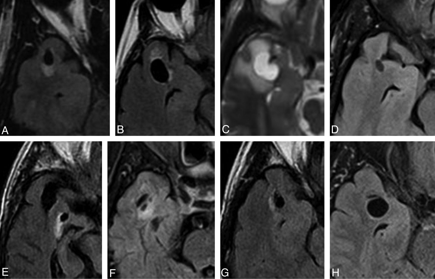
- Fig 1.
Selected axial FLAIR/T2 images of right-sided lesions within the anterior superior temporal gyrus as identified in case 1 (A), 2 (B), 7 (C), 8 (D), 9 (E), 10 (F), 12 (G), and 14 (H). All lesions were located adjacent to the SAS. The images illustrate the variability in the degree of surrounding signal change. Note the proximity to the adjacent middle cerebral artery and more prominent surrounding signal hyperintensity seen in case 7 (C).
- Fig 2.
Selected axial FLAIR images of left-sided lesions within the anterior superior temporal gyrus as identified in case 3 (A), 4 (B), 5 (C), 6 (D), 11 (E),13 (F), and 15 (G). All lesions were located adjacent to the SAS. Again, the images illustrate the variability in the degree of surrounding signal change.
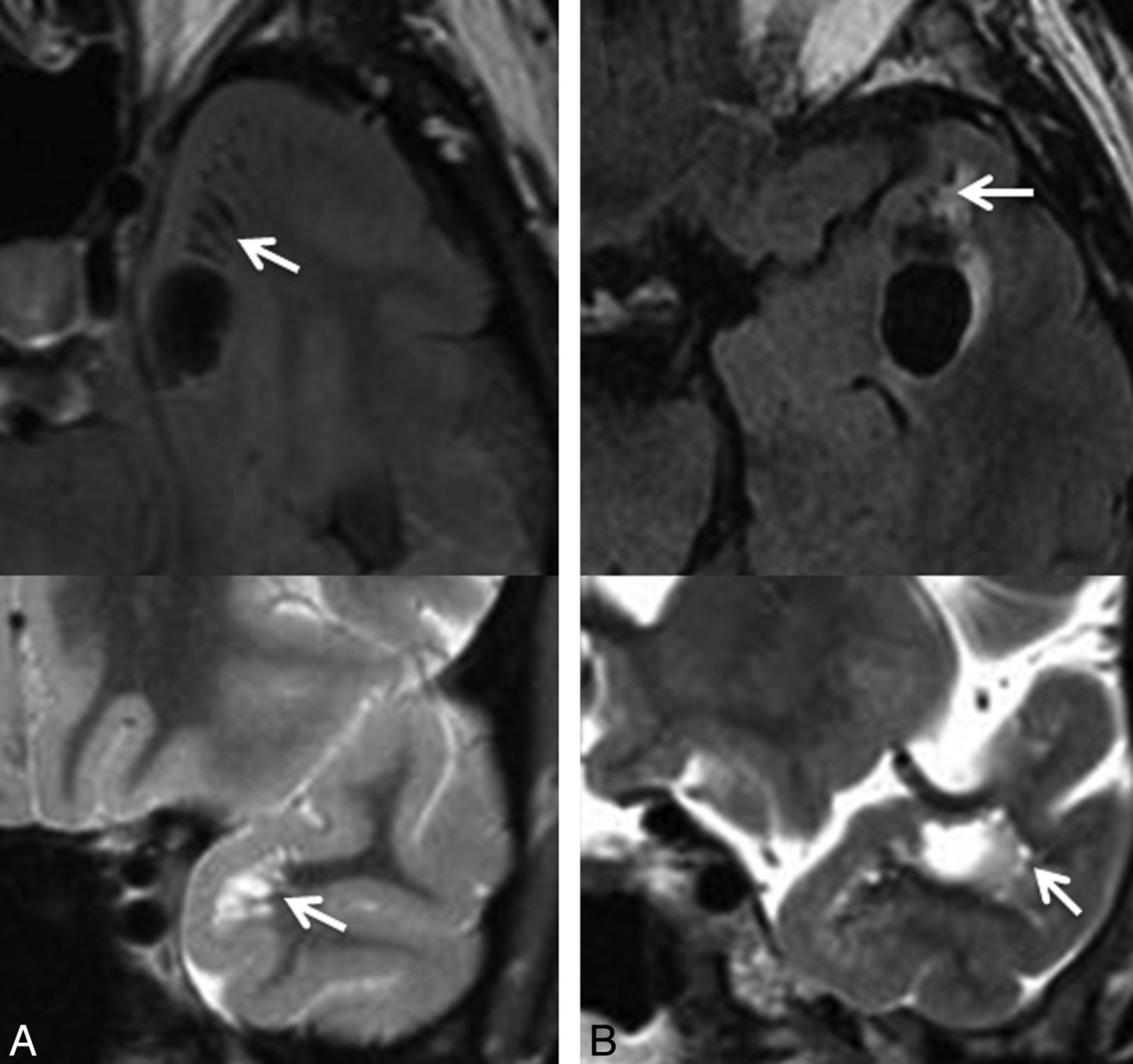
- Fig 3.
Selected axial FLAIR images demonstrating the presence of a cystic lesion within the anterior temporal gyrus as identified in case 6 (A) and case 11 (B), with adjacent smaller cystic lesions, suggestive of a dominant enlarged perivascular space with adjacent smaller prominent perivascular spaces (arrows). Corresponding coronal T2 images (below) through the region of interest confirm these findings (arrows). In case 11 (B), the proximity to the adjacent middle cerebral artery is identified on the coronal T2 image.
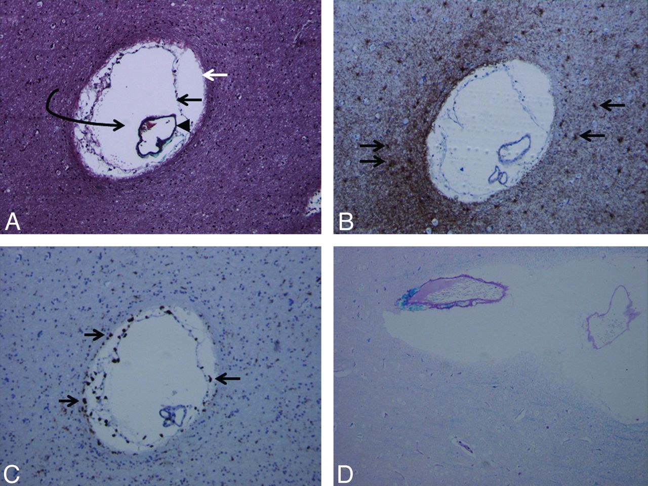
- Fig 4.
Trichrome stain (A) demonstrates the presence of an enlarged perivascular space (white arrow, glia limitans; black arrow, inner pial membrane; black arrowhead, vessel lined by outer pial membrane; curved arrow, perivascular space). Multiple such lesions were seen in the surgical specimen. Glial fibrillary acidic protein stain (B) and CD68 stain (C) demonstrate reactive astrocytes (arrows in B) and perivascular microglia (arrows in C) as multiple brown-staining dots, confirming chronicity of the pathophysiologic process. Luxol fast blue stain (D) demonstrates diffuse absence of blue staining (myelin staining), indicating demyelination and gliosis in the brain parenchyma surrounding the dilated perivascular space (magnification of all slides, 20×).


Tables
- Table 1:
MR imaging characteristics of lesions identified within the anterior superior temporal subcortical white matter
Case Location (R/L) Maximal Dimension (cm) Morphologic Feature Adjacent to SAS (Y/N) FLAIR Suppression (Y/N) DWI Restriction (Y/N) SWI/GRE Blooming (Y/N) Gadolinium Enhancement (Y/N) Perilesional FLAIR Signal Change Imaging Follow-Up (mo) Change over Follow-Up Period 1 R 1.0 Elongated Y Y N N N Mild 14 N 2 R 1.5 Elongated Y Y N N N Mild 38 N 3 L 0.9 Elongated Y Y N N N Mild 35 N 4 L 1.5 Elongated Y Y N N N Mild 22 N 5 L 0.9 Round Y Y N N N Mild 0 N/A 6 L 1.7 Elongated Y Y N N N None 11 N 7 R 1.8 Elongated Y Y N N N Extensive 0 N/A 8 R 0.7 Elongated Y Y N N N None 6 N 9 R 0.6 Elongated Y Y N N N Mild 99 N 10 R 0.5 Elongated Y Y N N N Mild 0 N/A 11 L 1.7 Elongated Y Y N N N Mild 7 N 12 R 1.1 Elongated Y Y N N N Mild 0 N/A 13 L 1.9 Elongated Y Y N N N None 0 N/A 14 R 1.3 Round Y Y N N N None 0 N/A 15 L 1.6 Elongated Y Y N N N Mild 112 N Note:—GRE indicates gradient-recalled echo; L, left; N, no; N/A, not applicable; R, right; Y, yes.


