
Article Figures & Data
Figures
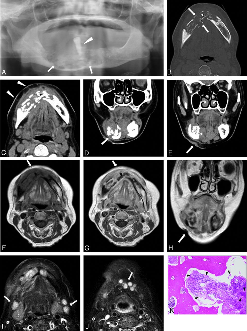
- Fig 1.
A 68-year-old woman with discharge of pus on the right side of the face. A, Panoramic radiograph reveals a large osteolytic region (arrows), with a floating tooth (arrowhead), in the right mandibular body crossing the midline. Axial CT in bone (B) and soft tissue (C) windows reveals an ill-defined osteolytic lesion in the right mandibular body crossing the midline (arrows). Note extensive demineralization of the buccal and lingual cortices and extensive soft tissue infiltrative change extending to the skin (arrowheads). D and E, Coronal CT in soft tissue window shows bone destruction and fistula from the mandible to the skin (arrows). F, Axial T1-weighted MR image shows heterogeneous, low signal intensity in the lesion involving the mandible and surrounding soft tissues (arrow). G, Contrast-enhanced axial T1-weighted MR image shows heterogeneous mass with moderate contrast enhancement in the lesion involving the mandible and surrounding soft tissues (arrow). H, Coronal T2-weighted MR image shows a fistula (arrow). I and J, Axial STIR MR images shows multiple mildly reactive nodes with increased signal intensity in levels IA and IB (arrows). K, Photomicrograph of a specimen shows actinomycotic granules (arrowheads) and presence of sequestra (arrows) (hematoxylin-eosin stain, original magnification × 200).
- Fig 2.
A 40-year-old man with discharge of pus on the right side of the face. A, Axial CT in soft tissue window reveals extensive soft tissue (outer layer fat around mandible) infiltrative change extending to the skin (arrow). B, Contrast-enhanced axial CT scan in soft tissue window reveals a heterogeneous, moderately enhancing mass in the lesion and extensive soft tissue (outer layer fat around mandible) infiltrative change extending to the skin (arrow). Axial T1-weighted (C) and contrast-enhanced axial T1-weighted (D) MR imaging reveals heterogeneous, low signal intensity in the lesion involving the mandible and surrounding soft tissues (arrow). Note this lesion shows diffuse and moderate contrast enhancement of the soft tissue and marrow space.
- Fig 3.
A 28-year-old man with swelling of the right mandibular region. A, Axial CT in bone window shows a heterogeneous osteolytic lesion with periosteal reaction in the posterior body to ramus of the right mandible (arrows). B, Axial CT in soft tissue window shows extensive soft tissue infiltrative change extending to the skin (arrow), masseter muscle (arrowhead), and parotid gland (thin arrow).
- Fig 4.
A 66-year-old woman with swelling of left mandibular region. A, Axial CT in soft tissue window demonstrates foci of air adjacent to the left mandible (arrow) and swelling of masseter and medial pterygoid muscle (arrowheads). B, Axial STIR MR image shows extensive inflammation in the left masseter muscle, medial pterygoid muscles, parotid gland (arrowheads), and mandibular bone marrow (arrow).


Tables
Patient No/Age (y)/Sex CT Imaging Findings Contrast Enhancement Margin Density Intralesional Gas Fistula Osteolysis Periosteal Reaction Sequestra Lymphadenopathy 1/40/M Mod. heterog. Irregular Low–intermed. − + + + + − 2/34/M Not done Irregular Low–intermed. + + + − − − 3/66/F Not done Irregular Low–intermed. + − + + + − 4/28/M Not done Irregular Low–intermed. − + + + + − 5/78/F Not done Irregular Low–intermed. + + + + + − 6/68/F Not done Irregular Low–intermed. + − + − − − Note:—F indicates female; M, male; Low–intermed., low to intermediate; Mod. heterog., moderate heterogeneous.
Patient No MRI Findings Gadolinium Enhancement Cellulitis Adjacent to the Facial Skin Inflammation of Masseter Muscle Inflammation of Pterygoid Muscle Lymph Adenopathy 1 Moderate heterogeneous + − − 2 Not done + + − − 3 Not done + + + − 4 Not done + + + − 5 Not done + + + − 6 Moderate heterogeneous + + + − Note:—+ indicates findings present; −, findings absent.


