Advertisement

Main menu

  • Home
  • Content
    • Current Issue
    • Accepted Manuscripts
    • Article Preview
    • Past Issue Archive
    • AJNR Case Collection
    • Case of the Week Archive
    • Classic Case Archive
    • Case of the Month Archive
  • Special Collections
    • Spinal CSF Leak Articles (Jan 2020-June 2024)
    • 2024 AJNR Journal Awards
    • Most Impactful AJNR Articles
  • Multimedia
    • AJNR Podcast
    • AJNR Scantastics
    • Video Articles
  • For Authors
    • Submit a Manuscript
    • Author Policies
    • Fast publishing of Accepted Manuscripts
    • Graphical Abstract Preparation
    • Manuscript Submission Guidelines
    • Imaging Protocol Submission
    • Submit a Case for the Case Collection
  • About Us
    • About AJNR
    • Editorial Board
  • More
    • Become a Reviewer/Academy of Reviewers
    • Subscribers
    • Permissions
    • Alerts
    • Feedback
    • Advertisers
    • ASNR Home
  • Other Publications
    • ajnr

User menu

  • Alerts
  • Log in

Search

  • Advanced search
American Journal of Neuroradiology

American Journal of Neuroradiology

ASHNR American Society of Functional Neuroradiology ASHNR American Society of Pediatric Neuroradiology ASSR
  • Alerts
  • Log in

Advanced Search

  • Home
  • Content
    • Current Issue
    • Accepted Manuscripts
    • Article Preview
    • Past Issue Archive
    • AJNR Case Collection
    • Case of the Week Archive
    • Classic Case Archive
    • Case of the Month Archive
  • Special Collections
    • Spinal CSF Leak Articles (Jan 2020-June 2024)
    • 2024 AJNR Journal Awards
    • Most Impactful AJNR Articles
  • Multimedia
    • AJNR Podcast
    • AJNR Scantastics
    • Video Articles
  • For Authors
    • Submit a Manuscript
    • Author Policies
    • Fast publishing of Accepted Manuscripts
    • Graphical Abstract Preparation
    • Manuscript Submission Guidelines
    • Imaging Protocol Submission
    • Submit a Case for the Case Collection
  • About Us
    • About AJNR
    • Editorial Board
  • More
    • Become a Reviewer/Academy of Reviewers
    • Subscribers
    • Permissions
    • Alerts
    • Feedback
    • Advertisers
    • ASNR Home
  • Follow AJNR on Twitter
  • Visit AJNR on Facebook
  • Follow AJNR on Instagram
  • Join AJNR on LinkedIn
  • RSS Feeds

Welcome to the new AJNR, Updated Hall of Fame, and more. Read the full announcements.


AJNR is seeking candidates for the position of Associate Section Editor, AJNR Case Collection. Read the full announcement.


Getting new auth cookie, if you see this message a lot, tell someone!
Research ArticlePediatrics

Bannayan-Riley-Ruvalcaba Syndrome: MRI Neuroimaging Features in a Series of 7 Patients

R. Bhargava, K.J. Au Yong and N. Leonard
American Journal of Neuroradiology February 2014, 35 (2) 402-406; DOI: https://doi.org/10.3174/ajnr.A3680
R. Bhargava
aFrom the Departments of Radiology and Diagnostic Imaging (R.B.)
  • Find this author on Google Scholar
  • Find this author on PubMed
  • Search for this author on this site
K.J. Au Yong
cDepartment of Radiology and Diagnostic Imaging (K.J.A.), McMaster University Medical Centre, McMaster Children's Hospital, Hamilton, Ontario, Canada.
  • Find this author on Google Scholar
  • Find this author on PubMed
  • Search for this author on this site
N. Leonard
bMedical Genetics (N.L.), University of Alberta Hospital, Edmonton, Alberta, Canada
  • Find this author on Google Scholar
  • Find this author on PubMed
  • Search for this author on this site
  • Article
  • Figures & Data
  • Info & Metrics
  • Responses
  • References
  • PDF
Loading

Article Figures & Data

Figures

  • Tables
  • Fig 1.
    • Download figure
    • Open in new tab
    • Download powerpoint
    Fig 1.

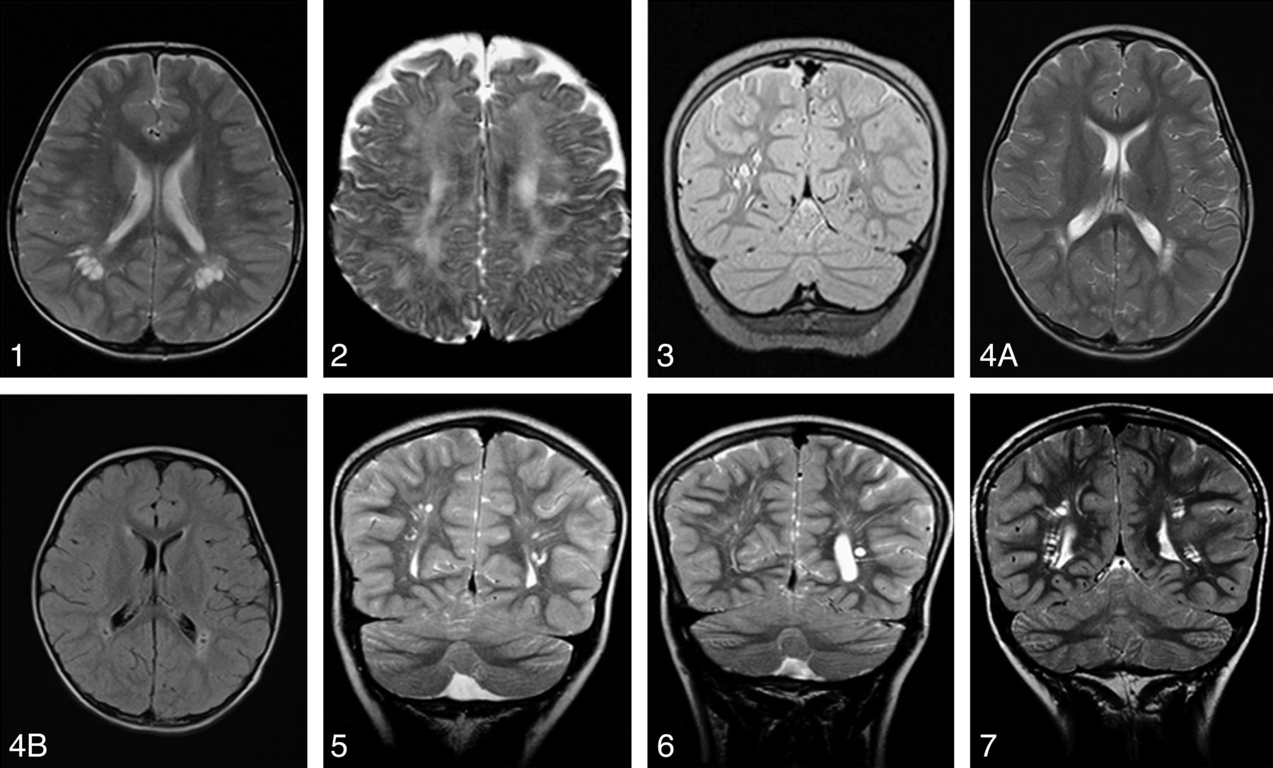
    1, Axial T2 image in patient 1 demonstrates bilateral cystic spaces in the biparietal periventricular white matter. 2, Axial T2 image in patient 2 demonstrating small cystic spaces in the parietal white matter associated with increased T2 signal of the periventricular white matter. 3, Coronal T2 image in patient 3 demonstrates cystic spaces in the periventricular white matter of the parietal lobes. 4A, Axial T2 image in patient 4 demonstrates bilateral small parietal periventricular cyst associated with white matter T2 hyperintensities predominantly around the cysts. 4B, An axial FLAIR image in patient 4 demonstrates pericyst hyperintensities and suppression of the VR space fluid. 5, Coronal T2 image of patient 5 demonstrates a well-defined small periventricular cyst in the parietal region. 6, Coronal T2 image in patient 6 demonstrates a well-defined small periventricular cyst in the left parietal region. 7, Coronal T2 image in patient 7 demonstrates small periventricular cysts in the biparietal region.

Tables

  • Figures
  • Summary of MRI and clinical characteristic of 7 patients diagnosed with BRRS

    Patient No.SexAge at MRI (y)MacrocephalyMutation PTENDevelopmentPhysical FeaturesWM CystCyst Location
    1M4YesYesAutisticAbdominal hemangiomaYesf,p,t
    2M0.65YesYesDelayCafé au lait spot, thyroid nodules, testicular hamartomas, rectal and gastric polypsYesp
    3M2.7YesNo proteinAutisticMotor speech disorderYesf,p
    4M3.590th centileNo proteinDelayCafé au lait spotYesp
    5M2.6YesNo proteinDelayMoles, thyroid nodules, intestinal polypsYesp
    6M7.7YesNo proteinDelayMoles, thyroid nodules, intestinal polypsYesp
    7M7.7YesYesDelayPigmentation of the glans penis, multiple thyroid nodules, and carcinomaYesf,p
    • Note:—f indicates frontal; p, parietal; t, temporal.

PreviousNext
Back to top

In this issue

American Journal of Neuroradiology: 35 (2)
American Journal of Neuroradiology
Vol. 35, Issue 2
1 Feb 2014
  • Table of Contents
  • Index by author
  • Complete Issue (PDF)
Advertisement
Print
Download PDF
Email Article

Thank you for your interest in spreading the word on American Journal of Neuroradiology.

NOTE: We only request your email address so that the person you are recommending the page to knows that you wanted them to see it, and that it is not junk mail. We do not capture any email address.

Enter multiple addresses on separate lines or separate them with commas.
Bannayan-Riley-Ruvalcaba Syndrome: MRI Neuroimaging Features in a Series of 7 Patients
(Your Name) has sent you a message from American Journal of Neuroradiology
(Your Name) thought you would like to see the American Journal of Neuroradiology web site.
CAPTCHA
This question is for testing whether or not you are a human visitor and to prevent automated spam submissions.
Cite this article
R. Bhargava, K.J. Au Yong, N. Leonard
Bannayan-Riley-Ruvalcaba Syndrome: MRI Neuroimaging Features in a Series of 7 Patients
American Journal of Neuroradiology Feb 2014, 35 (2) 402-406; DOI: 10.3174/ajnr.A3680

Citation Manager Formats

  • BibTeX
  • Bookends
  • EasyBib
  • EndNote (tagged)
  • EndNote 8 (xml)
  • Medlars
  • Mendeley
  • Papers
  • RefWorks Tagged
  • Ref Manager
  • RIS
  • Zotero
0 Responses
Respond to this article
Share
Bookmark this article
Bannayan-Riley-Ruvalcaba Syndrome: MRI Neuroimaging Features in a Series of 7 Patients
R. Bhargava, K.J. Au Yong, N. Leonard
American Journal of Neuroradiology Feb 2014, 35 (2) 402-406; DOI: 10.3174/ajnr.A3680
del.icio.us logo Twitter logo Facebook logo Mendeley logo
  • Tweet Widget
  • Facebook Like
  • Google Plus One
Purchase

Jump to section

  • Article
    • Abstract
    • ABBREVATIONS:
    • Materials and Methods
    • Results
    • Discussion
    • Conclusions
    • Footnotes
    • References
  • Figures & Data
  • Info & Metrics
  • Responses
  • References
  • PDF

Related Articles

  • No related articles found.
  • PubMed
  • Google Scholar

Cited By...

  • No citing articles found.
  • Crossref (27)
  • Google Scholar

This article has been cited by the following articles in journals that are participating in Crossref Cited-by Linking.

  • PTEN Hamartoma tumor syndrome in childhood: A review of the clinical literature
    William L. Macken, Marc Tischkowitz, Katherine L. Lachlan
    American Journal of Medical Genetics Part C: Seminars in Medical Genetics 2019 181 4
  • Clinical spectrum of PTEN mutation in pediatric patients. A bicenter experience
    Claudia Ciaccio, Veronica Saletti, Stefano D'Arrigo, Silvia Esposito, Enrico Alfei, Isabella Moroni, Davide Tonduti, Luisa Chiapparini, Chiara Pantaleoni, Donatella Milani
    European Journal of Medical Genetics 2019 62 12
  • A clinical review on megalencephaly
    Piero Pavone, Andrea Domenico Praticò, Renata Rizzo, Giovanni Corsello, Martino Ruggieri, Enrico Parano, Raffaele Falsaperla
    Medicine 2017 96 26
  • Behavioural and psychological features of PTEN mutations: a systematic review of the literature and meta-analysis of the prevalence of autism spectrum disorder characteristics
    Katherine Cummings, Alice Watkins, Chris Jones, Renuka Dias, Alice Welham
    Journal of Neurodevelopmental Disorders 2022 14 1
  • Broad spectrum of neuropsychiatric phenotypes associated with white matter disease in PTEN hamartoma tumor syndrome
    Tugce B. Balci, Jorge Davila, Denice Lewis, Addo Boafo, Erick Sell, Julie Richer, Sarah M. Nikkel, Christine M. Armour, Eva Tomiak, Matthew A. Lines, Sarah L. Sawyer
    American Journal of Medical Genetics Part B: Neuropsychiatric Genetics 2018 177 1
  • Clinical presentation of PTEN mutations in childhood in the absence of family history of Cowden syndrome
    T. Busa, M. Milh, N. Degardin, N. Girard, S. Sigaudy, M. Longy, S. Olshchwang, H. Sobol, B. Chabrol, N. Philip
    European Journal of Paediatric Neurology 2015 19 2
  • Vascular Anomaly Syndromes in the ISSVA Classification System: Imaging Findings and Role of Interventional Radiology in Management
    Mindy X. Wang, Serageldin Kamel, Khaled M. Elsayes, R. Paul Guillerman, Ahmed Habiba, Lauren Heng, Margarita Revzin, Vincent Mellnick, Ionela Iacobas, Alex Chau
    RadioGraphics 2022 42 6
  • Polymicrogyria is Associated With Pathogenic Variants in PTEN
    Diane D. Shao, Christelle M. Achkar, Abbe Lai, Siddharth Srivastava, Ryan N. Doan, Lance H. Rodan, Allen Y. Chen, Annapurna Poduri, Edward Yang, Christopher A. Walsh
    Annals of Neurology 2020 88 6
  • Distinctive facies, macrocephaly, and developmental delay are signs of a PTEN mutation in childhood
    Kohji Kato, Seiji Mizuno, Mie Inaba, Shinobu Fukumura, Naoko Kurahashi, Koichi Maruyama, Daisuke Ieda, Kei Ohashi, Ikumi Hori, Yutaka Negishi, Ayako Hattori, Shinji Saitoh
    Brain and Development 2018 40 8
  • Imaging of PTEN-related abnormalities in the central nervous system
    Radhika Dhamija, Joseph M. Hoxworth
    Clinical Imaging 2020 60 2

More in this TOC Section

  • Comparison of Image Quality and Radiation Dose in Pediatric Temporal Bone CT Using Photon-Counting Detector CT and Energy-Integrating Detector CT
  • SyMRI & MR Fingerprinting in Brainstem Myelination
  • ASL in PEDS Nontraumatic Orbital Lesions
Show more Pediatrics

Similar Articles

Advertisement

Indexed Content

  • Current Issue
  • Accepted Manuscripts
  • Article Preview
  • Past Issues
  • Editorials
  • Editors Choice
  • Fellow Journal Club
  • Letters to the Editor

Cases

  • Case Collection
  • Archive - Case of the Week
  • Archive - Case of the Month
  • Archive - Classic Case

Special Collections

  • Special Collections

Resources

  • News and Updates
  • Turn around Times
  • Submit a Manuscript
  • Author Policies
  • Manuscript Submission Guidelines
  • Evidence-Based Medicine Level Guide
  • Publishing Checklists
  • Graphical Abstract Preparation
  • Imaging Protocol Submission
  • Submit a Case
  • Become a Reviewer/Academy of Reviewers
  • Get Peer Review Credit from Publons

Multimedia

  • AJNR Podcast
  • AJNR SCANtastic
  • Video Articles

About Us

  • About AJNR
  • Editorial Board
  • Not an AJNR Subscriber? Join Now
  • Alerts
  • Feedback
  • Advertise with us
  • Librarian Resources
  • Permissions
  • Terms and Conditions

American Society of Neuroradiology

  • Not an ASNR Member? Join Now

© 2025 by the American Society of Neuroradiology All rights, including for text and data mining, AI training, and similar technologies, are reserved.
Print ISSN: 0195-6108 Online ISSN: 1936-959X

Powered by HighWire