
Article Figures & Data
Figures
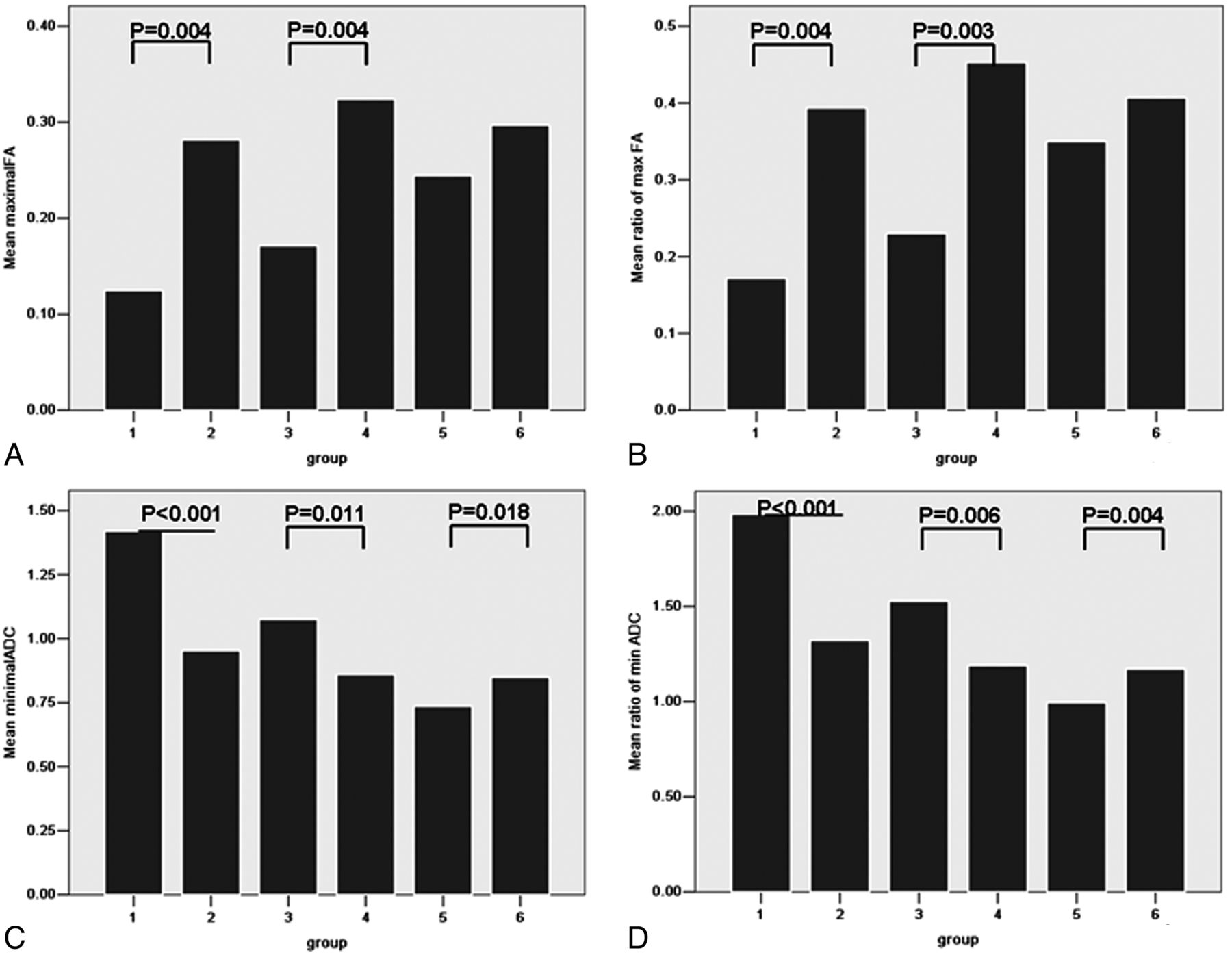
- Fig 1.
Bar graph of maximal fractional anisotropy (FA) (A), ratio of maximal FA (rmFA) (B), minimal ADC (C), and ratio of minimal ADC (rmADC) (D) in different groups. P values with statistical significance are shown. Maximal FA and rmFA could differentiate group 1 from group 2 and group 3 from group 4. Minimal ADC (unit: ×10–3 mm2/s) and rmADC showed statistical significance in groups 1 and 2, in groups 3 and 4, and in groups 5 and 6.
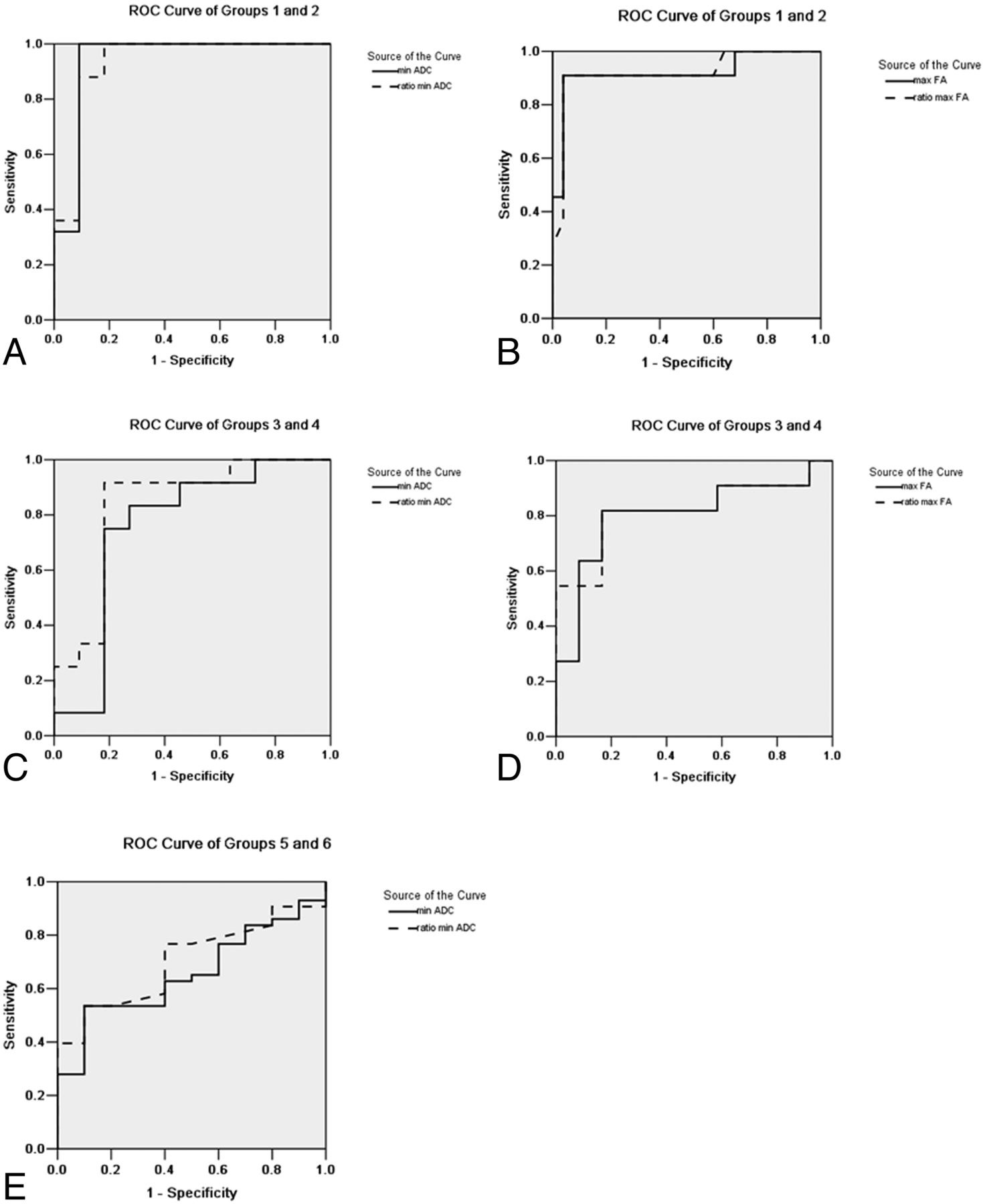
- Fig 2.
Receiver operating characteristic curves with statistical significance are shown. The area under the curve (AUC) showed a decreasing trend in imaging parameters from grade II to grade IV. Minimal ADC showed the largest AUC in group 1 and group 2, and the second largest parameter was the ratio of minimal ADC (rmADC). In groups 3, 4, 5, and 6, the largest AUC was rmADC.
- Fig 3.
A–C, A 43-year-old man with a grade II astrocytoma and IDH1 R132H antibody examination expressed positively. D–F, A 28-year-old woman with a grade II astrocytoma and IDH1 R132H expressed negatively. In A, the fractional anisotropy gray-scale mapping shows a homogeneous low signal; in D, a heterogeneous image is shown with a dotted high signal. The signal of ADC mapping in B was higher than that in E. C shows diffuse positivity on immunochemistry; F shows negativity on immunochemistry.
- Fig 4.
A–C, A 35-year-old woman with a grade III anaplastic astrocytoma and IDH1 R132H antibody examination expressed positively. D–F, A 17-year-old young man with grade III anaplastic astrocytoma and IDH1 R132H antibody examination expressed negatively. In A, fractional anisotropy gray-scale mapping showed a heterogeneous low signal with some iso- and hyper-signals; in D, a high signal was seen, except for the middle necrosis area. The signal of ADC mapping in B is higher than that in E. C and F, Immunochemistry results are shown.
- Fig 5.
A–C, A 33-year-old woman with a grade IV primary glioblastoma and IDH1 R132H antibody examination expressed positively. D–F, A 48-year-old man with a grade IV primary glioblastoma and IDH1 R132H antibody examination expressed negatively. In A and D, fractional anisotropy mappings show heterogeneous images with scattered high signals. The signal of ADC mapping in B is lower than that in E. Immunohistochemistry results of these patients are shown.


Tables
Group 1 Group 2 Group 3 Group 4 Group 5 Group 6 No. of patient cases 25 11 12 11 10 43 Sex, M/F 15/10 5/6 9/3 7/4 8/2 27/16 Age, y 39.68 ± 7.22 42.45 ± 18.38 41.92 ± 8.71 42.64 ± 17.90 41.70 ± 11.39 54.42 ± 12.63 Edema FA 0.26 ± 0.12 0.28 ± 0.14 0.18 ± 0.09 0.24 ± 0.11 0.18 ± 0.06 0.20 ± 0.09 Ratio of edema FA 0.37 ± 0.17 0.40 ± 0.20 0.24 ± 0.12 0.34 ± 0.16 0.25 ± 0.07 0.27 ± 0.13 Contralateral FA 0.72 ± 0.03 0.72 ± 0.03 0.74 ± 0.04 0.72 ± 0.04 0.70 ± 0.05 0.73 ± 0.04 Edema ADC (×10–3 mm2/s) 1.15 ± 0.37 1.13 ± 0.27 1.35 ± 0.31 1.33 ± 0.42 1.57 ± 0.22 1.53 ± 0.33 Ratio of edema ADC 1.63 ± 0.56 1.58 ± 0.46 1.93 ± 0.50 1.84 ± 0.62 2.12 ± 0.30 2.11 ± 0.47 Contralateral ADC 0.71 ± 0.03 0.72 ± 0.04 0.71 ± 0.05 0.74 ± 0.04 0.74 ± 0.04 0.73 ± 0.03 - Table 2:
Cutoff value, sensitivity, specificity, and AUC of imaging parameters in the differentiation of astroglioma with and without IDH1 R132H mutations
Cutoff Value Sensitivity Specificity AUC Maximal FA Groups 1 and 2 0.18 90.90% 96.00% 0.92 Groups 3 and 4 0.27 81.80% 83.30% 0.80 Ratio of maximal FA Groups 1 and 2 0.25 90.90% 96.00% 0.92 Groups 3 and 4 0.37 81.80% 83.30% 0.82 Minimal ADC (×10–3 mm2/s) Groups 1 and 2 1.07 100.00% 90.90% 0.94 Groups 3 and 4 0.99 75.00% 81.80% 0.76 Groups 5 and 6 0.81 53.50% 90.00% 0.66 Ratio of minimal ADC Groups 1 and 2 1.47 100.00% 81.80% 0.93 Groups 3 and 4 1.31 91.70% 81.80% 0.83 Groups 5 and 6 1.10 53.50% 90.00% 0.70


Jump to section
Related Articles
Cited By...
- Regional and Volumetric Parameters for Diffusion-Weighted WHO Grade II and III Glioma Genotyping: A Method Comparison
- Predicting Genotype and Survival in Glioma Using Standard Clinical MR Imaging Apparent Diffusion Coefficient Images: A Pilot Study from The Cancer Genome Atlas
- Prediction of IDH1-Mutation and 1p/19q-Codeletion Status Using Preoperative MR Imaging Phenotypes in Lower Grade Gliomas
- Noninvasive Assessment of IDH Mutational Status in World Health Organization Grade II and III Astrocytomas Using DWI and DSC-PWI Combined with Conventional MR Imaging