
Article Figures & Data
Figures
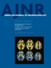
- Fig 1.
Comparison of imaging features between glioblastomas (A–C) and brain metastases (D–F). Both show ring enhancement and extensive edema on axial contrast-enhanced T1-weighted images (A and D) and restricted diffusion of the enhancing part on MD maps (B and E). However, for the FA map, the glioblastoma case demonstrates high FA values from the enhancing region. The high FA starts from the enhancing region and extends to the immediate peritumoral region, making an FA rim.
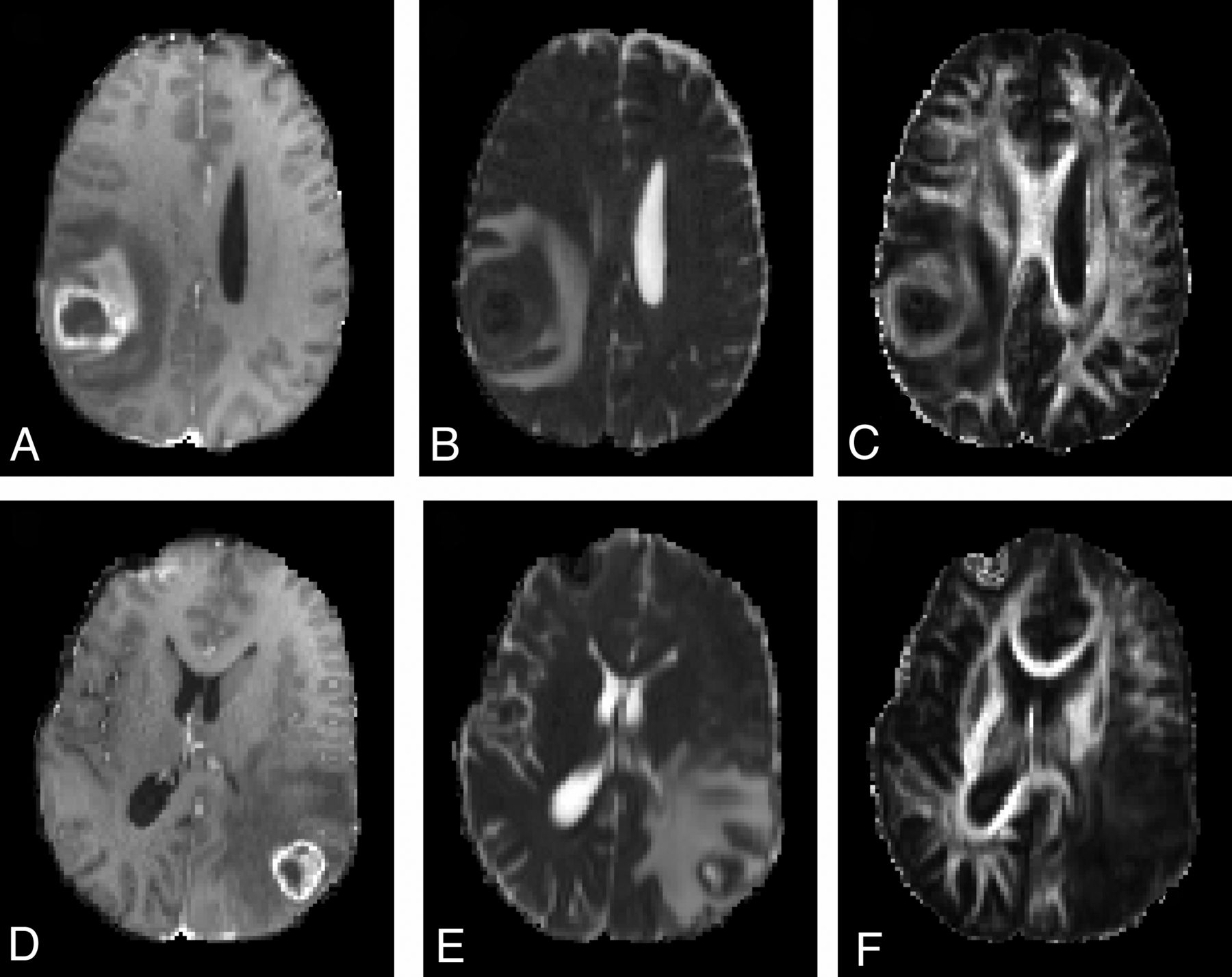
- Fig 2.
Boxplot of FA and MD from the enhancing region in glioblastomas (white) and brain metastases (gray) (A and B). The outliers are represented by circles. Asterisks indicate significant differences (P < .01). A scatterplot of FA and MD from the enhancing region of glioblastomas (blue square) and brain metastases (purple circle) (C) is shown. The green line represents the cutoff line of MD; the blue line, the cutoff line of FA; and the red line, the cutoff line of the combined model of FA and MD, which can successfully separate the glioblastomas and brain metastases. FA and MD regression lines for glioblastomas (D), FA and MD regression lines for brain metastases (E), and the dotted line indicate 95% confidence intervals. There is a negative correlation of FA and MD in glioblastomas (R = 0.51, P < .05).
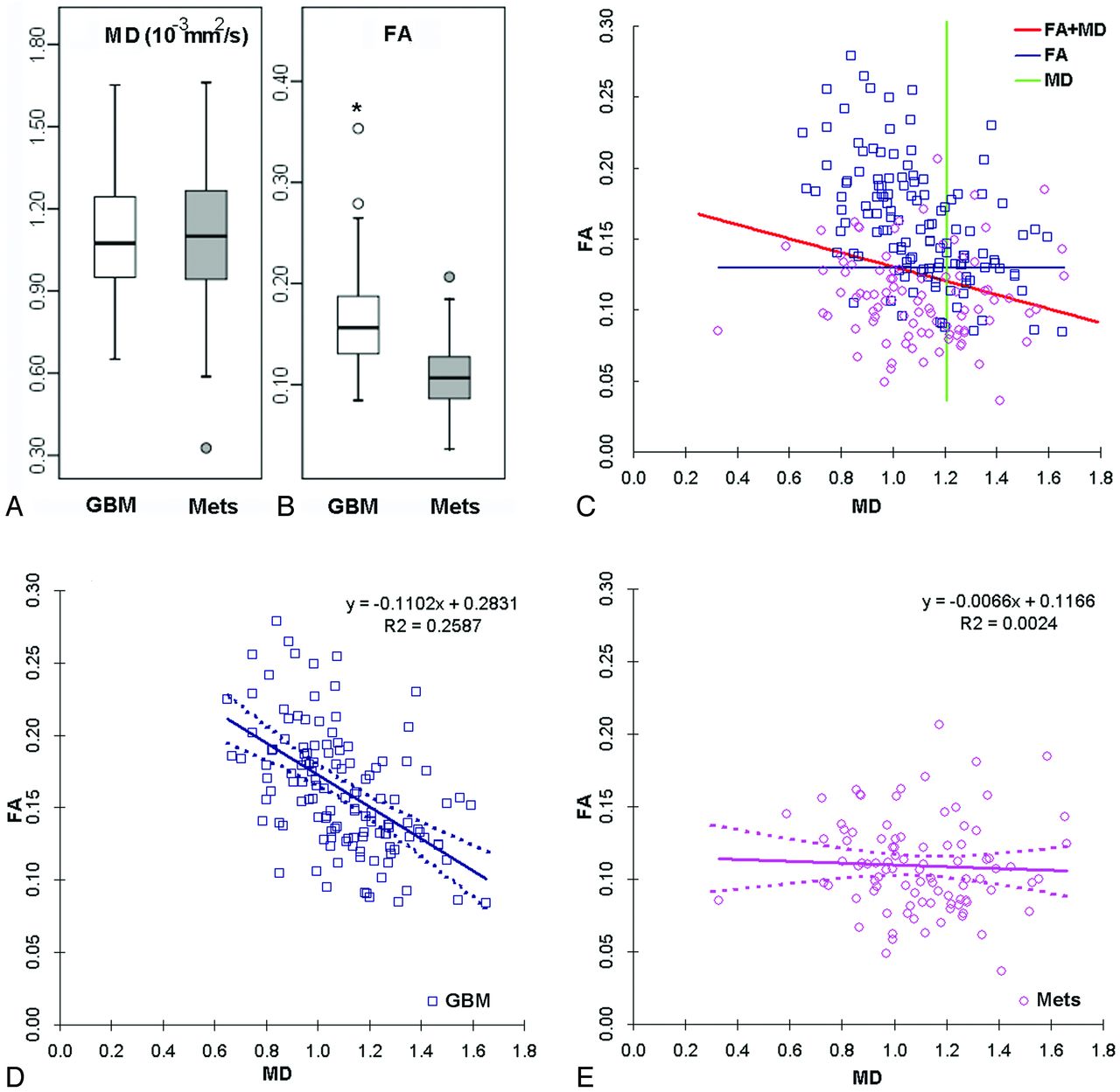
- Fig 3.
Scatterplot of 2 raters and the logistic regression model for glioblastomas (blue square) and brain metastases (purple circles) (A and B). Approximately half of cases were with low confidence levels (levels 2–4 for the raters and 0.2–0.8 for the LRM). PGBM represents the probability, predicted by the model, for glioblastoma (GBM). Receiver operative characteristic curve analysis from 2 raters and the LRM for the whole cases (C) and challenging cases for the raters with confidence level of 2–4 (D) are shown. The performance of the LRM is close to that of both raters. For challenging cases, the performance of the LRM remains at about the same level as those for all cases.
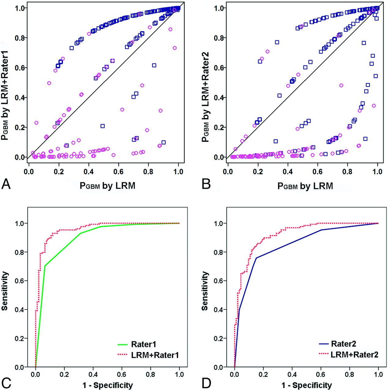
- Fig 4.
The diagnostic performance of rater 1 improves by combining the DTI logistic regression model (A and C). Most of the glioblastomas (blue squares) are above the diagonal line, and most of the brain metastases (purple circles) are below the diagonal line. PGBM represents the probability, predicted by the model, for glioblastoma (GBM). AUC improves from 0.90 to 0.960 for rater 1. The combined model with LRM plus rater 1 can also improve the results of rater 2 (B and D). AUC improves from 0.85 to 0.93 for rater 2.


Tables
Diagnostic performance of 2 raters and the logistic regression model of DTI parameters for all the cases
Sensitivity Specificity PPV NPV AUC Cutoff Value FAER 0.80 0.76 0.80 0.73 0.84 0.13 MDER (10−3 mm2/s) 0.73 0.34 0.60 0.47 0.51 1.21 FAIPR 0.65 0.68 0.73 0.58 0.69 0.17 LRM 0.84 0.77 0.83 0.78 0.86 0.50 Rater 1 0.70 0.93 0.75 0.94 0.90 4.5 LRM + rater 1 0.93 0.88 0.92 0.90 0.96 0.54 Rater 2 0.76 0.85 0.94 0.54 0.85 2.5 LRM + rater 2 0.85 0.85 0.88 0.81 0.93 0.54 Note:—PPV indicates positive predictive value; NPV, negative predictive value.


Jump to section
Related Articles
Cited By...
- Mesoscopic Assessment of Microstructure in Glioblastomas and Metastases by Merging Advanced Diffusion Imaging with Immunohistopathology
- Diffusion-Weighted Imaging and Diffusion Tensor Imaging for Differentiating High-Grade Glioma from Solitary Brain Metastasis: A Systematic Review and Meta-Analysis
- Differentiating Tumor Progression from Pseudoprogression in Patients with Glioblastomas Using Diffusion Tensor Imaging and Dynamic Susceptibility Contrast MRI