Advertisement

Main menu

  • Home
  • Content
    • Current Issue
    • Accepted Manuscripts
    • Article Preview
    • Past Issue Archive
    • AJNR Case Collection
    • Case of the Week Archive
    • Classic Case Archive
    • Case of the Month Archive
  • Special Collections
    • Spinal CSF Leak Articles (Jan 2020-June 2024)
    • 2024 AJNR Journal Awards
    • Most Impactful AJNR Articles
  • Multimedia
    • AJNR Podcast
    • AJNR Scantastics
    • Video Articles
  • For Authors
    • Submit a Manuscript
    • Author Policies
    • Fast publishing of Accepted Manuscripts
    • Graphical Abstract Preparation
    • Manuscript Submission Guidelines
    • Imaging Protocol Submission
    • Submit a Case for the Case Collection
  • About Us
    • About AJNR
    • Editorial Board
  • More
    • Become a Reviewer/Academy of Reviewers
    • Subscribers
    • Permissions
    • Alerts
    • Feedback
    • Advertisers
    • ASNR Home
  • Other Publications
    • ajnr

User menu

  • Alerts
  • Log in

Search

  • Advanced search
American Journal of Neuroradiology

American Journal of Neuroradiology

ASHNR American Society of Functional Neuroradiology ASHNR American Society of Pediatric Neuroradiology ASSR
  • Alerts
  • Log in

Advanced Search

  • Home
  • Content
    • Current Issue
    • Accepted Manuscripts
    • Article Preview
    • Past Issue Archive
    • AJNR Case Collection
    • Case of the Week Archive
    • Classic Case Archive
    • Case of the Month Archive
  • Special Collections
    • Spinal CSF Leak Articles (Jan 2020-June 2024)
    • 2024 AJNR Journal Awards
    • Most Impactful AJNR Articles
  • Multimedia
    • AJNR Podcast
    • AJNR Scantastics
    • Video Articles
  • For Authors
    • Submit a Manuscript
    • Author Policies
    • Fast publishing of Accepted Manuscripts
    • Graphical Abstract Preparation
    • Manuscript Submission Guidelines
    • Imaging Protocol Submission
    • Submit a Case for the Case Collection
  • About Us
    • About AJNR
    • Editorial Board
  • More
    • Become a Reviewer/Academy of Reviewers
    • Subscribers
    • Permissions
    • Alerts
    • Feedback
    • Advertisers
    • ASNR Home
  • Follow AJNR on Twitter
  • Visit AJNR on Facebook
  • Follow AJNR on Instagram
  • Join AJNR on LinkedIn
  • RSS Feeds

Welcome to the new AJNR, Updated Hall of Fame, and more. Read the full announcements.


AJNR is seeking candidates for the position of Associate Section Editor, AJNR Case Collection. Read the full announcement.


Getting new auth cookie, if you see this message a lot, tell someone!
Research ArticleBrain

Transition into Driven Equilibrium of the Balanced Steady-State Free Precession as an Ultrafast Multisection T2-Weighted Imaging of the Brain

Y.-C.K. Huang, T.-Y. Huang, H.-C. Chiu, T.-S. Kuo, C.-J. Hsueh, H.-W. Kao, C.-W. Wang, H.-H. Hsu and C.-J. Juan
American Journal of Neuroradiology June 2014, 35 (6) 1137-1144; DOI: https://doi.org/10.3174/ajnr.A3863
Y.-C.K. Huang
aFrom the Department of Electrical Engineering (Y.-C.K.H., T.-S.K.), National Taiwan University, Taipei, Taiwan, Republic of China
bDepartment of Radiology (Y.-C.K.H., C.-J.H., H.-W.K, C.-W.W., H.-H. H., C.-J.J.), Tri-Service General Hospital, Taipei, Taiwan, Republic of China
  • Find this author on Google Scholar
  • Find this author on PubMed
  • Search for this author on this site
T.-Y. Huang
cDepartment of Electrical Engineering (T.-Y.H.), National Taiwan University of Science and Technology, Taiwan, Republic of China
  • Find this author on Google Scholar
  • Find this author on PubMed
  • Search for this author on this site
H.-C. Chiu
dGraduate Institute of Design Science (H.-C.C.), Tatung University, Taipei, Taiwan, Republic of China
  • Find this author on Google Scholar
  • Find this author on PubMed
  • Search for this author on this site
T.-S. Kuo
aFrom the Department of Electrical Engineering (Y.-C.K.H., T.-S.K.), National Taiwan University, Taipei, Taiwan, Republic of China
  • Find this author on Google Scholar
  • Find this author on PubMed
  • Search for this author on this site
C.-J. Hsueh
bDepartment of Radiology (Y.-C.K.H., C.-J.H., H.-W.K, C.-W.W., H.-H. H., C.-J.J.), Tri-Service General Hospital, Taipei, Taiwan, Republic of China
eDepartment of Radiology (C.-J.H., H.-W.K, C.-W.W., H.-H. H., C.-J.J.), National Defense Medical Center, Taipei, Taiwan, Republic of China.
  • Find this author on Google Scholar
  • Find this author on PubMed
  • Search for this author on this site
H.-W. Kao
bDepartment of Radiology (Y.-C.K.H., C.-J.H., H.-W.K, C.-W.W., H.-H. H., C.-J.J.), Tri-Service General Hospital, Taipei, Taiwan, Republic of China
eDepartment of Radiology (C.-J.H., H.-W.K, C.-W.W., H.-H. H., C.-J.J.), National Defense Medical Center, Taipei, Taiwan, Republic of China.
  • Find this author on Google Scholar
  • Find this author on PubMed
  • Search for this author on this site
C.-W. Wang
bDepartment of Radiology (Y.-C.K.H., C.-J.H., H.-W.K, C.-W.W., H.-H. H., C.-J.J.), Tri-Service General Hospital, Taipei, Taiwan, Republic of China
eDepartment of Radiology (C.-J.H., H.-W.K, C.-W.W., H.-H. H., C.-J.J.), National Defense Medical Center, Taipei, Taiwan, Republic of China.
  • Find this author on Google Scholar
  • Find this author on PubMed
  • Search for this author on this site
H.-H. Hsu
bDepartment of Radiology (Y.-C.K.H., C.-J.H., H.-W.K, C.-W.W., H.-H. H., C.-J.J.), Tri-Service General Hospital, Taipei, Taiwan, Republic of China
eDepartment of Radiology (C.-J.H., H.-W.K, C.-W.W., H.-H. H., C.-J.J.), National Defense Medical Center, Taipei, Taiwan, Republic of China.
  • Find this author on Google Scholar
  • Find this author on PubMed
  • Search for this author on this site
C.-J. Juan
bDepartment of Radiology (Y.-C.K.H., C.-J.H., H.-W.K, C.-W.W., H.-H. H., C.-J.J.), Tri-Service General Hospital, Taipei, Taiwan, Republic of China
eDepartment of Radiology (C.-J.H., H.-W.K, C.-W.W., H.-H. H., C.-J.J.), National Defense Medical Center, Taipei, Taiwan, Republic of China.
  • Find this author on Google Scholar
  • Find this author on PubMed
  • Search for this author on this site
  • Article
  • Figures & Data
  • Supplemental
  • Info & Metrics
  • Responses
  • References
  • PDF
Loading

Article Figures & Data

Figures

  • Tables
  • Fig 1.
    • Download figure
    • Open in new tab
    • Download powerpoint
    Fig 1.

    TSE (A, D), TGSE (B, E), and TIDE (C, F) images of a 38-year-old healthy man. The results show similar T2 contrast of the neocortex (A–C). At the basal ganglia, iron-containing gray matter (eg, globus pallidus) has a much higher intensity in the TIDE image compared with the other 2 results (D–F). Ghosting artifacts appear in the TIDE image (arrow in C).
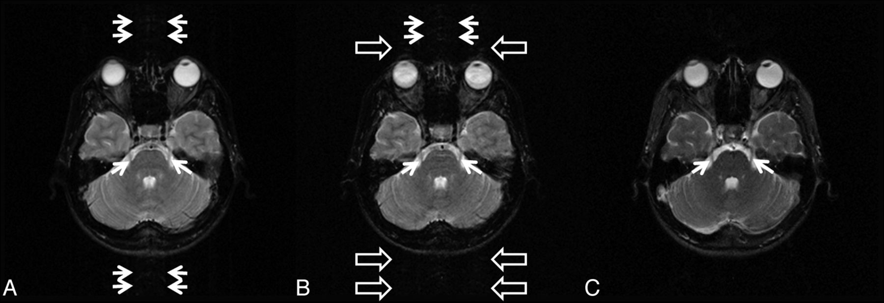
  • Fig 2.
    • Download figure
    • Open in new tab
    • Download powerpoint
    Fig 2.

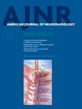
    TSE (A, D), TGSE (B, E), and TIDE (C, F) images of a 25-year-old healthy man. Motion artifacts can be found in the TSE and TGSE images, due to eyeball movement (open arrows in A and B) and CSF flow (open arrows in D and E). Magnetic susceptibility artifacts appear in the TIDE image (solid arrows in C). Occasional basilar artery flow artifacts are seen in the TIDE image (thin arrow in C).

  • Fig 3.
    • Download figure
    • Open in new tab
    • Download powerpoint
    Fig 3.

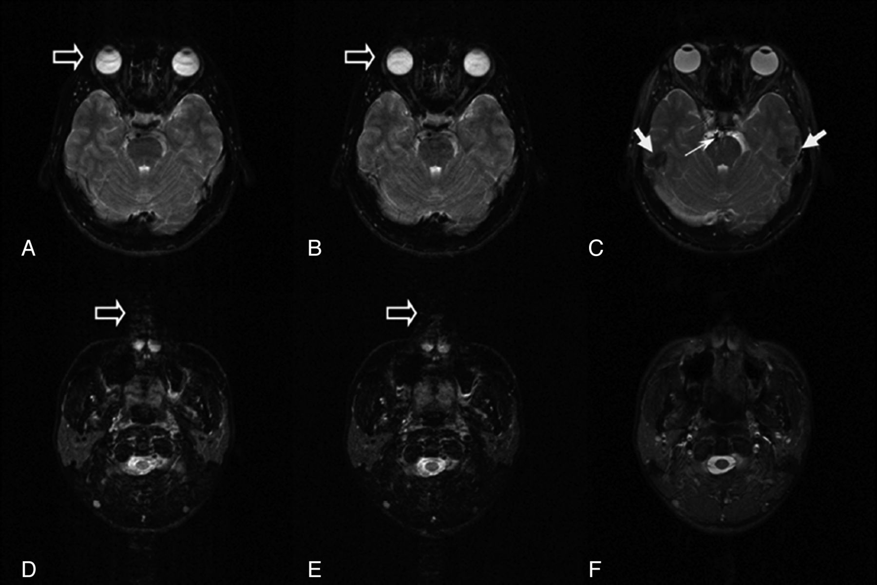
    TSE (A, D), TGSE (B, E), and TIDE (C, F) images of a 57-year-old woman with stroke history. A hyperintense lesion appears near the left lateral ventricle (white arrows in A–C). The degrees of hyperintensity are different in these images. Similar contrast of a late subacute hematoma involving the right corpus striatum is found in all sequences (black arrows in D–F).

  • Fig 4.
    • Download figure
    • Open in new tab
    • Download powerpoint
    Fig 4.

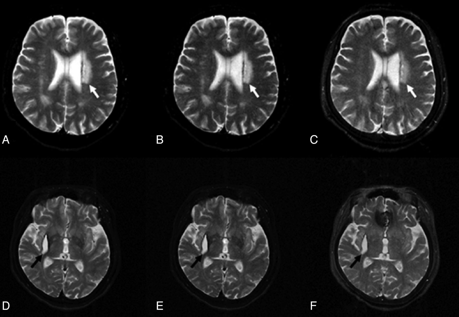
    A 9-day-old neonate with seizures, presenting with involuntary movements, was imagined with TSE (A) and TIDE (B) sequences. A motion artifacts-free T2-weighted image can be acquired with TIDE, depicting a clear brain anatomy.

  • Fig 5.
    • Download figure
    • Open in new tab
    • Download powerpoint
    Fig 5.

    TSE (A), TGSE (B), and TIDE (C) images of a 26-year-old female volunteer. CSF motion artifacts (double arrows) are seen on TSE and TGSE images, while eyeball motion artifacts (open arrows) are seen on the TGSE image. The trigeminal nerves (arrows) are clearly demonstrated on TIDE, which is free of the aforementioned motion artifacts but are somewhat blurred on TSE and TGSE.
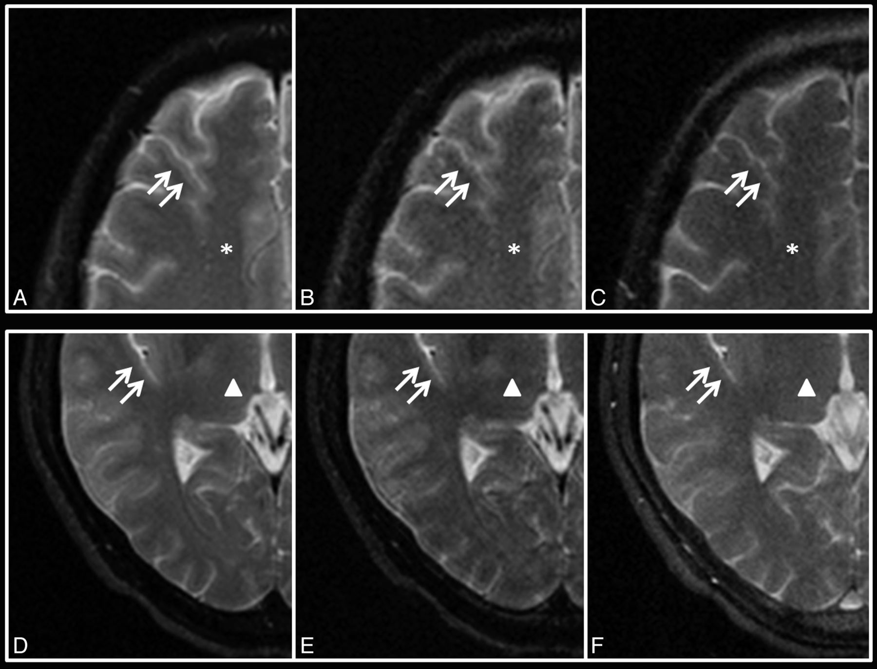
  • Fig 6.
    • Download figure
    • Open in new tab
    • Download powerpoint
    Fig 6.

    TSE (A, D), TGSE (B, E), and TIDE (C, F) images of a 40-year-old male volunteer at the levels of the centrum semiovale (star, A–C) and thalamus (triangle, D–F). The gray-white matter junctions (double arrows) are clearly demonstrated on TSE but are blurred on TGSE and TIDE.

Tables

  • Figures
  • SNRs and CNRs of TSE, TGSE, and TIDE sequences

    TSETGSETIDEPTSE,TGSEPTGSE,TIDEPTSE,TIDE
    SNR
        WM
            CSO56.14 ± 8.0529.29 ± 4.3625.02 ± 3.70–––
            CR56.48 ± 8.0529.71 ± 4.2825.86 ± 3.63–––
            aPVWM52.54 ± 6.9427.35 ± 3.5724.12 ± 3.25–––
            pPVWM58.58 ± 7.4831.05 ± 3.8528.20 ± 3.59–––
        GM
            CH68.38 ± 9.3535.80 ± 4.6932.39 ± 4.34–––
            PUT61.97 ± 8.5232.11 ± 4.2532.58 ± 4.35–.364–
            THA61.09 ± 8.1832.03 ± 4.1830.71 ± 3.76–.031–
            GP48.47 ± 6.5824.80 ± 3.4228.30 ± 3.95–––
    CNR
        NICGM-WM, THA-CSO4.95 ± 3.642.74 ± 1.855.69 ± 1.37––.169
        ICGM-WM, GP-CSO−7.67 ± 3.98−4.494 ± 2.353.275 ± 1.70–––
    • Note:—CSO indicates centrum semiovale; CR, corona radiata; aPVWM, anterior periventricular white matter; pPVWM, posterior periventricular white matter; CH, head of caudate nucleus; PUT, putamen; THA: thalamus; GP, globus pallidus; NICGM, non-iron-containing gray matter; ICGM, iron-containing gray matter; –, P < .0001.

PreviousNext
Back to top

In this issue

American Journal of Neuroradiology: 35 (6)
American Journal of Neuroradiology
Vol. 35, Issue 6
1 Jun 2014
  • Table of Contents
  • Index by author
  • Complete Issue (PDF)
Advertisement
Print
Download PDF
Email Article

Thank you for your interest in spreading the word on American Journal of Neuroradiology.

NOTE: We only request your email address so that the person you are recommending the page to knows that you wanted them to see it, and that it is not junk mail. We do not capture any email address.

Enter multiple addresses on separate lines or separate them with commas.
Transition into Driven Equilibrium of the Balanced Steady-State Free Precession as an Ultrafast Multisection T2-Weighted Imaging of the Brain
(Your Name) has sent you a message from American Journal of Neuroradiology
(Your Name) thought you would like to see the American Journal of Neuroradiology web site.
CAPTCHA
This question is for testing whether or not you are a human visitor and to prevent automated spam submissions.
Cite this article
Y.-C.K. Huang, T.-Y. Huang, H.-C. Chiu, T.-S. Kuo, C.-J. Hsueh, H.-W. Kao, C.-W. Wang, H.-H. Hsu, C.-J. Juan
Transition into Driven Equilibrium of the Balanced Steady-State Free Precession as an Ultrafast Multisection T2-Weighted Imaging of the Brain
American Journal of Neuroradiology Jun 2014, 35 (6) 1137-1144; DOI: 10.3174/ajnr.A3863

Citation Manager Formats

  • BibTeX
  • Bookends
  • EasyBib
  • EndNote (tagged)
  • EndNote 8 (xml)
  • Medlars
  • Mendeley
  • Papers
  • RefWorks Tagged
  • Ref Manager
  • RIS
  • Zotero
0 Responses
Respond to this article
Share
Bookmark this article
Transition into Driven Equilibrium of the Balanced Steady-State Free Precession as an Ultrafast Multisection T2-Weighted Imaging of the Brain
Y.-C.K. Huang, T.-Y. Huang, H.-C. Chiu, T.-S. Kuo, C.-J. Hsueh, H.-W. Kao, C.-W. Wang, H.-H. Hsu, C.-J. Juan
American Journal of Neuroradiology Jun 2014, 35 (6) 1137-1144; DOI: 10.3174/ajnr.A3863
del.icio.us logo Twitter logo Facebook logo Mendeley logo
  • Tweet Widget
  • Facebook Like
  • Google Plus One
Purchase

Jump to section

  • Article
    • Abstract
    • ABBREVIATIONS:
    • Materials and Methods
    • Results
    • Discussion
    • Conclusions
    • Footnotes
    • References
  • Figures & Data
  • Supplemental
  • Info & Metrics
  • Responses
  • References
  • PDF

Related Articles

  • PubMed
  • Google Scholar

Cited By...

  • No citing articles found.
  • Crossref
  • Google Scholar

This article has not yet been cited by articles in journals that are participating in Crossref Cited-by Linking.

More in this TOC Section

  • Evaluating the Effects of White Matter Multiple Sclerosis Lesions on the Volume Estimation of 6 Brain Tissue Segmentation Methods
  • White Matter Alterations in the Brains of Patients with Active, Remitted, and Cured Cushing Syndrome: A DTI Study
  • Enhanced Axonal Metabolism during Early Natalizumab Treatment in Relapsing-Remitting Multiple Sclerosis
Show more Brain

Similar Articles

Advertisement

Indexed Content

  • Current Issue
  • Accepted Manuscripts
  • Article Preview
  • Past Issues
  • Editorials
  • Editors Choice
  • Fellow Journal Club
  • Letters to the Editor

Cases

  • Case Collection
  • Archive - Case of the Week
  • Archive - Case of the Month
  • Archive - Classic Case

Special Collections

  • Special Collections

Resources

  • News and Updates
  • Turn around Times
  • Submit a Manuscript
  • Author Policies
  • Manuscript Submission Guidelines
  • Evidence-Based Medicine Level Guide
  • Publishing Checklists
  • Graphical Abstract Preparation
  • Imaging Protocol Submission
  • Submit a Case
  • Become a Reviewer/Academy of Reviewers
  • Get Peer Review Credit from Publons

Multimedia

  • AJNR Podcast
  • AJNR SCANtastic
  • Video Articles

About Us

  • About AJNR
  • Editorial Board
  • Not an AJNR Subscriber? Join Now
  • Alerts
  • Feedback
  • Advertise with us
  • Librarian Resources
  • Permissions
  • Terms and Conditions

American Society of Neuroradiology

  • Not an ASNR Member? Join Now

© 2025 by the American Society of Neuroradiology All rights, including for text and data mining, AI training, and similar technologies, are reserved.
Print ISSN: 0195-6108 Online ISSN: 1936-959X

Powered by HighWire