
Article Figures & Data
Figures
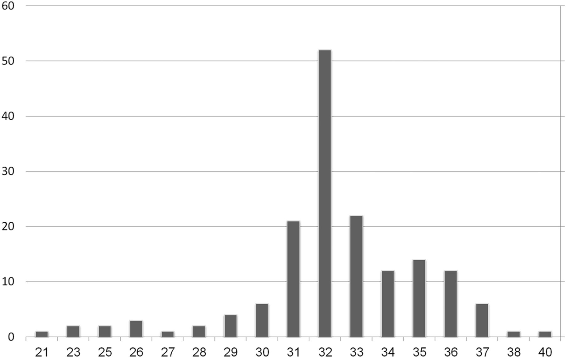
- Fig 1.
Distribution of number of fetuses providing data as a function of gestational age (n = 162).
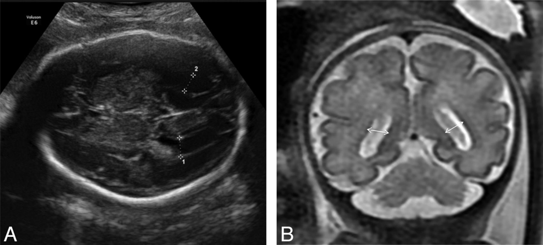
- Fig 2.
A, Sonographic measurement of the fetal lateral ventricles in the axial transventricular plane. B, Measurement of the fetal lateral ventricles in a T2-weighted MR image (coronal plane) at the level of the ventricles; ventricular diameter is measured at the midheight of the ventricle.
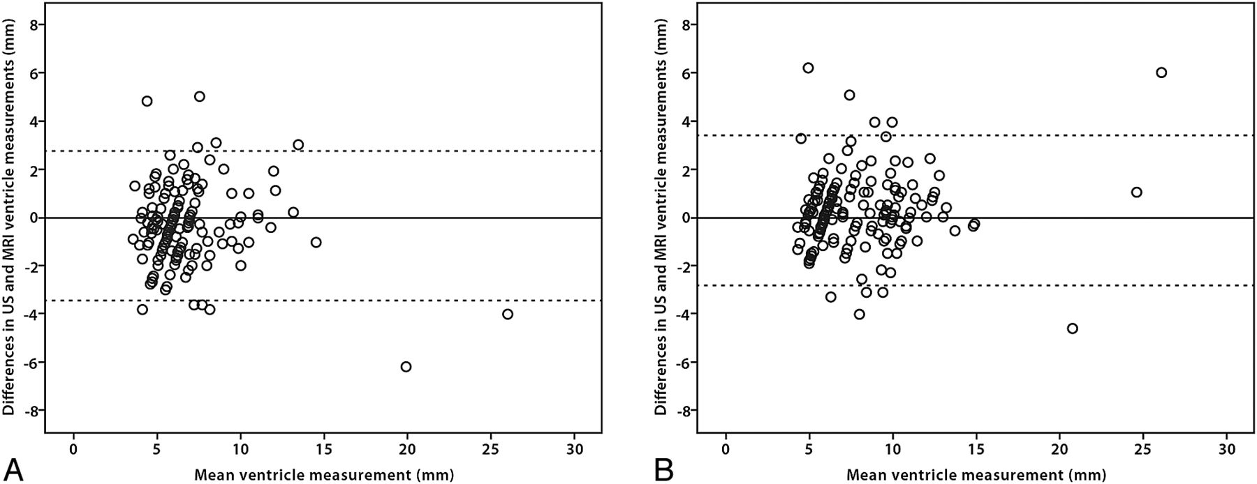
- Fig 3.
Plots of the difference between sonographic and MR imaging ventricular measurements against their mean (A, narrower ventricle; B, wider ventricle).
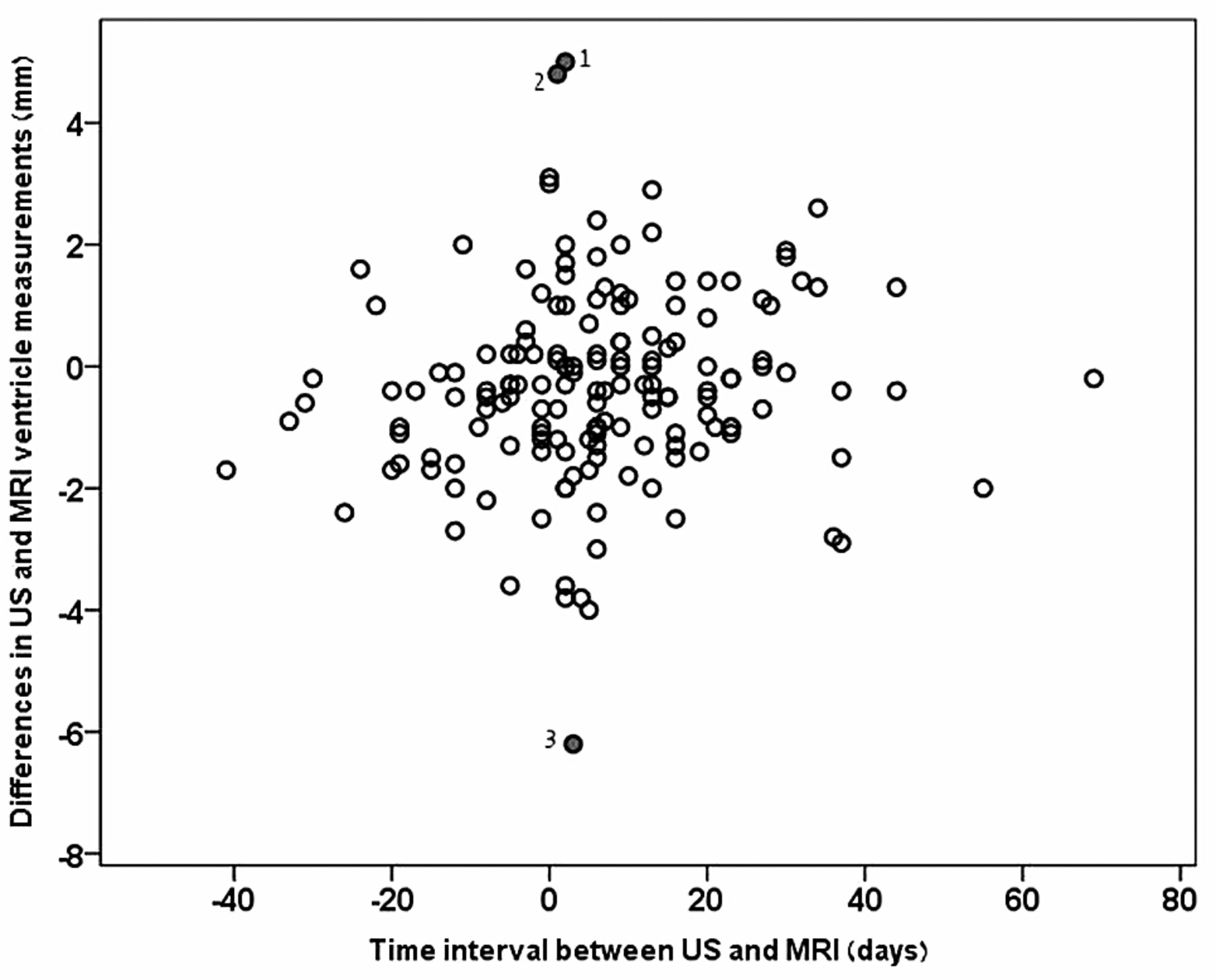
- Fig 4.
Plots of the difference between sonography and MR imaging ventricular measurements against the time interval between sonography and MR imaging. Cases of great differences between the 2 modalities: 1) very narrow ventricles on MR imaging, most likely below the measurement resolution by sonography (5 mm); 2) very wide ventricles (>20 mm) in both modalities; and 3) a rare case of disagreement between sonography and MR imaging.


Tables
- Table 1:
Distribution of numbers of fetuses providing data as a function of the indication for neurosonography
Indication No. Mean Gestational Week Min Max Median CMV seroconversion 35 33.63 30 40 33 Ventricular asymmetry 54 31.78 21 37 32 Abnormal head circumference 15 34.27 29 38 35 Abnormal posterior fossa 10 31.60 26 35 32 Cystic lesions 8 33.75 31 37 33 Family history of CNS illness 6 32.33 31 34 32 Midline abnormalities 6 31.67 23 37 32 Obstetric complications and extra-CNS anatomic findings 28 31.00 23 36 32 Note:—Min indicates minimum; Max, maximum; CMV, cytomegalovirus.
Differences in Measurement of Ventricles, Ultrasound-MRI (mm) Definition of Ventriculomegaly κ Coefficient Mean 95% Limits of Agreement ICC (95% CI) MRI radiologist 1 Ventricle (a) 0.33 ± 1.58 (−2.76–1.58) 0.91 (0.88–0.94) 93.8 Ventricle (b) −0.26 ± 1.58 (−3.35–1.58) 0.94 (0.91–0.95) 83.4 MRI radiologist 2 Ventricle (a) −0.88 ± 1.52 (−3.86–1.52) 0.9 (0.78–0.95) 93.8 Ventricle (b) −1.09 ± 1.5 (−4.05–1.5) 0.92 (0.75–0.96) 84.2 Note:—ICC indicates intraclass correlation coefficient.
- Table 3:
Comparison between ultrasonography and MRI data in the evaluation of ventricular diameter below and above the clinical cutoff of 10 mm
Ultrasonography Axial Plane Ventricle (a) Ventricle (b) <10 mm ≥10 mm <10 mm ≥10 mm MRI coronal plane, radiologist 1 <10 mm 138 5* 103 14* ≥10 mm 5* 14 13* 32 MRI coronal plane, radiologist 2 <10 mm 134 1** 108 20** ≥10 mm 9** 18 5** 25 Mean 95% Limits of Agreement ICC 95% CI Ventricle (a) −1.20 ± 1.11 −3.39–0.97 0.91 0.41–0.97 Ventricle (b) −0.82 ± 1.03 −2.84–1.19 0.96 0.84–0.98 Note:—ICC indicates intraclass correlation coefficient.


