Advertisement

Main menu

  • Home
  • Content
    • Current Issue
    • Accepted Manuscripts
    • Article Preview
    • Past Issue Archive
    • Video Articles
    • AJNR Case Collection
    • Case of the Week Archive
    • Case of the Month Archive
    • Classic Case Archive
  • Special Collections
    • AJNR Awards
    • Low-Field MRI
    • Alzheimer Disease
    • ASNR Foundation Special Collection
    • Photon-Counting CT
    • View All
  • Multimedia
    • AJNR Podcasts
    • AJNR SCANtastic
    • Trainee Corner
    • MRI Safety Corner
    • Imaging Protocols
  • For Authors
    • Submit a Manuscript
    • Submit a Video Article
    • Submit an eLetter to the Editor/Response
    • Manuscript Submission Guidelines
    • Statistical Tips
    • Fast Publishing of Accepted Manuscripts
    • Graphical Abstract Preparation
    • Imaging Protocol Submission
    • Author Policies
  • About Us
    • About AJNR
    • Editorial Board
    • Editorial Board Alumni
  • More
    • Become a Reviewer/Academy of Reviewers
    • Subscribers
    • Permissions
    • Alerts
    • Feedback
    • Advertisers
    • ASNR Home

User menu

  • Alerts
  • Log in

Search

  • Advanced search
American Journal of Neuroradiology

American Journal of Neuroradiology

ASHNR American Society of Functional Neuroradiology ASHNR American Society of Pediatric Neuroradiology ASSR
  • Alerts
  • Log in

Advanced Search

  • Home
  • Content
    • Current Issue
    • Accepted Manuscripts
    • Article Preview
    • Past Issue Archive
    • Video Articles
    • AJNR Case Collection
    • Case of the Week Archive
    • Case of the Month Archive
    • Classic Case Archive
  • Special Collections
    • AJNR Awards
    • Low-Field MRI
    • Alzheimer Disease
    • ASNR Foundation Special Collection
    • Photon-Counting CT
    • View All
  • Multimedia
    • AJNR Podcasts
    • AJNR SCANtastic
    • Trainee Corner
    • MRI Safety Corner
    • Imaging Protocols
  • For Authors
    • Submit a Manuscript
    • Submit a Video Article
    • Submit an eLetter to the Editor/Response
    • Manuscript Submission Guidelines
    • Statistical Tips
    • Fast Publishing of Accepted Manuscripts
    • Graphical Abstract Preparation
    • Imaging Protocol Submission
    • Author Policies
  • About Us
    • About AJNR
    • Editorial Board
    • Editorial Board Alumni
  • More
    • Become a Reviewer/Academy of Reviewers
    • Subscribers
    • Permissions
    • Alerts
    • Feedback
    • Advertisers
    • ASNR Home
  • Follow AJNR on Twitter
  • Visit AJNR on Facebook
  • Follow AJNR on Instagram
  • Join AJNR on LinkedIn
  • RSS Feeds

AJNR Awards, New Junior Editors, and more. Read the latest AJNR updates

Getting new auth cookie, if you see this message a lot, tell someone!
Research ArticleBrain
Open Access

Diffusion-Weighted Imaging with Dual-Echo Echo-Planar Imaging for Better Sensitivity to Acute Stroke

S.J. Holdsworth, K.W. Yeom, M.U. Antonucci, J.B. Andre, J. Rosenberg, M. Aksoy, M. Straka, N.J. Fischbein, R. Bammer, M.E. Moseley, G. Zaharchuk and S. Skare
American Journal of Neuroradiology July 2014, 35 (7) 1293-1302; DOI: https://doi.org/10.3174/ajnr.A3921
S.J. Holdsworth
aFrom the Department of Radiology (S.J.H., K.W.Y., M.U.A., J.R., M.A., M.S., N.J.F., R.B., M.E.M., G.Z.), Stanford University, Stanford, California
  • Find this author on Google Scholar
  • Find this author on PubMed
  • Search for this author on this site
K.W. Yeom
aFrom the Department of Radiology (S.J.H., K.W.Y., M.U.A., J.R., M.A., M.S., N.J.F., R.B., M.E.M., G.Z.), Stanford University, Stanford, California
  • Find this author on Google Scholar
  • Find this author on PubMed
  • Search for this author on this site
M.U. Antonucci
aFrom the Department of Radiology (S.J.H., K.W.Y., M.U.A., J.R., M.A., M.S., N.J.F., R.B., M.E.M., G.Z.), Stanford University, Stanford, California
  • Find this author on Google Scholar
  • Find this author on PubMed
  • Search for this author on this site
J.B. Andre
bDepartment of Radiology (J.B.A.), University of Washington, Seattle, Washington
  • Find this author on Google Scholar
  • Find this author on PubMed
  • Search for this author on this site
J. Rosenberg
aFrom the Department of Radiology (S.J.H., K.W.Y., M.U.A., J.R., M.A., M.S., N.J.F., R.B., M.E.M., G.Z.), Stanford University, Stanford, California
  • Find this author on Google Scholar
  • Find this author on PubMed
  • Search for this author on this site
M. Aksoy
aFrom the Department of Radiology (S.J.H., K.W.Y., M.U.A., J.R., M.A., M.S., N.J.F., R.B., M.E.M., G.Z.), Stanford University, Stanford, California
  • Find this author on Google Scholar
  • Find this author on PubMed
  • Search for this author on this site
M. Straka
aFrom the Department of Radiology (S.J.H., K.W.Y., M.U.A., J.R., M.A., M.S., N.J.F., R.B., M.E.M., G.Z.), Stanford University, Stanford, California
  • Find this author on Google Scholar
  • Find this author on PubMed
  • Search for this author on this site
N.J. Fischbein
aFrom the Department of Radiology (S.J.H., K.W.Y., M.U.A., J.R., M.A., M.S., N.J.F., R.B., M.E.M., G.Z.), Stanford University, Stanford, California
  • Find this author on Google Scholar
  • Find this author on PubMed
  • Search for this author on this site
R. Bammer
aFrom the Department of Radiology (S.J.H., K.W.Y., M.U.A., J.R., M.A., M.S., N.J.F., R.B., M.E.M., G.Z.), Stanford University, Stanford, California
  • Find this author on Google Scholar
  • Find this author on PubMed
  • Search for this author on this site
M.E. Moseley
aFrom the Department of Radiology (S.J.H., K.W.Y., M.U.A., J.R., M.A., M.S., N.J.F., R.B., M.E.M., G.Z.), Stanford University, Stanford, California
  • Find this author on Google Scholar
  • Find this author on PubMed
  • Search for this author on this site
G. Zaharchuk
aFrom the Department of Radiology (S.J.H., K.W.Y., M.U.A., J.R., M.A., M.S., N.J.F., R.B., M.E.M., G.Z.), Stanford University, Stanford, California
  • Find this author on Google Scholar
  • Find this author on PubMed
  • Search for this author on this site
S. Skare
cClinical Neuroscience (S.S.), Karolinska Institute, Stockholm, Sweden.
  • Find this author on Google Scholar
  • Find this author on PubMed
  • Search for this author on this site
  • Article
  • Figures & Data
  • Info & Metrics
  • Responses
  • References
  • PDF
Loading

Article Figures & Data

Figures

  • Tables
  • Fig 1.
    • Download figure
    • Open in new tab
    • Download powerpoint
    Fig 1.

    Dual-echo DWI pulse sequence showing the section-select module (blue); diffusion preparation period (yellow); and the first and second imaging echoes (echoes 1 and 2 acquired at 2 different TEs, TE1 and TE2) separated by a 180° refocusing pulse.

  • Fig 2.
    • Download figure
    • Open in new tab
    • Download powerpoint
    Fig 2.

    Assessment of readers A, B, and C of image quality, lesion conspicuity, and diagnostic confidence. Nominal values of 3–7 represent reader grading compared with the product DW-EPI (assigned as 4), represented by the percentage of total assigned values. Note that values of 1–2 (corresponding to “nondiagnostic” and “poor”) are not shown because these values were not assigned in this study.

  • Fig 3.
    • Download figure
    • Open in new tab
    • Download powerpoint
    Fig 3.

    Comparison of the vendor-supplied (product) DWI, echo 1, and echo 2 DWI acquired from the dual-echo sequence on 3 patients (from left to right). The ADC (calculated from echo 1) shows the presence of reduced diffusivity in each lesion (far right column). A, A 62-year-old female patient with stroke. B, A 49-year-old male patient with vasospasm and infarction post-aneurysm clipping. C, An 88-year-old woman presenting with strokelike symptoms. Note that the small infarct in the splenium of the corpus callosum present on the dual-echo DWIs was initially missed on the product DWI.

  • Fig 4.
    • Download figure
    • Open in new tab
    • Download powerpoint
    Fig 4.

    Examples of improved lesion detection of echo 2. A, A 56-year-old man status post posterior fossa tumor resection. Right cerebellar injury is more conspicuous on echo 2 and was confirmed to have reduced diffusion based on ADC. B, A 39-year-old man with known Moyamoya disease status post right superficial temporal artery to middle cerebral artery anastomosis presented with acute strokelike symptoms. Superficial temporal cortical lesion (closed arrow) and a punctate putaminal lesion (arrowhead) are confirmed with reduced diffusivity based on the ADC map. Also note improved delineation of a small subdural hematoma on echo 2 (open arrow).

  • Fig 5.
    • Download figure
    • Open in new tab
    • Download powerpoint
    Fig 5.

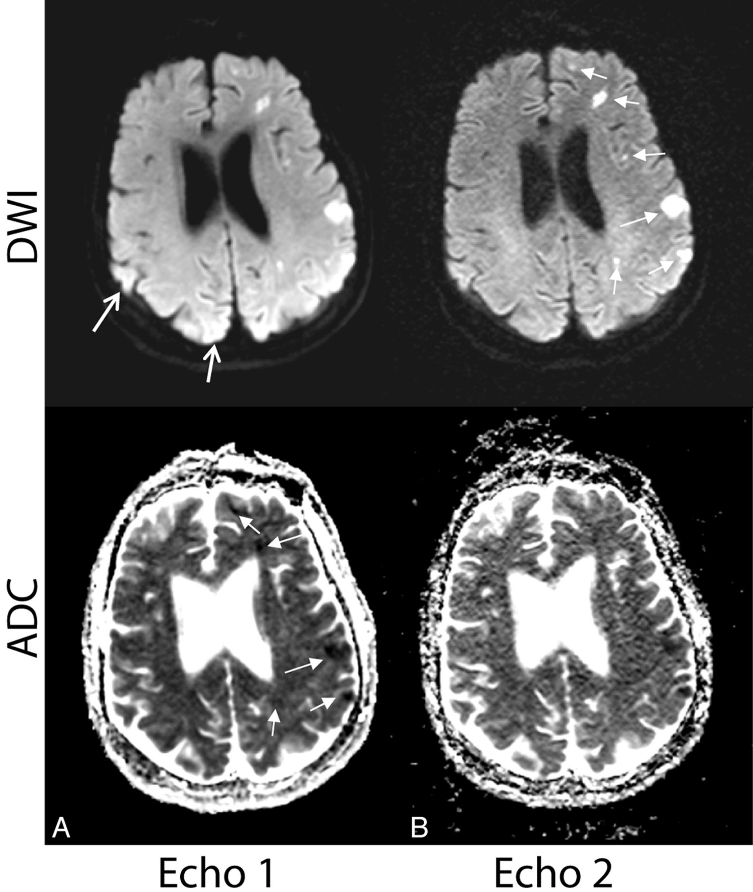
    Examples in which additional lesions identified by echo 2 suggested a potential underlying mechanism of stroke and potentially altered diagnostic impression and clinical management. A, A 60-year-old woman with vasospasm after subarachnoid hemorrhage. B, A 65-year-old woman with embolic infarcts who also had lesions in the right cerebral hemisphere more inferiorly (not shown). On both patients, echo 2 showed missed sites of reduced diffusivity on the contralateral hemisphere (arrows), suggesting multiple vascular distribution involvement (vasospasm in multiple vascular territories or embolic source). Note that the lesions were retrospectively observed on echo 1 (but not on the product DWI).

  • Fig 6.
    • Download figure
    • Open in new tab
    • Download powerpoint
    Fig 6.

    Examples in which the heightened sensitivity of echo 2 prompted further assessment by using ADC. A, A 61-year-old woman with hemorrhage from a cavernous malformation. Bright signal around the lesion was seen on echo 2 (and product) DWI and was confirmed as edema around a hemorrhage site (open arrows). B, A 69-year-old man with strokelike symptoms. Example of acute and subacute (closed arrows)/chronic stroke (open arrows) on echo 2 DWI, also present on the product DWI. Also note the arrowhead on echo 1 showing unwanted heightened coil sensitivity in the posterior brain region.

  • Fig 7.
    • Download figure
    • Open in new tab
    • Download powerpoint
    Fig 7.

    Case illustrating why echo 1 is more useful than echo 2 for calculating ADC maps used for the validation of acute infarct. Select images of a 66-year-old woman with embolic infarcts. DWI and ADC maps for echo 1 (A) and echo 2 (B) are shown. The DWI of echo 2 was found to have much higher sensitivity to acute lesions (confirmed on the ADC of echo 1) than echo 1. However, because the ADC of echo 2 is plagued by noise, echo 1 is considerably more useful for calculating ADC maps used for the validation of acute stroke (closed white arrows). The open white arrows represent areas where it can be difficult to rule out stroke from heightened coil sensitivity in (particularly posterior) regions of the brain.

  • Fig 8.
    • Download figure
    • Open in new tab
    • Download powerpoint
    Fig 8.

    A sample case showing the potential contribution of the R2 map. A 69-year-old female patient with vasospasm after subarachnoid hemorrhage. The low-R2 lesion is more conspicuous than the corresponding T2 hyperintensity on the FLAIR image. On the basis of DWI/ADC, this area represents acute right MCA territory infarction. The potential contribution of the R2 map with regard to timing of stroke and its evolution is unknown but prompts future investigation. The R2 map also shows more conspicuous mineralization in the basal ganglia than the gradient-recalled echo image (arrowhead).

Tables

  • Figures
    • View popup
    Table 1:

    Clinical history of the 50 patients who were suspected of stroke and scanned for this studya

    Clinical HistoryNo. of PatientsNo. of Patients with Lesions with Reduced Diffusivity
    Moyamoya disease55
    Transient ischemic attack165
    Stroke1414
    Cavernous malformation32
    Hemorrhage55
    Vasospasm post-subarachnoid hemorrhage33
    Metabolic disease10
    Brain tumor21
    Headache11
    Total No. of patients5036
    • ↵a The number of patients with lesions with reduced diffusivity present on ≥1 of the scanned DWI sequences is also shown.

    • View popup
    Table 2:

    Agreement among readers on specific ratings using a weighted κ statistic (N = 50)a

    Echo 1Echo 2
    Diag ConfConspicuityQualityDiag ConfConspicuityQuality
    A vs B−0.050.100.000.07−0.00−0.09
    A vs C0.100.130.310.160.13−0.09
    B vs C0.02−0.020.000.090.140.31
    • Note:—Diag Conf indicates diagnostic confidence.

    • ↵a All ratings are with P > .14.

    • View popup
    Table 3:

    Preference for the second echo over the first echo

    ReaderFractionPercentage95% CIP Value (1-tailed)
    Reader A35/5070%(55%–82%).003
    Reader B38/5076%(62%–87%)<.001
    Reader C34/5068%(53%–80%).008
    • View popup
    Table 4:

    Percentage of cases rated greater than the product DWI (N = 50)

    Echo 1Echo 2
    Diag ConfConspicuityQualityDiag ConfConspicuityQuality
    A vs B92%84%100%96%98%100%
    A vs C78%66%100%96%98%92%
    B vs C96%92%100%96%94%94%
    • Note:—Diag Conf indicates diagnostic confidence.

    • View popup
    Table 5:

    Mean (SD) of ratings (N = 50)

    Diagnostic ConfidenceConspicuityQuality
    Echo 1Echo 2Echo 1Echo 2Echo 1Echo 2
    Reader A5.7 (0.9)6.4 (0.7)5.1 (1.0)6.3 (0.9)6.3 (0.5)5.5 (0.6)
    Reader B4.8 (0.7)5.6 (0.6)4.4 (0.9)6.0 (0.7)5.8 (0.4)4.5 (1.0)a
    Reader C5.8 (0.6)6.4 (0.8)5.2 (0.7)6.1 (0.9)6.1 (0.3)4.8 (0.7)b
    • ↵a P = .004.

    • ↵b P = .002; all others, P < .0001.

PreviousNext
Back to top

In this issue

American Journal of Neuroradiology: 35 (7)
American Journal of Neuroradiology
Vol. 35, Issue 7
1 Jul 2014
  • Table of Contents
  • Index by author
  • Complete Issue (PDF)
Advertisement
Print
Download PDF
Email Article

Thank you for your interest in spreading the word on American Journal of Neuroradiology.

NOTE: We only request your email address so that the person you are recommending the page to knows that you wanted them to see it, and that it is not junk mail. We do not capture any email address.

Enter multiple addresses on separate lines or separate them with commas.
Diffusion-Weighted Imaging with Dual-Echo Echo-Planar Imaging for Better Sensitivity to Acute Stroke
(Your Name) has sent you a message from American Journal of Neuroradiology
(Your Name) thought you would like to see the American Journal of Neuroradiology web site.
CAPTCHA
This question is for testing whether or not you are a human visitor and to prevent automated spam submissions.
Cite this article
S.J. Holdsworth, K.W. Yeom, M.U. Antonucci, J.B. Andre, J. Rosenberg, M. Aksoy, M. Straka, N.J. Fischbein, R. Bammer, M.E. Moseley, G. Zaharchuk, S. Skare
Diffusion-Weighted Imaging with Dual-Echo Echo-Planar Imaging for Better Sensitivity to Acute Stroke
American Journal of Neuroradiology Jul 2014, 35 (7) 1293-1302; DOI: 10.3174/ajnr.A3921

Citation Manager Formats

  • BibTeX
  • Bookends
  • EasyBib
  • EndNote (tagged)
  • EndNote 8 (xml)
  • Medlars
  • Mendeley
  • Papers
  • RefWorks Tagged
  • Ref Manager
  • RIS
  • Zotero
0 Responses
Respond to this article
Share
Bookmark this article
Diffusion-Weighted Imaging with Dual-Echo Echo-Planar Imaging for Better Sensitivity to Acute Stroke
S.J. Holdsworth, K.W. Yeom, M.U. Antonucci, J.B. Andre, J. Rosenberg, M. Aksoy, M. Straka, N.J. Fischbein, R. Bammer, M.E. Moseley, G. Zaharchuk, S. Skare
American Journal of Neuroradiology Jul 2014, 35 (7) 1293-1302; DOI: 10.3174/ajnr.A3921
del.icio.us logo Twitter logo Facebook logo Mendeley logo
  • Tweet Widget
  • Facebook Like
  • Google Plus One
Purchase

Jump to section

  • Article
    • Abstract
    • ABBREVIATIONS:
    • Materials and Methods
    • Results
    • Discussion
    • Conclusions
    • Acknowledgments
    • Footnotes
    • References
  • Figures & Data
  • Info & Metrics
  • Responses
  • References
  • PDF

Related Articles

  • No related articles found.
  • PubMed
  • Google Scholar

Cited By...

  • No citing articles found.
  • Crossref
  • Google Scholar

This article has not yet been cited by articles in journals that are participating in Crossref Cited-by Linking.

More in this TOC Section

  • Quiet PROPELLER MRI Techniques Match the Quality of Conventional PROPELLER Brain Imaging Techniques
  • Predictors of Reperfusion in Patients with Acute Ischemic Stroke
  • Enhanced Axonal Metabolism during Early Natalizumab Treatment in Relapsing-Remitting Multiple Sclerosis
Show more Brain

Similar Articles

Advertisement

Indexed Content

  • Current Issue
  • Accepted Manuscripts
  • Article Preview
  • Past Issues
  • Editorials
  • Editor's Choice
  • Fellows' Journal Club
  • Letters to the Editor
  • Video Articles

Cases

  • Case Collection
  • Archive - Case of the Week
  • Archive - Case of the Month
  • Archive - Classic Case

More from AJNR

  • Trainee Corner
  • Imaging Protocols
  • MRI Safety Corner
  • Book Reviews

Multimedia

  • AJNR Podcasts
  • AJNR Scantastics

Resources

  • Turnaround Time
  • Submit a Manuscript
  • Submit a Video Article
  • Submit an eLetter to the Editor/Response
  • Manuscript Submission Guidelines
  • Statistical Tips
  • Fast Publishing of Accepted Manuscripts
  • Graphical Abstract Preparation
  • Imaging Protocol Submission
  • Evidence-Based Medicine Level Guide
  • Publishing Checklists
  • Author Policies
  • Become a Reviewer/Academy of Reviewers
  • News and Updates

About Us

  • About AJNR
  • Editorial Board
  • Editorial Board Alumni
  • Alerts
  • Permissions
  • Not an AJNR Subscriber? Join Now
  • Advertise with Us
  • Librarian Resources
  • Feedback
  • Terms and Conditions
  • AJNR Editorial Board Alumni

American Society of Neuroradiology

  • Not an ASNR Member? Join Now

© 2025 by the American Society of Neuroradiology All rights, including for text and data mining, AI training, and similar technologies, are reserved.
Print ISSN: 0195-6108 Online ISSN: 1936-959X

Powered by HighWire