Advertisement

Main menu

  • Home
  • Content
    • Current Issue
    • Accepted Manuscripts
    • Article Preview
    • Past Issue Archive
    • AJNR Case Collection
    • Case of the Week Archive
    • Classic Case Archive
    • Case of the Month Archive
  • Special Collections
    • Spinal CSF Leak Articles (Jan 2020-June 2024)
    • 2024 AJNR Journal Awards
    • Most Impactful AJNR Articles
  • Multimedia
    • AJNR Podcast
    • AJNR Scantastics
    • Video Articles
  • For Authors
    • Submit a Manuscript
    • Author Policies
    • Fast publishing of Accepted Manuscripts
    • Graphical Abstract Preparation
    • Manuscript Submission Guidelines
    • Imaging Protocol Submission
    • Submit a Case for the Case Collection
  • About Us
    • About AJNR
    • Editorial Board
  • More
    • Become a Reviewer/Academy of Reviewers
    • Subscribers
    • Permissions
    • Alerts
    • Feedback
    • Advertisers
    • ASNR Home
  • Other Publications
    • ajnr

User menu

  • Alerts
  • Log in

Search

  • Advanced search
American Journal of Neuroradiology

American Journal of Neuroradiology

ASHNR American Society of Functional Neuroradiology ASHNR American Society of Pediatric Neuroradiology ASSR
  • Alerts
  • Log in

Advanced Search

  • Home
  • Content
    • Current Issue
    • Accepted Manuscripts
    • Article Preview
    • Past Issue Archive
    • AJNR Case Collection
    • Case of the Week Archive
    • Classic Case Archive
    • Case of the Month Archive
  • Special Collections
    • Spinal CSF Leak Articles (Jan 2020-June 2024)
    • 2024 AJNR Journal Awards
    • Most Impactful AJNR Articles
  • Multimedia
    • AJNR Podcast
    • AJNR Scantastics
    • Video Articles
  • For Authors
    • Submit a Manuscript
    • Author Policies
    • Fast publishing of Accepted Manuscripts
    • Graphical Abstract Preparation
    • Manuscript Submission Guidelines
    • Imaging Protocol Submission
    • Submit a Case for the Case Collection
  • About Us
    • About AJNR
    • Editorial Board
  • More
    • Become a Reviewer/Academy of Reviewers
    • Subscribers
    • Permissions
    • Alerts
    • Feedback
    • Advertisers
    • ASNR Home
  • Follow AJNR on Twitter
  • Visit AJNR on Facebook
  • Follow AJNR on Instagram
  • Join AJNR on LinkedIn
  • RSS Feeds

Welcome to the new AJNR, Updated Hall of Fame, and more. Read the full announcements.


AJNR is seeking candidates for the position of Associate Section Editor, AJNR Case Collection. Read the full announcement.


Research ArticleInterventional

Role of C-Arm VasoCT in the Use of Endovascular WEB Flow Disruption in Intracranial Aneurysm Treatment

J. Caroff, C. Mihalea, H. Neki, D. Ruijters, L. Ikka, N. Benachour, J. Moret and L. Spelle
American Journal of Neuroradiology July 2014, 35 (7) 1353-1357; DOI: https://doi.org/10.3174/ajnr.A3860
J. Caroff
aFrom the Department of Interventional Neuroradiology (J.C., C.M., H.N., L.I., N.B., J.M., L.S.), Beaujon Medical Centre, Clichy, France
  • Find this author on Google Scholar
  • Find this author on PubMed
  • Search for this author on this site
C. Mihalea
aFrom the Department of Interventional Neuroradiology (J.C., C.M., H.N., L.I., N.B., J.M., L.S.), Beaujon Medical Centre, Clichy, France
  • Find this author on Google Scholar
  • Find this author on PubMed
  • Search for this author on this site
H. Neki
aFrom the Department of Interventional Neuroradiology (J.C., C.M., H.N., L.I., N.B., J.M., L.S.), Beaujon Medical Centre, Clichy, France
  • Find this author on Google Scholar
  • Find this author on PubMed
  • Search for this author on this site
D. Ruijters
bInterventional X-Ray Innovation (D.R.), Philips Healthcare, Best, the Netherlands.
  • Find this author on Google Scholar
  • Find this author on PubMed
  • Search for this author on this site
L. Ikka
aFrom the Department of Interventional Neuroradiology (J.C., C.M., H.N., L.I., N.B., J.M., L.S.), Beaujon Medical Centre, Clichy, France
  • Find this author on Google Scholar
  • Find this author on PubMed
  • Search for this author on this site
N. Benachour
aFrom the Department of Interventional Neuroradiology (J.C., C.M., H.N., L.I., N.B., J.M., L.S.), Beaujon Medical Centre, Clichy, France
  • Find this author on Google Scholar
  • Find this author on PubMed
  • Search for this author on this site
J. Moret
aFrom the Department of Interventional Neuroradiology (J.C., C.M., H.N., L.I., N.B., J.M., L.S.), Beaujon Medical Centre, Clichy, France
  • Find this author on Google Scholar
  • Find this author on PubMed
  • Search for this author on this site
L. Spelle
aFrom the Department of Interventional Neuroradiology (J.C., C.M., H.N., L.I., N.B., J.M., L.S.), Beaujon Medical Centre, Clichy, France
  • Find this author on Google Scholar
  • Find this author on PubMed
  • Search for this author on this site
  • Article
  • Figures & Data
  • Info & Metrics
  • Responses
  • References
  • PDF
Loading

Article Figures & Data

Figures

  • Fig 1.
    • Download figure
    • Open in new tab
    • Download powerpoint
    Fig 1.

    A, Photograph of the WEB device. B, Corresponding nonenhanced VasoCT clearly depicts the 2 different compartments and the 3 markers (arrows).
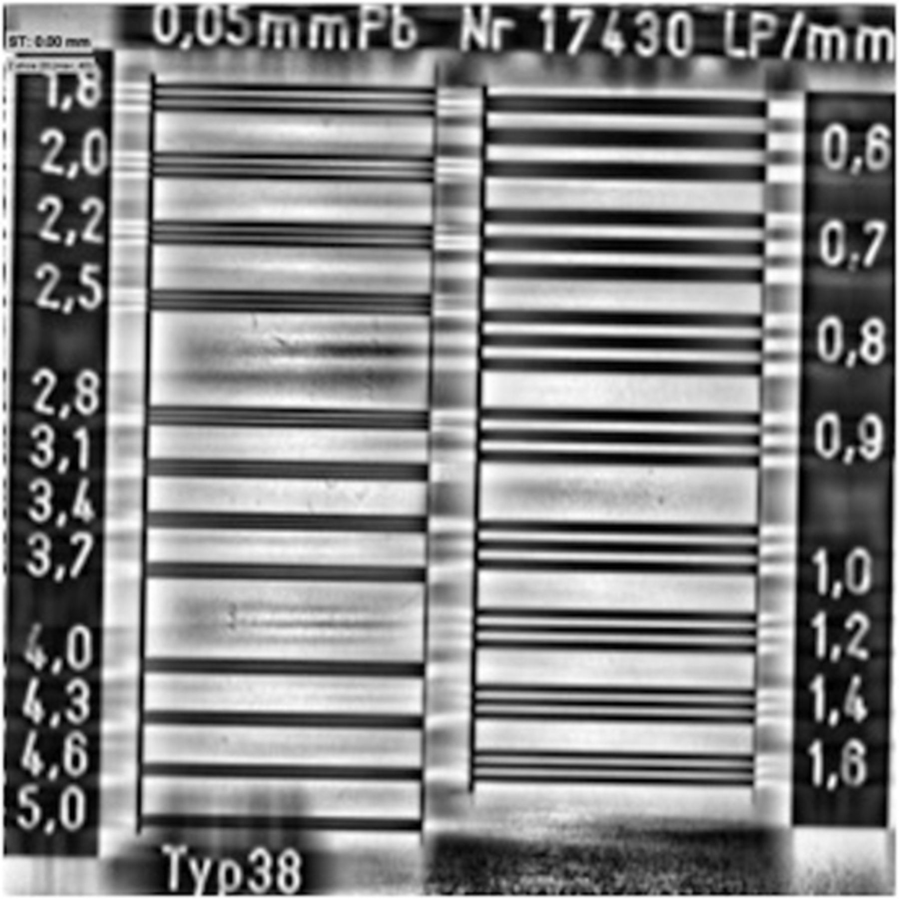
  • Fig 2.
    • Download figure
    • Open in new tab
    • Download powerpoint
    Fig 2.

    Central section of the VasoCT conebeam reconstruction of a line-pair phantom. The numbers at the left and right side indicate the line pairs per millimeter.

  • Fig 3.
    • Download figure
    • Open in new tab
    • Download powerpoint
    Fig 3.

    A, DSA before detachment of a WEB positioned in a left MCA bifurcation aneurysm. It is impossible to depict any protrusion of the device in the parent artery because only the 3 markers are seen and the mesh is almost not visible. B, Corresponding unsubstracted image. C, VasoCT confirms correct positioning of the WEB without any protrusion. D, Three-month control VasoCT shows residual flow in the proximal compartment (arrow).
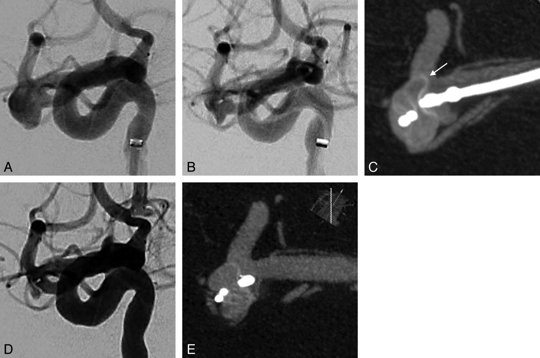
  • Fig 4.
    • Download figure
    • Open in new tab
    • Download powerpoint
    Fig 4.

    A, DSA of a broad-neck right MCA aneurysm. First, a 7 × 4 cm WEB was deployed. It is nearly undetectable with DSA (B) and there is no flow modification. However, VasoCT (C) clearly shows the device protrusion in the superior bifurcation branch (arrow). Thus, the WEB is retrieved and replaced by a 6 × 4 mm device. D and E, Final DSA and VasoCT after detachment confirm perfect positioning of the WEB without any protrusion.

  • Fig 5.
    • Download figure
    • Open in new tab
    • Download powerpoint
    Fig 5.

    A, DSA anteroposterior view of a basilar tip aneurysm. B, Control angiogram and corresponding unsubtracted view (C) prior to detachment of the WEB. Some contrast agent stagnation is visible in the second layer. It is impossible to analyze the good positioning of the device; no flow abnormality is depicted in the left P1 segment. D, VasoCT clearly shows the protrusion in the left P1 segment. The WEB was then retrieved and replaced by a smaller one.

  • Fig 6.
    • Download figure
    • Open in new tab
    • Download powerpoint
    Fig 6.

    A, DSA working projection of a large-neck anterior communicating artery aneurysm. B, After placement of a 9 × 7 mm WEB, the control DSA shows the occlusion of the right A2 segment and a narrowing of the left A2. C, VasoCT confirms the occlusion and that the WEB is not properly deployed. It is in a heart-shaped configuration with a distal trough; the 2 distal markers are attached (arrow). This “heart sign” depicts an inappropriate width of the WEB regarding aneurysm dimensions. It was then replaced by a correctly deployed 7 × 6 cm WEB.

  • Fig 7.
    • Download figure
    • Open in new tab
    • Download powerpoint
    Fig 7.

    Nonenhanced VasoCT. It is very important to consider the acquisition plane to avoid the projection of any metal artifacts over the region of interest. Here, the angulation of the head of the patient was modified so that coil artifacts from a previously treated basilar tip aneurysm do not superimpose on the MCA bifurcation.
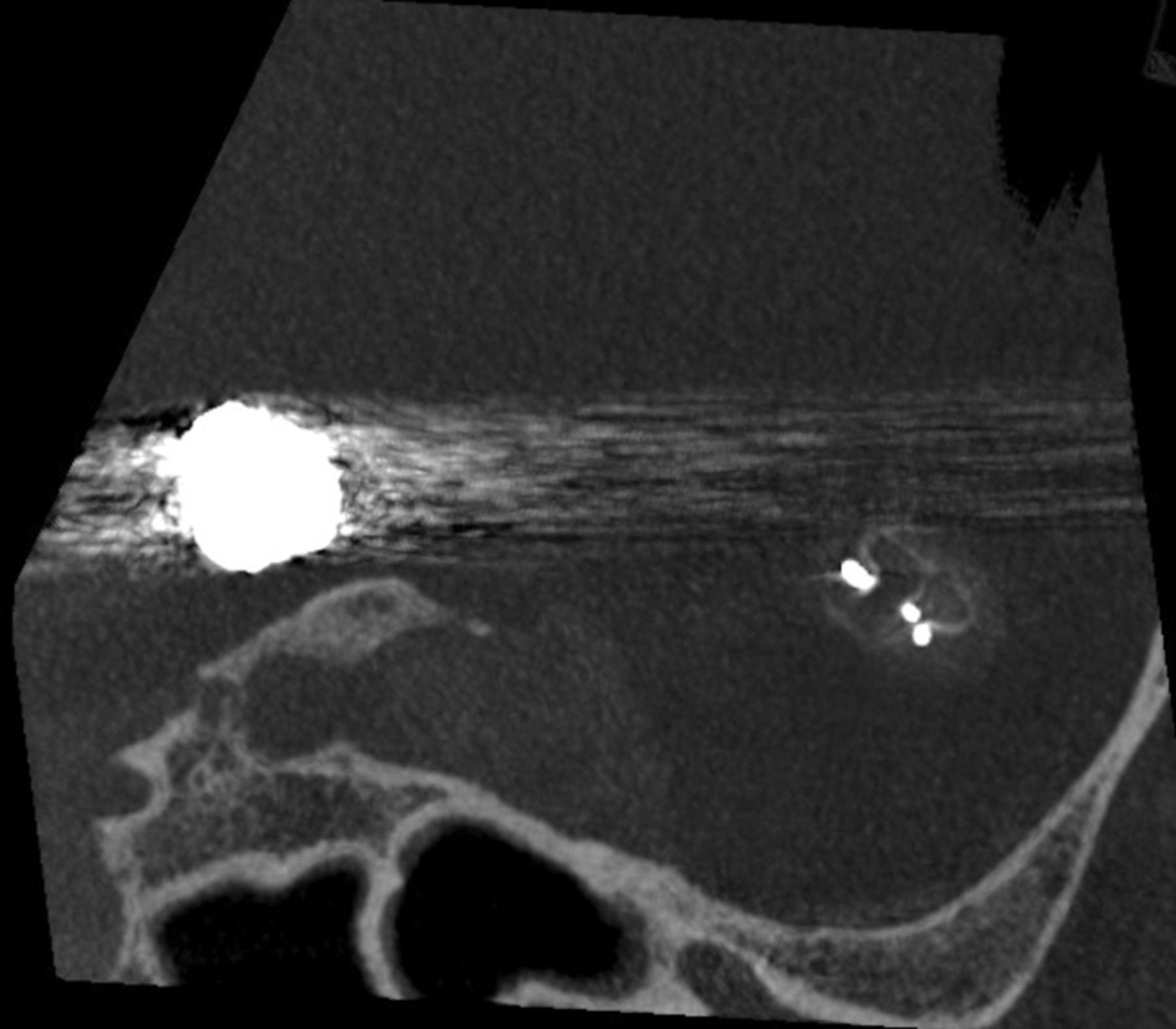
  • Fig 8.
    • Download figure
    • Open in new tab
    • Download powerpoint
    Fig 8.

    A, VasoCT of an MCA aneurysm treated with a WEB. Marker artifacts are projecting over the parent vessels. B, A new reconstruction with a metal-artifacts-reduction algorithm clearly improves the image quality and the visualization of the bifurcation.

PreviousNext
Back to top

In this issue

American Journal of Neuroradiology: 35 (7)
American Journal of Neuroradiology
Vol. 35, Issue 7
1 Jul 2014
  • Table of Contents
  • Index by author
  • Complete Issue (PDF)
Advertisement
Print
Download PDF
Email Article

Thank you for your interest in spreading the word on American Journal of Neuroradiology.

NOTE: We only request your email address so that the person you are recommending the page to knows that you wanted them to see it, and that it is not junk mail. We do not capture any email address.

Enter multiple addresses on separate lines or separate them with commas.
Role of C-Arm VasoCT in the Use of Endovascular WEB Flow Disruption in Intracranial Aneurysm Treatment
(Your Name) has sent you a message from American Journal of Neuroradiology
(Your Name) thought you would like to see the American Journal of Neuroradiology web site.
CAPTCHA
This question is for testing whether or not you are a human visitor and to prevent automated spam submissions.
Cite this article
J. Caroff, C. Mihalea, H. Neki, D. Ruijters, L. Ikka, N. Benachour, J. Moret, L. Spelle
Role of C-Arm VasoCT in the Use of Endovascular WEB Flow Disruption in Intracranial Aneurysm Treatment
American Journal of Neuroradiology Jul 2014, 35 (7) 1353-1357; DOI: 10.3174/ajnr.A3860

Citation Manager Formats

  • BibTeX
  • Bookends
  • EasyBib
  • EndNote (tagged)
  • EndNote 8 (xml)
  • Medlars
  • Mendeley
  • Papers
  • RefWorks Tagged
  • Ref Manager
  • RIS
  • Zotero
0 Responses
Respond to this article
Share
Bookmark this article
Role of C-Arm VasoCT in the Use of Endovascular WEB Flow Disruption in Intracranial Aneurysm Treatment
J. Caroff, C. Mihalea, H. Neki, D. Ruijters, L. Ikka, N. Benachour, J. Moret, L. Spelle
American Journal of Neuroradiology Jul 2014, 35 (7) 1353-1357; DOI: 10.3174/ajnr.A3860
del.icio.us logo Twitter logo Facebook logo Mendeley logo
  • Tweet Widget
  • Facebook Like
  • Google Plus One
Purchase

Jump to section

  • Article
    • Abstract
    • ABBREVIATIONS:
    • Materials and Methods
    • Results
    • Discussion
    • Conclusions
    • Footnotes
    • References
  • Figures & Data
  • Info & Metrics
  • Responses
  • References
  • PDF

Related Articles

  • No related articles found.
  • PubMed
  • Google Scholar

Cited By...

  • 15 years of WEB embolization: a transformative journey in aneurysm treatment
  • Intraoperative contrast-enhanced cone beam CT allows visualization of the 'dark side of the clot and improves mechanical thrombectomy performance
  • WEB shape modifications: angiography-histopathology correlations in rabbits
  • WEB shape modifications: angiography-histopathology correlations in rabbits
  • Determinants of cerebral aneurysm occlusion after embolization with the WEB device: a single-institution series of 215 cases with angiographic follow-up
  • Persistent Opacification of the Woven EndoBridge Device: A Conebeam CT Analysis of the Bicetre Occlusion Scale Score 1 Phenomenon
  • Determinants of cerebral aneurysm occlusion after embolization with the WEB device: a single-institution series of 215 cases with angiographic follow-up
  • WEB device for treatment of posterior communicating artery aneurysms
  • Woven EndoBridge device shape modification can be mitigated with an appropriate oversizing strategy: a VasoCT based study
  • Impact of A1 Asymmetry on the Woven EndoBridge Device in Anterior Communicating Artery Aneurysms
  • Endovascular Treatment of Small and Very Small Intracranial Aneurysms with the Woven EndoBridge Device
  • Residual Flow Inside the Woven EndoBridge Device at Follow-Up: Potential Predictors of the Bicetre Occlusion Scale Score 1 Phenomenon
  • How to WEB: a practical review of methodology for the use of the Woven EndoBridge
  • Application of High-Resolution C-Arm CT Combined with Streak Metal Artifact Removal Technology for the Stent-Assisted Embolization of Intracranial Aneurysms
  • The occurrence of neointimal hyperplasia after flow-diverter implantation is associated with cardiovascular risks factors and the stent design
  • Safety and efficiency of the fifth generation Woven EndoBridge device: technical note
  • Balloon remodeling-assisted Woven EndoBridge technique: description and feasibility for complex bifurcation aneurysms
  • Multiparametric MRI of intracranial aneurysms treated with the Woven EndoBridge (WEB): a case of Faradays cage?
  • WEB Device: Ready for Ruptured Aneurysms?
  • Flow changes in the posterior communicating artery related to flow-diverter stents in carotid siphon aneurysms
  • Large Basilar Apex Aneurysms Treated with Flow-Diverter Stents
  • Wall Apposition Is a Key Factor for Aneurysm Occlusion after Flow Diversion: A Histologic Evaluation in 41 Rabbits
  • Metal artifact reduction for flat panel detector intravenous CT angiography in patients with intracranial metallic implants after endovascular and surgical treatment
  • Follow-up of intracranial aneurysms treated by a WEB flow disrupter: a comparative study of DSA and contrast-enhanced MR angiography
  • Flow-Diverter Stents for the Treatment of Saccular Middle Cerebral Artery Bifurcation Aneurysms
  • Single-Layer WEBs: Intrasaccular Flow Disrupters for Aneurysm Treatment--Feasibility Results from a European Study
  • Intrasaccular Flow Disruption in Acutely Ruptured Aneurysms: A Multicenter Retrospective Review of the Use of the WEB
  • Crossref (48)
  • Google Scholar

This article has been cited by the following articles in journals that are participating in Crossref Cited-by Linking.

  • How to WEB: a practical review of methodology for the use of the Woven EndoBridge
    Nitin Goyal, Daniel Hoit, Julie DiNitto, Lucas Elijovich, David Fiorella, Laurent Pierot, Saleh Lamin, Laurent Spelle, Isil Saatci, Saru Cekirge, Adam S Arthur
    Journal of NeuroInterventional Surgery 2020 12 5
  • Remnants and Recurrences After the Use of the WEB Intrasaccular Device in Large-Neck Bifurcation Aneurysms
    Christophe Cognard, Anne Christine Januel
    Neurosurgery 2015 76 5
  • Intrasaccular Flow Disruption in Acutely Ruptured Aneurysms: A Multicenter Retrospective Review of the Use of the WEB
    T. Liebig, C. Kabbasch, C. Strasilla, A. Berlis, W. Weber, L. Pierot, T. Patankar, X. Barreau, J. Dervin, A. Kuršumović, S. Rath, B. Lubicz, J. Klisch
    American Journal of Neuroradiology 2015 36 9
  • Wall Apposition Is a Key Factor for Aneurysm Occlusion after Flow Diversion: A Histologic Evaluation in 41 Rabbits
    A. Rouchaud, C. Ramana, W. Brinjikji, Y.-H. Ding, D. Dai, T. Gunderson, J. Cebral, D.F. Kallmes, R. Kadirvel
    American Journal of Neuroradiology 2016 37 11
  • Flow-Diverter Stents for the Treatment of Saccular Middle Cerebral Artery Bifurcation Aneurysms
    J. Caroff, H. Neki, C. Mihalea, F. D'Argento, H. Abdel Khalek, L. Ikka, J. Moret, L. Spelle
    American Journal of Neuroradiology 2016 37 2
  • Woven Endobridge (WEB) Device for endovascular treatment of ruptured intracranial wide-neck aneurysms: a single-center experience
    Jildaz Caroff, Cristian Mihalea, Francesco Dargento, Hiroaki Neki, Léon Ikka, Nidhal Benachour, Jacques Moret, Laurent Spelle
    Neuroradiology 2014 56 9
  • Single-Layer WEBs: Intrasaccular Flow Disrupters for Aneurysm Treatment—Feasibility Results from a European Study
    J. Caroff, C. Mihalea, J. Klisch, C. Strasilla, A. Berlis, T. Patankar, W. Weber, D. Behme, E.A. Jacobsen, T. Liebig, S. Prothmann, C. Cognard, T. Finkenzeller, J. Moret, L. Spelle
    American Journal of Neuroradiology 2015 36 10
  • Occlusion assessment of intracranial aneurysms treated with the WEB device
    Jildaz Caroff, Cristian Mihalea, Titien Tuilier, Xavier Barreau, Christophe Cognard, Hubert Desal, Laurent Pierot, Armelle Arnoux, Jacques Moret, Laurent Spelle
    Neuroradiology 2016 58 9
  • Safety and efficiency of the fifth generation Woven EndoBridge device: technical note
    Cristian Mihalea, Jildaz Caroff, Igor Pagiola, Leon Ikka, Gelareh Bani Hashemi, Soheil Naderi, Vanessa Chalumeau, Horia Ples, Bogdan Valeriu Popa, Thomas Yasuda, Joaquin Jose Marenco de la Torre, Marta Iacobucci, Augustin Ozanne, Sophie Gallas, Aymeric Rouchaud, Sorin Pescariu, Jacques Moret, Laurent Spelle
    Journal of NeuroInterventional Surgery 2019 11 5
  • Ruptured Intracranial Aneurysms Treated with Woven Endobridge Intrasaccular Flow Disruptor: A Multicenter Experience
    Valerio Da Ros, Alessio Bozzi, Chiara Comelli, Vittorio Semeraro, Simone Comelli, Nicola Lucarelli, Nicola Burdi, Roberto Gandini
    World Neurosurgery 2019 122

More in this TOC Section

  • SAVE vs. Solumbra Techniques for Thrombectomy
  • CT Perfusion&Reperfusion in Acute Ischemic Stroke
  • Delayed Reperfusion Post-Thrombectomy&Thrombolysis
Show more Interventional

Similar Articles

Advertisement

Indexed Content

  • Current Issue
  • Accepted Manuscripts
  • Article Preview
  • Past Issues
  • Editorials
  • Editors Choice
  • Fellow Journal Club
  • Letters to the Editor

Cases

  • Case Collection
  • Archive - Case of the Week
  • Archive - Case of the Month
  • Archive - Classic Case

Special Collections

  • Special Collections

Resources

  • News and Updates
  • Turn around Times
  • Submit a Manuscript
  • Author Policies
  • Manuscript Submission Guidelines
  • Evidence-Based Medicine Level Guide
  • Publishing Checklists
  • Graphical Abstract Preparation
  • Imaging Protocol Submission
  • Submit a Case
  • Become a Reviewer/Academy of Reviewers
  • Get Peer Review Credit from Publons

Multimedia

  • AJNR Podcast
  • AJNR SCANtastic
  • Video Articles

About Us

  • About AJNR
  • Editorial Board
  • Not an AJNR Subscriber? Join Now
  • Alerts
  • Feedback
  • Advertise with us
  • Librarian Resources
  • Permissions
  • Terms and Conditions

American Society of Neuroradiology

  • Not an ASNR Member? Join Now

© 2025 by the American Society of Neuroradiology All rights, including for text and data mining, AI training, and similar technologies, are reserved.
Print ISSN: 0195-6108 Online ISSN: 1936-959X

Powered by HighWire