Advertisement

Main menu

  • Home
  • Content
    • Current Issue
    • Accepted Manuscripts
    • Article Preview
    • Past Issue Archive
    • Video Articles
    • AJNR Case Collection
    • Case of the Week Archive
    • Case of the Month Archive
    • Classic Case Archive
  • Special Collections
    • AJNR Awards
    • Low-Field MRI
    • Alzheimer Disease
    • ASNR Foundation Special Collection
    • Photon-Counting CT
    • View All
  • Multimedia
    • AJNR Podcasts
    • AJNR SCANtastic
    • Trainee Corner
    • MRI Safety Corner
    • Imaging Protocols
  • For Authors
    • Submit a Manuscript
    • Submit a Video Article
    • Submit an eLetter to the Editor/Response
    • Manuscript Submission Guidelines
    • Statistical Tips
    • Fast Publishing of Accepted Manuscripts
    • Graphical Abstract Preparation
    • Imaging Protocol Submission
    • Author Policies
  • About Us
    • About AJNR
    • Editorial Board
    • Editorial Board Alumni
  • More
    • Become a Reviewer/Academy of Reviewers
    • Subscribers
    • Permissions
    • Alerts
    • Feedback
    • Advertisers
    • ASNR Home

User menu

  • Alerts
  • Log in

Search

  • Advanced search
American Journal of Neuroradiology

American Journal of Neuroradiology

ASHNR American Society of Functional Neuroradiology ASHNR American Society of Pediatric Neuroradiology ASSR
  • Alerts
  • Log in

Advanced Search

  • Home
  • Content
    • Current Issue
    • Accepted Manuscripts
    • Article Preview
    • Past Issue Archive
    • Video Articles
    • AJNR Case Collection
    • Case of the Week Archive
    • Case of the Month Archive
    • Classic Case Archive
  • Special Collections
    • AJNR Awards
    • Low-Field MRI
    • Alzheimer Disease
    • ASNR Foundation Special Collection
    • Photon-Counting CT
    • View All
  • Multimedia
    • AJNR Podcasts
    • AJNR SCANtastic
    • Trainee Corner
    • MRI Safety Corner
    • Imaging Protocols
  • For Authors
    • Submit a Manuscript
    • Submit a Video Article
    • Submit an eLetter to the Editor/Response
    • Manuscript Submission Guidelines
    • Statistical Tips
    • Fast Publishing of Accepted Manuscripts
    • Graphical Abstract Preparation
    • Imaging Protocol Submission
    • Author Policies
  • About Us
    • About AJNR
    • Editorial Board
    • Editorial Board Alumni
  • More
    • Become a Reviewer/Academy of Reviewers
    • Subscribers
    • Permissions
    • Alerts
    • Feedback
    • Advertisers
    • ASNR Home
  • Follow AJNR on Twitter
  • Visit AJNR on Facebook
  • Follow AJNR on Instagram
  • Join AJNR on LinkedIn
  • RSS Feeds

AJNR Awards, New Junior Editors, and more. Read the latest AJNR updates

Research ArticlePediatric Neuroimaging

Hydrocephalus Decreases Arterial Spin-Labeled Cerebral Perfusion

K.W. Yeom, R.M. Lober, A. Alexander, S.H. Cheshier and M.S.B. Edwards
American Journal of Neuroradiology July 2014, 35 (7) 1433-1439; DOI: https://doi.org/10.3174/ajnr.A3891
K.W. Yeom
aFrom the Departments of Radiology (K.W.Y.)
  • Find this author on Google Scholar
  • Find this author on PubMed
  • Search for this author on this site
R.M. Lober
bNeurosurgery (R.M.L., A.A., S.H.C., M.S.B.E.)
  • Find this author on Google Scholar
  • Find this author on PubMed
  • Search for this author on this site
A. Alexander
bNeurosurgery (R.M.L., A.A., S.H.C., M.S.B.E.)
  • Find this author on Google Scholar
  • Find this author on PubMed
  • Search for this author on this site
S.H. Cheshier
bNeurosurgery (R.M.L., A.A., S.H.C., M.S.B.E.)
cDivision of Pediatric Neurosurgery (S.H.C., M.S.B.E.), Lucile Packard Children's Hospital at Stanford University, Palo Alto, California.
  • Find this author on Google Scholar
  • Find this author on PubMed
  • Search for this author on this site
M.S.B. Edwards
bNeurosurgery (R.M.L., A.A., S.H.C., M.S.B.E.)
cDivision of Pediatric Neurosurgery (S.H.C., M.S.B.E.), Lucile Packard Children's Hospital at Stanford University, Palo Alto, California.
  • Find this author on Google Scholar
  • Find this author on PubMed
  • Search for this author on this site
  • Article
  • Figures & Data
  • Supplemental
  • Info & Metrics
  • Responses
  • References
  • PDF
Loading

Article Figures & Data

Figures

  • Tables
  • Fig 1.
    • Download figure
    • Open in new tab
    • Download powerpoint
    Fig 1.

    Brain region-of-interest placement and ventricular measurement. A, ROIs were placed in the 3 axial levels of the cerebrum near the vertex and just above the centrum semiovale. B and C. Additional ROIs were placed in bilateral periventricular white matter, and the deep gray matter, including the bilateral thalami and putamina. D, Ventricular measurement of the bilateral frontal horns was performed at the level of the caudate heads.

  • Fig 2.
    • Download figure
    • Open in new tab
    • Download powerpoint
    Fig 2.

    Comparison of ASL-CBF among controls, asymptomatic patients, and patients with hydrocephalus.

  • Fig 3.
    • Download figure
    • Open in new tab
    • Download powerpoint
    Fig 3.

    ASL-CBF values before and after alleviation of obstructive hydrocephalus.

  • Fig 4.
    • Download figure
    • Open in new tab
    • Download powerpoint
    Fig 4.

    ASL perfusion of a 6-year-old girl presenting with hydrocephalus from a diffuse intrinsic pontine glioma. A, The patient presented with acute symptoms, including headache, nausea/vomiting, and somnolence. Enlarged ventricles and periventricular edema were noted and low CBF of 9, 13, 8, and 6 mL/100 g/min at the cerebral vertex, putamina, thalami, and periventricular white matter, respectively. A shunt was placed a day after the MR imaging. B, Improved CBF (30, 40, 36, 27 mL/100 g/min) and resolution of acute symptoms were noted in the respective brain regions a month later. Note the shunt catheter in place (arrow) and residual ventricular enlargement and edema.
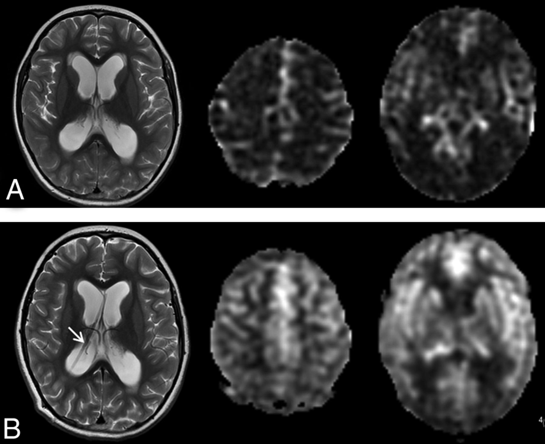
  • Fig 5.
    • Download figure
    • Open in new tab
    • Download powerpoint
    Fig 5.

    ASL perfusion of a 5-year-old boy presenting with acute hydrocephalus due to aqueduct obstruction from a tectal glioma. A, The patient presented with acute gait abnormality, nausea/vomiting, and somnolence. Enlarged ventricles and low global CBF of 16, 20, 17, and 8 mL/100 g/mL were seen at the cerebral vertex, putamina, thalami, and periventricular white matter, respectively. The patient underwent third ventriculostomy a day later and subsequently showed symptomatic relief. B, Two months later, improved CBF was seen, with CBF of 43, 58, 50, and 32 mL/100 g/min in the respective brain regions. Note a CSF jet at the patent third ventriculostomy site (arrow).

  • Fig 6.
    • Download figure
    • Open in new tab
    • Download powerpoint
    Fig 6.

    Serial ASL perfusion of a 10-year-old boy with hydrocephalus from medulloblastoma. A, The patient presented acutely with headaches and nausea/vomiting. Relatively low CBF of 29, 50, 45, 20 mL/100 g/min, compared with controls, was noted in the cerebral vertex, putamina, thalami, and periventricular white matter. The patient underwent PF tumor resection and extraventricular drain placement 2 days later. B, One day after the surgery, the patient continued to have high ICP despite extraventricular drain placement and showed low CBF of 22, 34, 30, and 12 mL/100 g/min in the respective brain regions. C, The symptoms continued to worsen with high ICP and further decrease in CBF of 17, 31, 18, and 10 mL/100 g/min 2 days later. Due to extraventricular drain malfunction, a new ventriculoperitoneal shunt was subsequently placed. D, Several months later, the patient presented for routine surveillance of medulloblastoma with a functioning ventriculoperitoneal shunt and decreased ventricular size and was clinically asymptomatic at this time. While some perfusion alteration may be expected from interval chemotherapy and radiation, it has, nevertheless, improved, with a CBF of 36, 44, 34, 21 mL/100 g/min in the respective brain regions.

Tables

  • Figures
    • View popup
    Table 1:

    Patient characteristics (n = 23)

    Characteristic
    Age at initial diagnosis (yr)
        Median6.2
        Range0.9–18
    Sex
        Male17 (74%)
        Female6 (26%)
    Diagnosis (in order of frequency)
        Medulloblastoma8 (35%)
        Pilocytic astrocytoma5 (22%)
        Ependymoma5 (22%)
        Choroid plexus papilloma2 (9%)
        Tectal astrocytoma2 (9%)
        Diffuse intrinsic pontine glioma1 (5%)
    Hydrocephalus at presentation19 (83%)
    • View popup
    Table 2:

    Cerebral blood flow in healthy controls (n = 16) and patients with PF Tumor (n = 23)

    Median Cerebral Blood Flow (mL/100 g/min) (Range)
    CerebrumBasal GangliaThalamusWhite Matter
    Healthy control62.3 (50.4–68.9)57.3 (44.6–70.8)56.4 (45.4–67.8)37.3 (26–43.4)
    PF tumor without hydrocephalus56.7 (46.4–63.1)57.4 (49.7–60.0)59.7 (53.2–65.4)32.3 (20.8–35.8)
    PF tumor with hydrocephalus34.3 (8.6–59.0)39.8 (12.5–59.6)32.4 (8.1–50.5)17.7 (6.0–28.1)
    • View popup
    Table 3:

    Cerebral blood flow before and after alleviation of obstructive hydrocephalus (n = 16)a

    Median Cerebral Blood Flow (mL/100 mg/min) (Range)
    CerebrumBasal GangliaThalamusWhite Matter
    Before alleviation of hydrocephalus35.8 (8.6–59)38.0 (12.5–59.6)29.9 (8.1–50.3)17.5 (6.0–28.1)
    After alleviation of hydrocephalus52.6 (29.8–69.7)53.9 (40.2–68.8)55.3 (29.1–63.3)31 (23.4–45.4)
    • ↵a For each site, CBF was increased following alleviation of hydrocephalus (Wilcoxon signed rank test for related samples, P < .002).

PreviousNext
Back to top

In this issue

American Journal of Neuroradiology: 35 (7)
American Journal of Neuroradiology
Vol. 35, Issue 7
1 Jul 2014
  • Table of Contents
  • Index by author
  • Complete Issue (PDF)
Advertisement
Print
Download PDF
Email Article

Thank you for your interest in spreading the word on American Journal of Neuroradiology.

NOTE: We only request your email address so that the person you are recommending the page to knows that you wanted them to see it, and that it is not junk mail. We do not capture any email address.

Enter multiple addresses on separate lines or separate them with commas.
Hydrocephalus Decreases Arterial Spin-Labeled Cerebral Perfusion
(Your Name) has sent you a message from American Journal of Neuroradiology
(Your Name) thought you would like to see the American Journal of Neuroradiology web site.
CAPTCHA
This question is for testing whether or not you are a human visitor and to prevent automated spam submissions.
Cite this article
K.W. Yeom, R.M. Lober, A. Alexander, S.H. Cheshier, M.S.B. Edwards
Hydrocephalus Decreases Arterial Spin-Labeled Cerebral Perfusion
American Journal of Neuroradiology Jul 2014, 35 (7) 1433-1439; DOI: 10.3174/ajnr.A3891

Citation Manager Formats

  • BibTeX
  • Bookends
  • EasyBib
  • EndNote (tagged)
  • EndNote 8 (xml)
  • Medlars
  • Mendeley
  • Papers
  • RefWorks Tagged
  • Ref Manager
  • RIS
  • Zotero
0 Responses
Respond to this article
Share
Bookmark this article
Hydrocephalus Decreases Arterial Spin-Labeled Cerebral Perfusion
K.W. Yeom, R.M. Lober, A. Alexander, S.H. Cheshier, M.S.B. Edwards
American Journal of Neuroradiology Jul 2014, 35 (7) 1433-1439; DOI: 10.3174/ajnr.A3891
del.icio.us logo Twitter logo Facebook logo Mendeley logo
  • Tweet Widget
  • Facebook Like
  • Google Plus One
Purchase

Jump to section

  • Article
    • Abstract
    • ABBREVIATIONS:
    • Materials and Methods
    • Results
    • Discussion
    • Conclusions
    • Footnotes
    • References
  • Figures & Data
  • Supplemental
  • Info & Metrics
  • Responses
  • References
  • PDF

Related Articles

  • PubMed
  • Google Scholar

Cited By...

  • Arterial Spin-Labeling Perfusion Metrics in Pediatric Posterior Fossa Tumor Surgery
  • Gray Matter Growth Is Accompanied by Increasing Blood Flow and Decreasing Apparent Diffusion Coefficient during Childhood
  • Crossref
  • Google Scholar

This article has not yet been cited by articles in journals that are participating in Crossref Cited-by Linking.

More in this TOC Section

  • FRACTURE MR in Congenital Vertebral Anomalies
  • Comparing MRI Perfusion in Pediatric Brain Tumors
  • Sodium MRI in Pediatric Brain Tumors
Show more Pediatric Neuroimaging

Similar Articles

Advertisement

Indexed Content

  • Current Issue
  • Accepted Manuscripts
  • Article Preview
  • Past Issues
  • Editorials
  • Editor's Choice
  • Fellows' Journal Club
  • Letters to the Editor
  • Video Articles

Cases

  • Case Collection
  • Archive - Case of the Week
  • Archive - Case of the Month
  • Archive - Classic Case

More from AJNR

  • Trainee Corner
  • Imaging Protocols
  • MRI Safety Corner
  • Book Reviews

Multimedia

  • AJNR Podcasts
  • AJNR Scantastics

Resources

  • Turnaround Time
  • Submit a Manuscript
  • Submit a Video Article
  • Submit an eLetter to the Editor/Response
  • Manuscript Submission Guidelines
  • Statistical Tips
  • Fast Publishing of Accepted Manuscripts
  • Graphical Abstract Preparation
  • Imaging Protocol Submission
  • Evidence-Based Medicine Level Guide
  • Publishing Checklists
  • Author Policies
  • Become a Reviewer/Academy of Reviewers
  • News and Updates

About Us

  • About AJNR
  • Editorial Board
  • Editorial Board Alumni
  • Alerts
  • Permissions
  • Not an AJNR Subscriber? Join Now
  • Advertise with Us
  • Librarian Resources
  • Feedback
  • Terms and Conditions
  • AJNR Editorial Board Alumni

American Society of Neuroradiology

  • Not an ASNR Member? Join Now

© 2025 by the American Society of Neuroradiology All rights, including for text and data mining, AI training, and similar technologies, are reserved.
Print ISSN: 0195-6108 Online ISSN: 1936-959X

Powered by HighWire