
Article Figures & Data
Figures
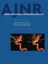
- Fig 1.
A 67-year-old woman with left hemiparesis and dysarthria who presented to the hospital 2 hours after onset. CTA identified a right intracranial ICA occlusion. CTP-SI showed that the contrast (black circles) was first detected at 20 seconds in the left Sylvian fissure (A) and at 27 seconds in the affected right hemisphere (B). The relative filling time delay was, therefore, 7 seconds.
- Fig 2.
Correlation between rFTD and baseline NIHSS scores, 24-hour ASPECTS, and 90-day mRS scores.
- Fig 3.
Longer median rFTD was significantly associated with poor functional and radiologic outcomes.


Tables
mRS (0–2) (n = 23) mRS (3–6) (n = 32) P Value Alive (n = 43) Dead (n = 12) P Value ASPECTS (>7) (n = 21) ASPECTS (≤7) (n = 39) P Value Age (median) (yr) 69 (41–85) 73.5 (54–88) .014 69 (41–85) 79 (66–88) .005 72 (52–83) 73 (41–88) .877 Female (%) 26 56.2 .026 37.2 66.6 .101 33.3 51.2 .277 Hypertension (%) 52.1 62.5 .444 60.4 50 .516 56.4 61.9 .786 Diabetes mellitus (%) 26 28.1 .867 23.2 41.6 .205 33.3 25.6 .560 Hyperlipidemia (%) 60.8 46.8 .305 48.8 66.6 .274 61.9 51.2 .587 Current or past smoker (%) 26 9.3 .143 20.9 0 .181 14.2 20.5 .731 Atrial fibrillation (%) 34.3 34.7 .975 32.5 41.7 .733 42.8 35.8 .781 Baseline NIHSS (median) 13 (6–20) 19 (6–27) .001 15 (6–27) 21 (15–26) .001 12 (3–24) 18 (6–27) .003 OTT (median) (min) 120 (61–255) 140 (75–265) .366 140 (61–265) 124 (77–200) .683 137 (61–250) 140 (76–265) .364 IV-tPA (%) 60.9 71.9 .561 67.4 66.7 1.000 69.2 66.7 1.000 IA-tPA/UK (%) 13 6.2 .639 7.3 16.7 .298 11.7 8.3 1.000 Clot retrieval (%) 26.1 21.9 .717 25.5 16.7 .709 23.1 23.8 1.000 rFTD (median) (sec) 3.5 (0–7.5) 5.75 (0–10) .001 4 (0–17) 6.5 (3–20) .002 3.5 (0–6.5) 5.5 (2–20) .002 Note:—IA indicates intra-arterial; rFTD, relative filling delay time; UK, urokinase; OTT, onset to treatment time.
Poor Outcomes Predictors β SE P Value 90-Day mRS 3–6 Age 0.097 0.043 .024 Sex 0.548 0.816 .501 Baseline NIHSS 0.207 0.091 .023 rFTD 0.354 0.178 .047 90-Day death Age 0.185 0.074 .01 Baseline NIHSS 0.239 0.116 .028 rFTD 0.302 0.158 .039 24-Hour ASPECTS ≤7 Baseline NIHSS 0.114 0.065 .082 rFTD 0.456 0.162 .005


Jump to section
Related Articles
Cited By...
- Quantitative Collateral Assessment on CTP in the Prediction of Stroke Etiology
- Assessment of Ischemic Volumes by Using Relative Filling Time Delay on CTP Source Image in Patients with Acute Stroke with Anterior Circulation Large Vessel Occlusions
- Arterial Spin Labeling Magnetic Resonance Imaging Estimation of Antegrade and Collateral Flow in Unilateral Middle Cerebral Artery Stenosis