Advertisement

Main menu

  • Home
  • Content
    • Current Issue
    • Accepted Manuscripts
    • Article Preview
    • Past Issue Archive
    • AJNR Case Collection
    • Case of the Week Archive
    • Classic Case Archive
    • Case of the Month Archive
  • Special Collections
    • Spinal CSF Leak Articles (Jan 2020-June 2024)
    • 2024 AJNR Journal Awards
    • Most Impactful AJNR Articles
  • Multimedia
    • AJNR Podcast
    • AJNR Scantastics
    • Video Articles
  • For Authors
    • Submit a Manuscript
    • Author Policies
    • Fast publishing of Accepted Manuscripts
    • Graphical Abstract Preparation
    • Manuscript Submission Guidelines
    • Imaging Protocol Submission
    • Submit a Case for the Case Collection
  • About Us
    • About AJNR
    • Editorial Board
  • More
    • Become a Reviewer/Academy of Reviewers
    • Subscribers
    • Permissions
    • Alerts
    • Feedback
    • Advertisers
    • ASNR Home
  • Other Publications
    • ajnr

User menu

  • Alerts
  • Log in

Search

  • Advanced search
American Journal of Neuroradiology

American Journal of Neuroradiology

ASHNR American Society of Functional Neuroradiology ASHNR American Society of Pediatric Neuroradiology ASSR
  • Alerts
  • Log in

Advanced Search

  • Home
  • Content
    • Current Issue
    • Accepted Manuscripts
    • Article Preview
    • Past Issue Archive
    • AJNR Case Collection
    • Case of the Week Archive
    • Classic Case Archive
    • Case of the Month Archive
  • Special Collections
    • Spinal CSF Leak Articles (Jan 2020-June 2024)
    • 2024 AJNR Journal Awards
    • Most Impactful AJNR Articles
  • Multimedia
    • AJNR Podcast
    • AJNR Scantastics
    • Video Articles
  • For Authors
    • Submit a Manuscript
    • Author Policies
    • Fast publishing of Accepted Manuscripts
    • Graphical Abstract Preparation
    • Manuscript Submission Guidelines
    • Imaging Protocol Submission
    • Submit a Case for the Case Collection
  • About Us
    • About AJNR
    • Editorial Board
  • More
    • Become a Reviewer/Academy of Reviewers
    • Subscribers
    • Permissions
    • Alerts
    • Feedback
    • Advertisers
    • ASNR Home
  • Follow AJNR on Twitter
  • Visit AJNR on Facebook
  • Follow AJNR on Instagram
  • Join AJNR on LinkedIn
  • RSS Feeds

Welcome to the new AJNR, Updated Hall of Fame, and more. Read the full announcements.


AJNR is seeking candidates for the position of Associate Section Editor, AJNR Case Collection. Read the full announcement.


Research ArticleBrain
Open Access

Mapping p53 Mutations in Low-Grade Glioma: A Voxel-Based Neuroimaging Analysis

Y.Y. Wang, T. Zhang, S.W. Li, T.Y. Qian, X. Fan, X.X. Peng, J. Ma, L. Wang and T. Jiang
American Journal of Neuroradiology January 2015, 36 (1) 70-76; DOI: https://doi.org/10.3174/ajnr.A4065
Y.Y. Wang
aFrom the Beijing Neurosurgical Institute (Y.Y.W., T.J.)
cDepartments of Neurosurgery (Y.Y.W., T.Z., X.F., L.W., T.J.)
  • Find this author on Google Scholar
  • Find this author on PubMed
  • Search for this author on this site
T. Zhang
cDepartments of Neurosurgery (Y.Y.W., T.Z., X.F., L.W., T.J.)
  • Find this author on Google Scholar
  • Find this author on PubMed
  • Search for this author on this site
S.W. Li
dNeuroradiology (S.W.L., J.M.), Beijing Tiantan Hospital, Capital Medical University, Beijing, China
  • Find this author on Google Scholar
  • Find this author on PubMed
  • Search for this author on this site
T.Y. Qian
eSiemens Healthcare (T.Y.Q.), MR Collaboration NE Asia, Beijing, China
  • Find this author on Google Scholar
  • Find this author on PubMed
  • Search for this author on this site
X. Fan
cDepartments of Neurosurgery (Y.Y.W., T.Z., X.F., L.W., T.J.)
  • Find this author on Google Scholar
  • Find this author on PubMed
  • Search for this author on this site
X.X. Peng
bDepartment of Epidemiology and Biostatistics (X.X.P.), School of Public Health and Family Medicine, Capital Medical University, Beijing, China
  • Find this author on Google Scholar
  • Find this author on PubMed
  • Search for this author on this site
J. Ma
dNeuroradiology (S.W.L., J.M.), Beijing Tiantan Hospital, Capital Medical University, Beijing, China
  • Find this author on Google Scholar
  • Find this author on PubMed
  • Search for this author on this site
L. Wang
cDepartments of Neurosurgery (Y.Y.W., T.Z., X.F., L.W., T.J.)
fChina National Clinical Research Center for Neurological Diseases (L.W.), Beijing, China
  • Find this author on Google Scholar
  • Find this author on PubMed
  • Search for this author on this site
T. Jiang
aFrom the Beijing Neurosurgical Institute (Y.Y.W., T.J.)
cDepartments of Neurosurgery (Y.Y.W., T.Z., X.F., L.W., T.J.)
gBeijing Institute for Brain Disorders (T.J.), Beijing, China.
  • Find this author on Google Scholar
  • Find this author on PubMed
  • Search for this author on this site
  • Article
  • Figures & Data
  • Supplemental
  • Info & Metrics
  • Responses
  • References
  • PDF
Loading

Article Figures & Data

Figures

  • Tables
  • Fig 1.
    • Download figure
    • Open in new tab
    • Download powerpoint
    Fig 1.

    Overlap of tumor lesions. Overlap of all 182 patients with low-grade gliomas included in our study (A), overlap of a subgroup of 116 patients with mutant p53 gliomas (B), and overlap of a subgroup of 66 patients with wild-type p53 gliomas (C). The color range indicates the proportion of overlap of different cohorts, from violet (1 case) to red (>25% of cases overlap). Brain sections are displayed from z-coordinates −32 to +58 in the MNI space.

  • Fig 2.
    • Download figure
    • Open in new tab
    • Download powerpoint
    Fig 2.

    Power map for voxel-based lesion-symptom mapping analysis. The color map on the MNI space shows the distribution of power, ranging from 0 (violet) to 1 (red), with α set to P < .05. Only voxels with high-power values (>0.8) were included in the VLSM analysis.

  • Fig 3.
    • Download figure
    • Open in new tab
    • Download powerpoint
    Fig 3.

    Voxel-based lesion-symptom mapping–defined p53 high-mutant regions. VLSM analysis shows regions associated with high expression of mutant p53 in low-grade gliomas. The left-medial temporal lobe and right-anterior temporal lobes are significantly correlated to high mutant p53 expression. The color range indicates the level of t values from red to yellow (least to most significant). Only significant voxels are rendered on the basis of a critical threshold determined by permutation testing (n = 500, P < .05).
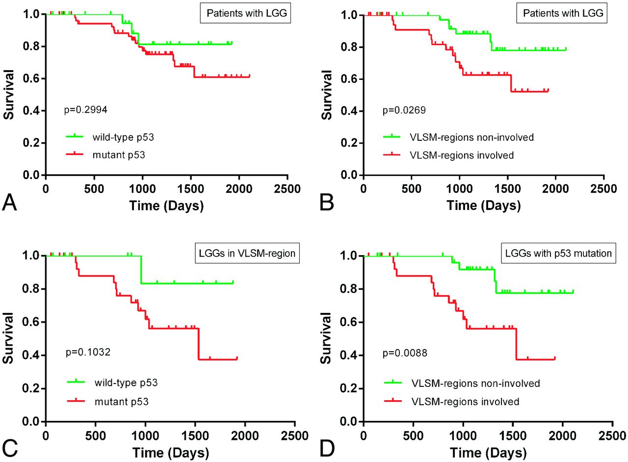
  • Fig 4.
    • Download figure
    • Open in new tab
    • Download powerpoint
    Fig 4.

    Kaplan-Meier curves showing the PFS for patients with low-grade gliomas. A, No significant difference was observed between patients with wild-type p53 tumors and those with mutant p53 tumors (log-rank, P = .2994). B, A statistically significant difference in PFS was observed between patients with tumors in VLSM-identified regions and patients with tumors located outside VLSM-identified regions (log-rank, P = .0269). C, Among patients with tumors located in VLSM-identified regions, a trend toward a difference in PFS was observed between patients with wild-type p53 tumors and those with mutant p53 tumors (log-rank, P = .1032). D, Among patients with mutant p53 tumors, a significantly worse PFS was observed in patients with tumors located in VLSM-identified regions compared with patients with tumors outside these regions (log-rank, P = .0088).

Tables

  • Figures
  • Clinical characteristics of patients

    CharacteristicsStatus of p53P Value
    Mutated (%) (n = 116)Wild-Type (%) (n = 66)
    Age
        40 yr and older41 (55)33 (45).028
        Younger than 40 yr75 (69)33 (31)
    Sex
        Male70 (65)37 (35).469
        Female46 (61)29 (39)
    History of seizures
        Yes70 (62)42 (38).378
        No46 (66)24 (34)
    Preoperative KPS
        ≥80109 (65)58 (35).499
        <807 (47)8 (53)
    Pathology
        Oligodendroglioma10 (36)18 (64).003
        Astrocytoma48 (76)15 (24)
        Oligoastrocytoma58 (64)33 (36)
    • Note:—KPS indicates Karnofsky Performance Status Scale.

PreviousNext
Back to top

In this issue

American Journal of Neuroradiology: 36 (1)
American Journal of Neuroradiology
Vol. 36, Issue 1
1 Jan 2015
  • Table of Contents
  • Index by author
  • Complete Issue (PDF)
Advertisement
Print
Download PDF
Email Article

Thank you for your interest in spreading the word on American Journal of Neuroradiology.

NOTE: We only request your email address so that the person you are recommending the page to knows that you wanted them to see it, and that it is not junk mail. We do not capture any email address.

Enter multiple addresses on separate lines or separate them with commas.
Mapping p53 Mutations in Low-Grade Glioma: A Voxel-Based Neuroimaging Analysis
(Your Name) has sent you a message from American Journal of Neuroradiology
(Your Name) thought you would like to see the American Journal of Neuroradiology web site.
CAPTCHA
This question is for testing whether or not you are a human visitor and to prevent automated spam submissions.
Cite this article
Y.Y. Wang, T. Zhang, S.W. Li, T.Y. Qian, X. Fan, X.X. Peng, J. Ma, L. Wang, T. Jiang
Mapping p53 Mutations in Low-Grade Glioma: A Voxel-Based Neuroimaging Analysis
American Journal of Neuroradiology Jan 2015, 36 (1) 70-76; DOI: 10.3174/ajnr.A4065

Citation Manager Formats

  • BibTeX
  • Bookends
  • EasyBib
  • EndNote (tagged)
  • EndNote 8 (xml)
  • Medlars
  • Mendeley
  • Papers
  • RefWorks Tagged
  • Ref Manager
  • RIS
  • Zotero
0 Responses
Respond to this article
Share
Bookmark this article
Mapping p53 Mutations in Low-Grade Glioma: A Voxel-Based Neuroimaging Analysis
Y.Y. Wang, T. Zhang, S.W. Li, T.Y. Qian, X. Fan, X.X. Peng, J. Ma, L. Wang, T. Jiang
American Journal of Neuroradiology Jan 2015, 36 (1) 70-76; DOI: 10.3174/ajnr.A4065
del.icio.us logo Twitter logo Facebook logo Mendeley logo
  • Tweet Widget
  • Facebook Like
  • Google Plus One
Purchase

Jump to section

  • Article
    • Abstract
    • ABBREVIATIONS:
    • Materials and Methods
    • Results
    • Discussion
    • Conclusions
    • Acknowledgments
    • Footnotes
    • References
  • Figures & Data
  • Supplemental
  • Info & Metrics
  • Responses
  • References
  • PDF

Related Articles

  • PubMed
  • Google Scholar

Cited By...

  • No citing articles found.
  • Crossref (28)
  • Google Scholar

This article has been cited by the following articles in journals that are participating in Crossref Cited-by Linking.

  • Noninvasive IDH1 mutation estimation based on a quantitative radiomics approach for grade II glioma
    Jinhua Yu, Zhifeng Shi, Yuxi Lian, Zeju Li, Tongtong Liu, Yuan Gao, Yuanyuan Wang, Liang Chen, Ying Mao
    European Radiology 2017 27 8
  • MRI features can predict EGFR expression in lower grade gliomas: A voxel-based radiomic analysis
    Yiming Li, Xing Liu, Kaibin Xu, Zenghui Qian, Kai Wang, Xing Fan, Shaowu Li, Yinyan Wang, Tao Jiang
    European Radiology 2018 28 1
  • MRI features predict p53 status in lower-grade gliomas via a machine-learning approach
    Yiming Li, Zenghui Qian, Kaibin Xu, Kai Wang, Xing Fan, Shaowu Li, Tao Jiang, Xing Liu, Yinyan Wang
    NeuroImage: Clinical 2018 17
  • IDH mutation and 1p19q codeletion distinguish two radiological patterns of diffuse low-grade gliomas
    Amélie Darlix, Jérémy Deverdun, Nicolas Menjot de Champfleur, Florence Castan, Sonia Zouaoui, Valérie Rigau, Michel Fabbro, Yordanka Yordanova, Emmanuelle Le Bars, Luc Bauchet, Catherine Gozé, Hugues Duffau
    Journal of Neuro-Oncology 2017 133 1
  • Genomic profiling of long non-coding RNA and mRNA expression associated with acquired temozolomide resistance in glioblastoma cells
    Huijun Zeng, Ningbo Xu, Yanting Liu, Boyang Liu, Zhao Yang, Zhao Fu, Changlin Lian, Hongbo Guo
    International Journal of Oncology 2017 51 2
  • Predicting the Type of Tumor-Related Epilepsy in Patients With Low-Grade Gliomas: A Radiomics Study
    Yinyan Wang, Wei Wei, Zhenyu Liu, Yuchao Liang, Xing Liu, Yiming Li, Zhenchao Tang, Tao Jiang, Jie Tian
    Frontiers in Oncology 2020 10
  • Detection and Correlation of Single and Concomitant TP53, PTEN, and CDKN2A Alterations in Gliomas
    Igor Andrade Pessôa, Carolina Koury Amorim, Wallax Augusto Silva Ferreira, Fernanda Sagica, José Reginaldo Brito, Moneeb Othman, Britta Meyer, Thomas Liehr, Edivaldo Herculano C. de Oliveira
    International Journal of Molecular Sciences 2019 20 11
  • Anatomic mapping of molecular subtypes in diffuse glioma
    Qisheng Tang, Yuxi Lian, Jinhua Yu, Yuanyuan Wang, Zhifeng Shi, Liang Chen
    BMC Neurology 2017 17 1
  • Advanced Diagnosis of Glioma by Using Emerging Magnetic Resonance Sequences
    Ruo-Lun Wei, Xin-Ting Wei
    Frontiers in Oncology 2021 11
  • Clinical Targeted Next-Generation sequencing Panels for Detection of Somatic Variants in Gliomas
    Hyemi Shin, Jason K. Sa, Joon Seol Bae, Harim Koo, Seonwhee Jin, Hee Jin Cho, Seung Won Choi, Jong Min Kyoung, Ja Yeon Kim, Yun Jee Seo, Je-Gun Joung, Nayoung K. D. Kim, Dae-Soon Son, Jongsuk Chung, Taeseob Lee, Doo-Sik Kong, Jung Won Choi, Ho Jun Seol, Jung-Il Lee, Yeon-Lim Suh, Woong-Yang Park, Do-Hyun Nam
    Cancer Research and Treatment 2020 52 1

More in this TOC Section

  • Statin Therapy Does Not Affect the Radiographic and Clinical Profile of Patients with TIA and Minor Stroke
  • Optimal MRI Sequence for Identifying Occlusion Location in Acute Stroke: Which Value of Time-Resolved Contrast-Enhanced MRA?
  • SWI or T2*: Which MRI Sequence to Use in the Detection of Cerebral Microbleeds? The Karolinska Imaging Dementia Study
Show more Brain

Similar Articles

Advertisement

Indexed Content

  • Current Issue
  • Accepted Manuscripts
  • Article Preview
  • Past Issues
  • Editorials
  • Editors Choice
  • Fellow Journal Club
  • Letters to the Editor

Cases

  • Case Collection
  • Archive - Case of the Week
  • Archive - Case of the Month
  • Archive - Classic Case

Special Collections

  • Special Collections

Resources

  • News and Updates
  • Turn around Times
  • Submit a Manuscript
  • Author Policies
  • Manuscript Submission Guidelines
  • Evidence-Based Medicine Level Guide
  • Publishing Checklists
  • Graphical Abstract Preparation
  • Imaging Protocol Submission
  • Submit a Case
  • Become a Reviewer/Academy of Reviewers
  • Get Peer Review Credit from Publons

Multimedia

  • AJNR Podcast
  • AJNR SCANtastic
  • Video Articles

About Us

  • About AJNR
  • Editorial Board
  • Not an AJNR Subscriber? Join Now
  • Alerts
  • Feedback
  • Advertise with us
  • Librarian Resources
  • Permissions
  • Terms and Conditions

American Society of Neuroradiology

  • Not an ASNR Member? Join Now

© 2025 by the American Society of Neuroradiology All rights, including for text and data mining, AI training, and similar technologies, are reserved.
Print ISSN: 0195-6108 Online ISSN: 1936-959X

Powered by HighWire