
Article Figures & Data
Figures
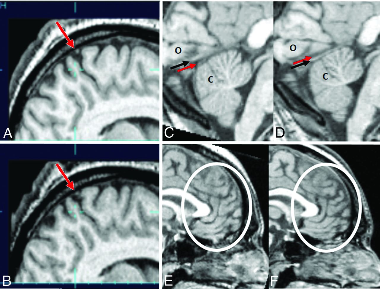
- Fig 1.
Structural images before and after bed rest. Sagittal images of the brain (subject H in On-line Tables 1–4) at the vertex before (A) and after (B) bed rest show a shift of the brain toward the vertex with contraction of the extra-axial spaces at the vertex and crowding of adjacent structures, including a cortical vein (arrow). Sagittal images of the posterior fossa before (C) and after (D) bed rest (subject A in On-line Tables 1–4). Before bed rest, the occipital lobes (o) lie against the tentorium cerebelli (black arrow). Below the tentorium, there is a thin layer of CSF (red arrow) between the tentorium and the upper aspect of the cerebellum (c). After bed rest, there is an upward shift of the occipital lobes away from the tentorium, now with a thin layer of CSF (red arrow) between the occipital lobes and the tentorium. In the posterior fossa, a layer of CSF is no longer visible between the tentorium and the cerebellum. Instead, the cerebellum now appears compressed against the tentorium. Sagittal images of the frontal lobes before (E) and after (F) bed rest show subtle expansion of the frontal lobe sulci after bed rest (subject A in On-line Tables 1–4).
- Fig 2.
Changes in ventricular volume before and after bed rest. Axial images of the brain of the subject with the largest change in ventricular size on the post–bed rest scan (B) compared with the pre–bed rest scan (A). Compared with pre–bed rest, there was a 22.4% reduction in ventricular size post–bed rest in this subject, best appreciated at the level of the atrium of the lateral ventricles (arrow) (subject D in On-line Tables 1–4). Axial images of the brain of the subject with the largest increase in ventricular size on the post–bed rest scan (D) compared with the pre–bed rest scan (C). Compared with pre–bed rest, there was a 10.4% increase in ventricular size post–bed rest in this subject, best appreciated at the level of the frontal horns of the lateral ventricles (arrow) (subject C in On-line Tables 1–4).
- Fig 3.
Brain translation and rotation in reference to the skull following bed rest. Parameters (translation and rotation on x, y, z-axes) were estimated on the basis of a rigid-body assumption. The arrow indicates the direction of movement that corresponds to positive values on the graph.
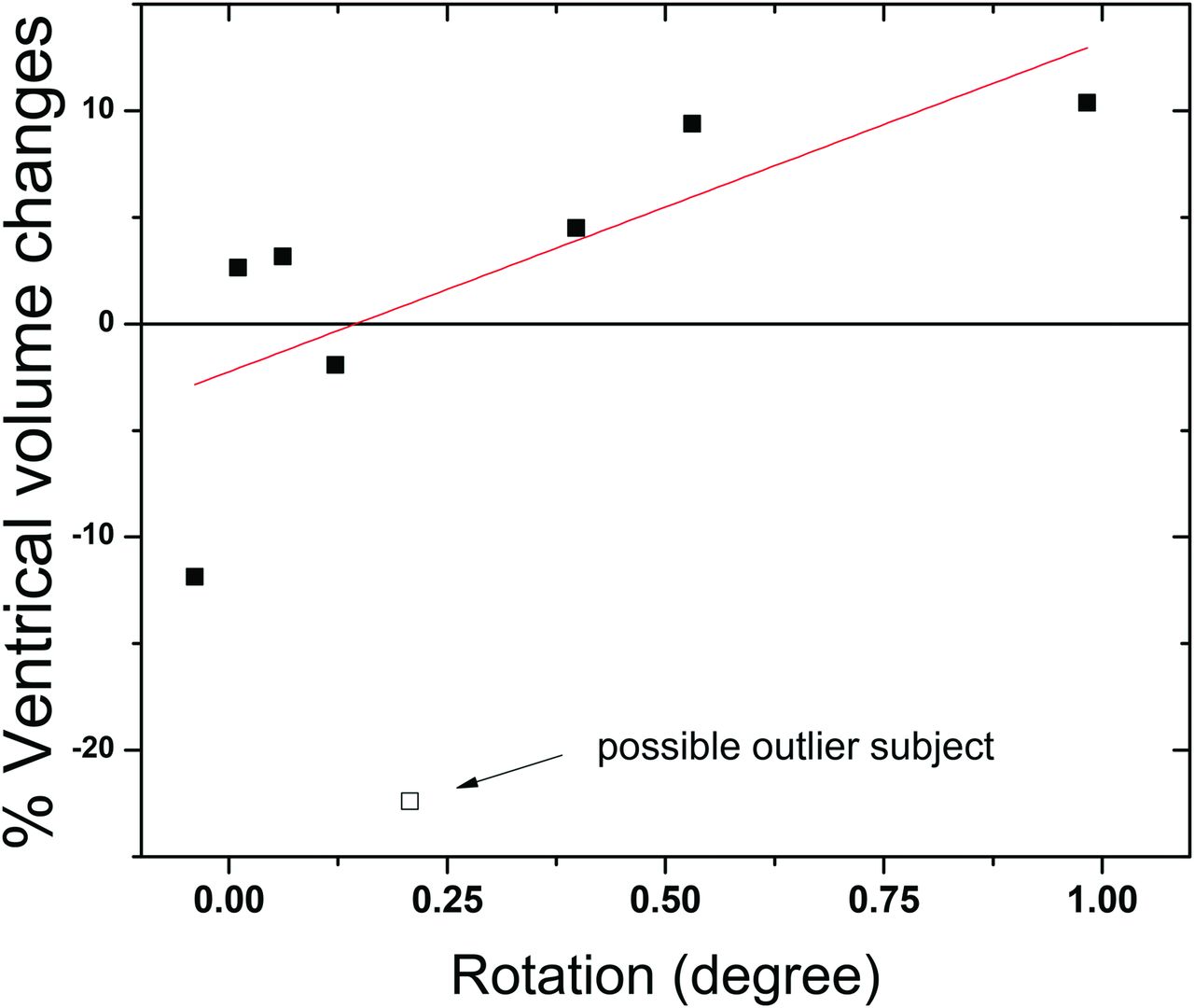
- Fig 4.
Correlation between brain rotation and ventricle volume changes. Without the potential outlier, which was the subject that demonstrated the largest ventricles before bed rest, the Spearman correlation is r = 0.893, P = .007. Including the outlier, the Spearman correlation is r = 0.690, P = .058.
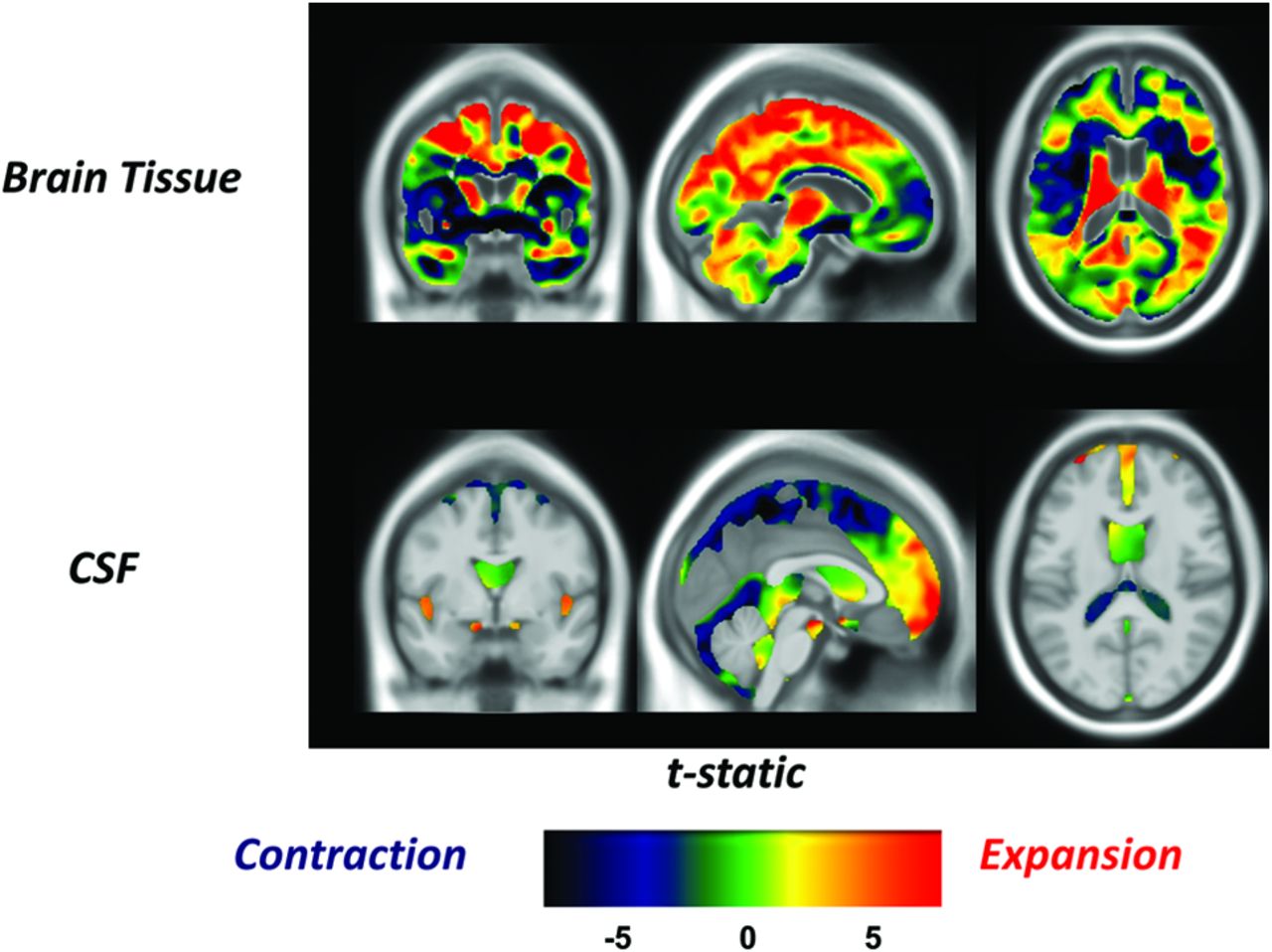
- Fig 5.
Regions of the brain most significantly affected by bed rest. There is increased brain tissue density (top row of images) at the vertex, particularly affecting the central frontoparietal lobes, with contraction of the adjacent CSF spaces (bottom row of images). There is decreased brain tissue density along the base of the brain, including the orbitofrontal cortex, with expansion of the adjacent CSF spaces.


Tables
- Table 1:
Pre- and post–bed rest volumetric MRI and physiologic measurements (group mean and SD)
Measurements Pre-bed rest Post-bed rest P Value ICV (mL) 1426 ± 105 1425 ± 105 .91 Gray matter (mL) 603.8 ± 40.8 615.1 ± 50.4 .18 White matter (mL) 546.0 ± 58.4 540.8 ± 51.5 .48 CSF (mL) 185.8 ± 15.0 186.6 ± 16.1 .83 Ventricle volume (mL) 15.45 ± 5.52 15.0 ± 4.31 .62 Plasma volume (mL) 2.63 ± 0.38 2.30 ± 0.46 .006 Blood volume (mL) 4.57 ± 1.02 4.08 ± 1.11 .004 Urine cortisol (μg/dL) 2.63 ± 0.97 3.58 ± 1.06 .14 IIH VIIP Finding Increased intracranial pressure ✓ Mildly elevated postflight; in-flight ICP unknown Papilledema ✓ ✓ Globe flattening ✓ ✓ Choroidal folds ✓ ✓ Hyperopic shifts ✓ ✓ Chronic headaches ✓ Diplopia ✓ Transient visual obscurations ✓ Pulse-synchronous tinnitus ✓ Sixth cranial nerve palsy ✓ Patient characteristics Obesity ✓ Sex predominance Female Male Imaging findings Optic nerve head protrusion ✓ ✓ Flattened posterior globe ✓ ✓ Enlarged optic nerve sheath ✓ ✓ Optic nerve tortuosity ✓ ✓ Empty sella ✓ ✓ Note:—✓ indicates clinical feature present.


