Advertisement

Main menu

  • Home
  • Content
    • Current Issue
    • Accepted Manuscripts
    • Article Preview
    • Past Issue Archive
    • Video Articles
    • AJNR Case Collection
    • Case of the Week Archive
    • Case of the Month Archive
    • Classic Case Archive
  • Special Collections
    • AJNR Awards
    • Low-Field MRI
    • Alzheimer Disease
    • ASNR Foundation Special Collection
    • Photon-Counting CT
    • View All
  • Multimedia
    • AJNR Podcasts
    • AJNR SCANtastic
    • Trainee Corner
    • MRI Safety Corner
    • Imaging Protocols
  • For Authors
    • Submit a Manuscript
    • Submit a Video Article
    • Submit an eLetter to the Editor/Response
    • Manuscript Submission Guidelines
    • Statistical Tips
    • Fast Publishing of Accepted Manuscripts
    • Graphical Abstract Preparation
    • Imaging Protocol Submission
    • Author Policies
  • About Us
    • About AJNR
    • Editorial Board
    • Editorial Board Alumni
  • More
    • Become a Reviewer/Academy of Reviewers
    • Subscribers
    • Permissions
    • Alerts
    • Feedback
    • Advertisers
    • ASNR Home

User menu

  • Alerts
  • Log in

Search

  • Advanced search
American Journal of Neuroradiology

American Journal of Neuroradiology

ASHNR American Society of Functional Neuroradiology ASHNR American Society of Pediatric Neuroradiology ASSR
  • Alerts
  • Log in

Advanced Search

  • Home
  • Content
    • Current Issue
    • Accepted Manuscripts
    • Article Preview
    • Past Issue Archive
    • Video Articles
    • AJNR Case Collection
    • Case of the Week Archive
    • Case of the Month Archive
    • Classic Case Archive
  • Special Collections
    • AJNR Awards
    • Low-Field MRI
    • Alzheimer Disease
    • ASNR Foundation Special Collection
    • Photon-Counting CT
    • View All
  • Multimedia
    • AJNR Podcasts
    • AJNR SCANtastic
    • Trainee Corner
    • MRI Safety Corner
    • Imaging Protocols
  • For Authors
    • Submit a Manuscript
    • Submit a Video Article
    • Submit an eLetter to the Editor/Response
    • Manuscript Submission Guidelines
    • Statistical Tips
    • Fast Publishing of Accepted Manuscripts
    • Graphical Abstract Preparation
    • Imaging Protocol Submission
    • Author Policies
  • About Us
    • About AJNR
    • Editorial Board
    • Editorial Board Alumni
  • More
    • Become a Reviewer/Academy of Reviewers
    • Subscribers
    • Permissions
    • Alerts
    • Feedback
    • Advertisers
    • ASNR Home
  • Follow AJNR on Twitter
  • Visit AJNR on Facebook
  • Follow AJNR on Instagram
  • Join AJNR on LinkedIn
  • RSS Feeds

AJNR Awards, New Junior Editors, and more. Read the latest AJNR updates

Research ArticlePediatric Neuroimaging
Open Access

White Matter Development is Potentially Influenced in Adolescents with Vertically Transmitted HIV Infections: A Tract-Based Spatial Statistics Study

J. Li, G. Wu, Z. Wen, J. Zhang, H. Lei, X. Gui and F. Lin
American Journal of Neuroradiology November 2015, 36 (11) 2163-2169; DOI: https://doi.org/10.3174/ajnr.A4417
J. Li
aFrom the Departments of Magnetic Resonance Imaging (J.L., G.W., Z.W., J.Z.)
  • Find this author on Google Scholar
  • Find this author on PubMed
  • Search for this author on this site
  • ORCID record for J. Li
G. Wu
aFrom the Departments of Magnetic Resonance Imaging (J.L., G.W., Z.W., J.Z.)
  • Find this author on Google Scholar
  • Find this author on PubMed
  • Search for this author on this site
Z. Wen
aFrom the Departments of Magnetic Resonance Imaging (J.L., G.W., Z.W., J.Z.)
  • Find this author on Google Scholar
  • Find this author on PubMed
  • Search for this author on this site
J. Zhang
aFrom the Departments of Magnetic Resonance Imaging (J.L., G.W., Z.W., J.Z.)
  • Find this author on Google Scholar
  • Find this author on PubMed
  • Search for this author on this site
H. Lei
cNational Center for Magnetic Resonance in Wuhan (H.L., F.L.), State Key Laboratory of Magnetic Resonance and Atomic and Molecular Physics, Wuhan Institute of Physics and Mathematics, Chinese Academy of Sciences, Wuhan, China.
  • Find this author on Google Scholar
  • Find this author on PubMed
  • Search for this author on this site
X. Gui
bInfectious Diseases (X.G.), Zhongnan Hospital of Wuhan University, Wuhan, China
  • Find this author on Google Scholar
  • Find this author on PubMed
  • Search for this author on this site
F. Lin
cNational Center for Magnetic Resonance in Wuhan (H.L., F.L.), State Key Laboratory of Magnetic Resonance and Atomic and Molecular Physics, Wuhan Institute of Physics and Mathematics, Chinese Academy of Sciences, Wuhan, China.
  • Find this author on Google Scholar
  • Find this author on PubMed
  • Search for this author on this site
  • ORCID record for F. Lin
  • Article
  • Figures & Data
  • Info & Metrics
  • Responses
  • References
  • PDF
Loading

Article Figures & Data

Figures

  • Tables
  • Fig 1.
    • Download figure
    • Open in new tab
    • Download powerpoint
    Fig 1.

    TBSS analysis of fractional anisotropy maps. Areas in red are brain regions where FA is significantly reduced (P < .05, corrected by multiple comparison) in HIV-positive subjects relative to HIV-negative controls. The results are shown overlaid on the Montreal Neurological Institute 152-T1 template and the mean FA skeleton (green). The left side of the image corresponds to the right hemisphere of the brain. FA changes occur in the region with Montreal Neurological Institute coordinates in the z-direction between z = 6 and z = 63.

  • Fig 2.
    • Download figure
    • Open in new tab
    • Download powerpoint
    Fig 2.

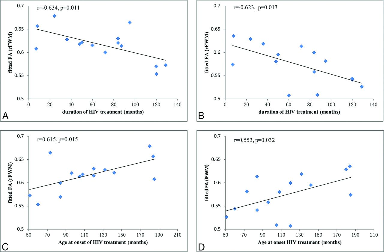
    Correlation results between FA alterations and clinical measures in the HIV-positive subjects. A, FA values in the right frontal WM (rFWM) are negatively correlated with the duration of HAART (r = −0.634, P = .011). B, FA values in the left frontal WM (lFWM) are negatively correlated with the duration of HAART (r = −0.623, P = .013). C, FA values in the rFWM are positively correlated with the age at onset of HAART (r = 0.615, P = .015). D, FA values in the lFWM are positively correlated with the age at onset of HAART (r = 0.553, P = .032).

Tables

  • Figures
    • View popup
    Table 1:

    Demographic information for subjects in each group

    HIV(+)HIV(−)P Value
    Age (yr)15.3 ± 1.315.0 ± 1.6.54
    Sex (male/female)8:713:13.84
    Education level (yr)7.5 ± 1.28.7 ± 1.9.05
    Current CD4 (cells/mL)605.8 ± 345.0NANA
    Age at first HIV treatment (yr)9.5 ± 3.4NANA
    HIV treatment duration (mo)68.3 ± 39.5NANA
    % Treated at younger than 2 yr13.3 (n = 2)NANA
    Plasma viral load (copies/mL)0–50NANA
    MoCA total score25.7 ± 3.827.1 ± 3.1.21
    MMSE total score25.8 ± 2.127.3 ± 2.4.05
    • Note:—NA indicates not applicable or available; MoCa, Montreal Cognitive Assessment; MMSE, Mini-Mental State Examination.

    • View popup
    Table 2:

    Neuroanatomic regions with decreased FA in HIV-positive subjects relative to HIV-negative controlsa

    Anatomic RegionHemisphereMNI Coordinates (mm)Cluster Size (mm3)
    XYZ
    Genu of corpus callosumBilateral−132024530
    Body of corpus callosumBilateral−1116242228
    SpleniumBilateral−11−3524564
    Superior corona radiataLeft−20−2539497
    Superior corona radiataRight20−2437386
    Posterior corona radiataLeft−21−2834262
    Posterior corona radiataRight25−2733249
    Frontal WMLeft−18543164
    Frontal WMRight171944399
    Pre-/postcentral gyrus WMLeft−21−2542269
    Pre-/postcentral gyrus WMRight21−2442524
    Parietal WMLeft−22−3140149
    Parietal WMRight17−5428437
    SLFLeft−30−173485
    • Note:—MNI indicates Montreal Neurological Institute; SLF, superior longitudinal fasciculus.

    • ↵a P < .05, threshold-free cluster enhancement multiple comparison–corrected. Coordinates for the peak voxels are displayed.

    • View popup
    Table 3:

    Group differences in diffusivity indices from VOIs

    Anatomic RegionHemisphereAD (×10−3mm2/s) (Mean)RD (×10−3mm2/s) (Mean)MD (×10−3mm2/s) (Mean)
    HIV−HIV+P ValueHIV−HIV+P ValueHIV−HIV+P Value
    Genu of corpus callosumBilateral1.62 ± 0.081.58 ± 0.08.160.36 ± 0.030.40 ± 0.06.0040.78 ± 0.030.79 ± 0.05.22
    Body of corpus callosumBilateral1.78 ± 0.061.78 ± 0.06.990.41 ± 0.050.49 ± 0.07.0003a0.87 ± 0.040.92 ± 0.05.002a
    SpleniumBilateral1.66 ± 0.061.67 ± 0.05.410.33 ± 0.030.37 ± 0.04.0003a0.77 ± 0.020.80 ± 0.03.001a
    Superior corona radiataLeft1.21 ± 0.041.20 ± 0.03.330.53 ± 0.020.56 ± 0.04.0003a0.76 ± 0.020.78 ± 0.03.01
    Superior corona radiataRight1.26 ± 0.051.26 ± 0.04.780.49 ± 0.020.53 ± 0.03.00009a0.74 ± 0.020.77 ± 0.02.001a
    Posterior corona radiataLeft1.23 ± 0.061.23 ± 0.03.810.59 ± 0.030.62 ± 0.03.002a0.80 ± 0.030.82 ± 0.02.02
    Posterior corona radiataRight1.26 ± 0.041.25 ± 0.03.320.57 ± 0.030.61 ± 0.04.002a0.80 ± 0.030.82 ± 0.04.05
    Frontal WMLeft1.35 ± 0.071.32 ± 0.05.220.44 ± 0.030.48 ± 0.04.001a0.74 ± 0.030.76 ± 0.03.09
    Frontal WMRight1.37 ± 0.061.32 ± 0.07.020.40 ± 0.030.48 ± 0.03.0003a0.72 ± 0.030.73 ± 0.03.21
    Pre-/postcentral gyrus WMLeft1.32 ± 0.061.29 ± 0.06.140.47 ± 0.030.50 ± 0.03.0007a0.75 ± 0.020.76 ± 0.02.06
    Pre-/postcentral gyrus WMRight1.30 ± 0.041.27 ± 0.04.010.44 ± 0.020.48 ± 0.03.00009a0.73 ± 0.020.74 ± 0.02.08
    Parietal WMLeft1.31 ± 0.051.31 ± 0.05.940.57 ± 0.040.61 ± 0.03.001a0.82 ± 0.030.84 ± 0.03.02
    Parietal WMRight1.29 ± 0.061.27 ± 0.04.280.58 ± 0.030.62 ± 0.05.002a0.81 ± 0.030.84 ± 0.04.07
    SLFLeft1.11 ± 0.041.09 ± 0.03.130.54 ± 0.030.57 ± 0.04.0040.73 ± 0.020.75 ± 0.03.05
    • Note:—HIV- indicates HIV-negative controls; HIV+, HIV-positive subjects.

    • ↵a P < .05/14 ≈ .0035 (Bonferroni-corrected for multiple comparisons).

PreviousNext
Back to top

In this issue

American Journal of Neuroradiology: 36 (11)
American Journal of Neuroradiology
Vol. 36, Issue 11
1 Nov 2015
  • Table of Contents
  • Index by author
  • Complete Issue (PDF)
Advertisement
Print
Download PDF
Email Article

Thank you for your interest in spreading the word on American Journal of Neuroradiology.

NOTE: We only request your email address so that the person you are recommending the page to knows that you wanted them to see it, and that it is not junk mail. We do not capture any email address.

Enter multiple addresses on separate lines or separate them with commas.
White Matter Development is Potentially Influenced in Adolescents with Vertically Transmitted HIV Infections: A Tract-Based Spatial Statistics Study
(Your Name) has sent you a message from American Journal of Neuroradiology
(Your Name) thought you would like to see the American Journal of Neuroradiology web site.
CAPTCHA
This question is for testing whether or not you are a human visitor and to prevent automated spam submissions.
Cite this article
J. Li, G. Wu, Z. Wen, J. Zhang, H. Lei, X. Gui, F. Lin
White Matter Development is Potentially Influenced in Adolescents with Vertically Transmitted HIV Infections: A Tract-Based Spatial Statistics Study
American Journal of Neuroradiology Nov 2015, 36 (11) 2163-2169; DOI: 10.3174/ajnr.A4417

Citation Manager Formats

  • BibTeX
  • Bookends
  • EasyBib
  • EndNote (tagged)
  • EndNote 8 (xml)
  • Medlars
  • Mendeley
  • Papers
  • RefWorks Tagged
  • Ref Manager
  • RIS
  • Zotero
0 Responses
Respond to this article
Share
Bookmark this article
White Matter Development is Potentially Influenced in Adolescents with Vertically Transmitted HIV Infections: A Tract-Based Spatial Statistics Study
J. Li, G. Wu, Z. Wen, J. Zhang, H. Lei, X. Gui, F. Lin
American Journal of Neuroradiology Nov 2015, 36 (11) 2163-2169; DOI: 10.3174/ajnr.A4417
del.icio.us logo Twitter logo Facebook logo Mendeley logo
  • Tweet Widget
  • Facebook Like
  • Google Plus One
Purchase

Jump to section

  • Article
    • Abstract
    • ABBREVIATIONS:
    • Materials and Methods
    • Results
    • Discussion
    • Conclusions
    • Footnotes
    • References
  • Figures & Data
  • Info & Metrics
  • Responses
  • References
  • PDF

Related Articles

  • No related articles found.
  • PubMed
  • Google Scholar

Cited By...

  • Brain structure of perinatally HIV-infected patients on long-term treatment: A systematic review
  • Early Antiretroviral Therapy in HIV-Infected Children Is Associated with Diffuse White Matter Structural Abnormality and Corpus Callosum Sparing
  • Crossref
  • Google Scholar

This article has not yet been cited by articles in journals that are participating in Crossref Cited-by Linking.

More in this TOC Section

  • Sodium MRI in Pediatric Brain Tumors
  • FRACTURE MR in Congenital Vertebral Anomalies
  • Comparing MRI Perfusion in Pediatric Brain Tumors
Show more Pediatric Neuroimaging

Similar Articles

Advertisement

Indexed Content

  • Current Issue
  • Accepted Manuscripts
  • Article Preview
  • Past Issues
  • Editorials
  • Editor's Choice
  • Fellows' Journal Club
  • Letters to the Editor
  • Video Articles

Cases

  • Case Collection
  • Archive - Case of the Week
  • Archive - Case of the Month
  • Archive - Classic Case

More from AJNR

  • Trainee Corner
  • Imaging Protocols
  • MRI Safety Corner
  • Book Reviews

Multimedia

  • AJNR Podcasts
  • AJNR Scantastics

Resources

  • Turnaround Time
  • Submit a Manuscript
  • Submit a Video Article
  • Submit an eLetter to the Editor/Response
  • Manuscript Submission Guidelines
  • Statistical Tips
  • Fast Publishing of Accepted Manuscripts
  • Graphical Abstract Preparation
  • Imaging Protocol Submission
  • Evidence-Based Medicine Level Guide
  • Publishing Checklists
  • Author Policies
  • Become a Reviewer/Academy of Reviewers
  • News and Updates

About Us

  • About AJNR
  • Editorial Board
  • Editorial Board Alumni
  • Alerts
  • Permissions
  • Not an AJNR Subscriber? Join Now
  • Advertise with Us
  • Librarian Resources
  • Feedback
  • Terms and Conditions
  • AJNR Editorial Board Alumni

American Society of Neuroradiology

  • Not an ASNR Member? Join Now

© 2025 by the American Society of Neuroradiology All rights, including for text and data mining, AI training, and similar technologies, are reserved.
Print ISSN: 0195-6108 Online ISSN: 1936-959X

Powered by HighWire