Advertisement

Main menu

  • Home
  • Content
    • Current Issue
    • Accepted Manuscripts
    • Article Preview
    • Past Issue Archive
    • AJNR Case Collection
    • Case of the Week Archive
    • Classic Case Archive
    • Case of the Month Archive
  • Special Collections
    • Spinal CSF Leak Articles (Jan 2020-June 2024)
    • 2024 AJNR Journal Awards
    • Most Impactful AJNR Articles
  • Multimedia
    • AJNR Podcast
    • AJNR Scantastics
    • Video Articles
  • For Authors
    • Submit a Manuscript
    • Author Policies
    • Fast publishing of Accepted Manuscripts
    • Graphical Abstract Preparation
    • Manuscript Submission Guidelines
    • Imaging Protocol Submission
    • Submit a Case for the Case Collection
  • About Us
    • About AJNR
    • Editorial Board
  • More
    • Become a Reviewer/Academy of Reviewers
    • Subscribers
    • Permissions
    • Alerts
    • Feedback
    • Advertisers
    • ASNR Home
  • Other Publications
    • ajnr

User menu

  • Alerts
  • Log in

Search

  • Advanced search
American Journal of Neuroradiology

American Journal of Neuroradiology

ASHNR American Society of Functional Neuroradiology ASHNR American Society of Pediatric Neuroradiology ASSR
  • Alerts
  • Log in

Advanced Search

  • Home
  • Content
    • Current Issue
    • Accepted Manuscripts
    • Article Preview
    • Past Issue Archive
    • AJNR Case Collection
    • Case of the Week Archive
    • Classic Case Archive
    • Case of the Month Archive
  • Special Collections
    • Spinal CSF Leak Articles (Jan 2020-June 2024)
    • 2024 AJNR Journal Awards
    • Most Impactful AJNR Articles
  • Multimedia
    • AJNR Podcast
    • AJNR Scantastics
    • Video Articles
  • For Authors
    • Submit a Manuscript
    • Author Policies
    • Fast publishing of Accepted Manuscripts
    • Graphical Abstract Preparation
    • Manuscript Submission Guidelines
    • Imaging Protocol Submission
    • Submit a Case for the Case Collection
  • About Us
    • About AJNR
    • Editorial Board
  • More
    • Become a Reviewer/Academy of Reviewers
    • Subscribers
    • Permissions
    • Alerts
    • Feedback
    • Advertisers
    • ASNR Home
  • Follow AJNR on Twitter
  • Visit AJNR on Facebook
  • Follow AJNR on Instagram
  • Join AJNR on LinkedIn
  • RSS Feeds

Welcome to the new AJNR, Updated Hall of Fame, and more. Read the full announcements.


AJNR is seeking candidates for the position of Associate Section Editor, AJNR Case Collection. Read the full announcement.


Getting new auth cookie, if you see this message a lot, tell someone!
Research ArticleHead & Neck

Detection of Calcifications in Retinoblastoma Using Gradient-Echo MR Imaging Sequences: Comparative Study between In Vivo MR Imaging and Ex Vivo High-Resolution CT

F. Rodjan, P. de Graaf, P. van der Valk, T. Hadjistilianou, A. Cerase, P. Toti, M.C. de Jong, A.C. Moll, J.A. Castelijns and P. Galluzzi on behalf of the European Retinoblastoma Imaging Collaboration
American Journal of Neuroradiology February 2015, 36 (2) 355-360; DOI: https://doi.org/10.3174/ajnr.A4163
F. Rodjan
aFrom the Departments of Radiology (F.R., P.d.G., M.C.d.J., J.A.C.)
  • Find this author on Google Scholar
  • Find this author on PubMed
  • Search for this author on this site
P. de Graaf
aFrom the Departments of Radiology (F.R., P.d.G., M.C.d.J., J.A.C.)
  • Find this author on Google Scholar
  • Find this author on PubMed
  • Search for this author on this site
P. van der Valk
bPathology (P.v.d.V.)
  • Find this author on Google Scholar
  • Find this author on PubMed
  • Search for this author on this site
T. Hadjistilianou
dDepartments of Ophthalmology (T.H.)
  • Find this author on Google Scholar
  • Find this author on PubMed
  • Search for this author on this site
A. Cerase
eNeuroimaging and Neurointerventional Unit (A.C., P.G.)
  • Find this author on Google Scholar
  • Find this author on PubMed
  • Search for this author on this site
P. Toti
fPathology (P.T.), Azienda Ospedaliera e Universitaria, Santa Maria alle Scotte, Siena, Italy.
  • Find this author on Google Scholar
  • Find this author on PubMed
  • Search for this author on this site
M.C. de Jong
aFrom the Departments of Radiology (F.R., P.d.G., M.C.d.J., J.A.C.)
  • Find this author on Google Scholar
  • Find this author on PubMed
  • Search for this author on this site
A.C. Moll
cOphthalmology (A.C.M.), VU University Medical Center, Amsterdam, the Netherlands
  • Find this author on Google Scholar
  • Find this author on PubMed
  • Search for this author on this site
J.A. Castelijns
aFrom the Departments of Radiology (F.R., P.d.G., M.C.d.J., J.A.C.)
  • Find this author on Google Scholar
  • Find this author on PubMed
  • Search for this author on this site
P. Galluzzi
eNeuroimaging and Neurointerventional Unit (A.C., P.G.)
  • Find this author on Google Scholar
  • Find this author on PubMed
  • Search for this author on this site
  • Article
  • Figures & Data
  • Info & Metrics
  • Responses
  • References
  • PDF
Loading

Article Figures & Data

Figures

  • Tables
  • Fig 1.
    • Download figure
    • Open in new tab
    • Download powerpoint
    Fig 1.

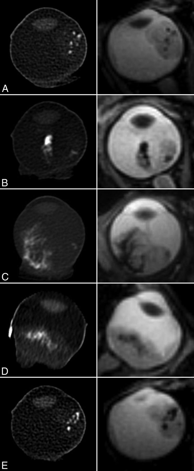
    Excellent-matching hyperattenuated calcifications on ex vivo high-resolution CT (left column) with signal-intensity void spots on gradient-echo T2*-weighted MR images (right column) in patients 12 (A), 16 (B), 18 (C), 19 (D), and 14 (E).

  • Fig 2.
    • Download figure
    • Open in new tab
    • Download powerpoint
    Fig 2.

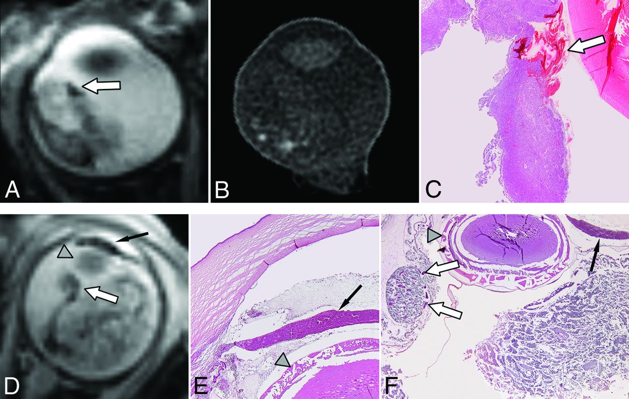
    Examples of additional signal-intensity voids on T2*-weighted MR images without correspondence with ex vivo high-resolution CT. Patient 13 shows a hypointense nodular structure (A) in the anterior part of the eye (arrow) on T2*-weighted imaging without corresponding hyperattenuation on ex vivo high-resolution CT (B). Histopathology demonstrates a hemorrhage (arrow) precisely matching this additional SIV (C). Adjacent to this hemorrhage multiple linear-arranged spots match hyperattenuated spots on CT. In patient 11 (D), a linear band of SIV on T2*WI is shown outside the tumor along the detached retina (white arrow) and in the iris (black arrow). The gray arrowhead indicates the lens, which is dislocated. Histology (E and F) shows necrotic tumor with dilated vessels (venous congestion) (white arrow) and hemorrhagic necrosis of the iris combined with venous congestion (black arrow). The anterior chamber is infiltrated by neoplastic cells and cellular debris. (H&E staining, ×20 magnification.)

  • Fig 3.
    • Download figure
    • Open in new tab
    • Download powerpoint
    Fig 3.

    Extra signal-intensity void spots in the anterior part of the eye on gradient-echo T2*-weighted images (arrow, A) were observed in patient 21, with excellent correspondence with HRCT (B). However, a band of additional signal-intensity void spots was present in the anterior part of the tumor on the gradient-echo T2*-weighted MR image (arrow, A) without correspondence on ex vivo high-resolution CT. Histopathologic correlation (C) shows multiple foci of calcifications in the anterior part of the tumor (arrow). (H&E staining, ×20 magnification.)

  • Fig 4.
    • Download figure
    • Open in new tab
    • Download powerpoint
    Fig 4.

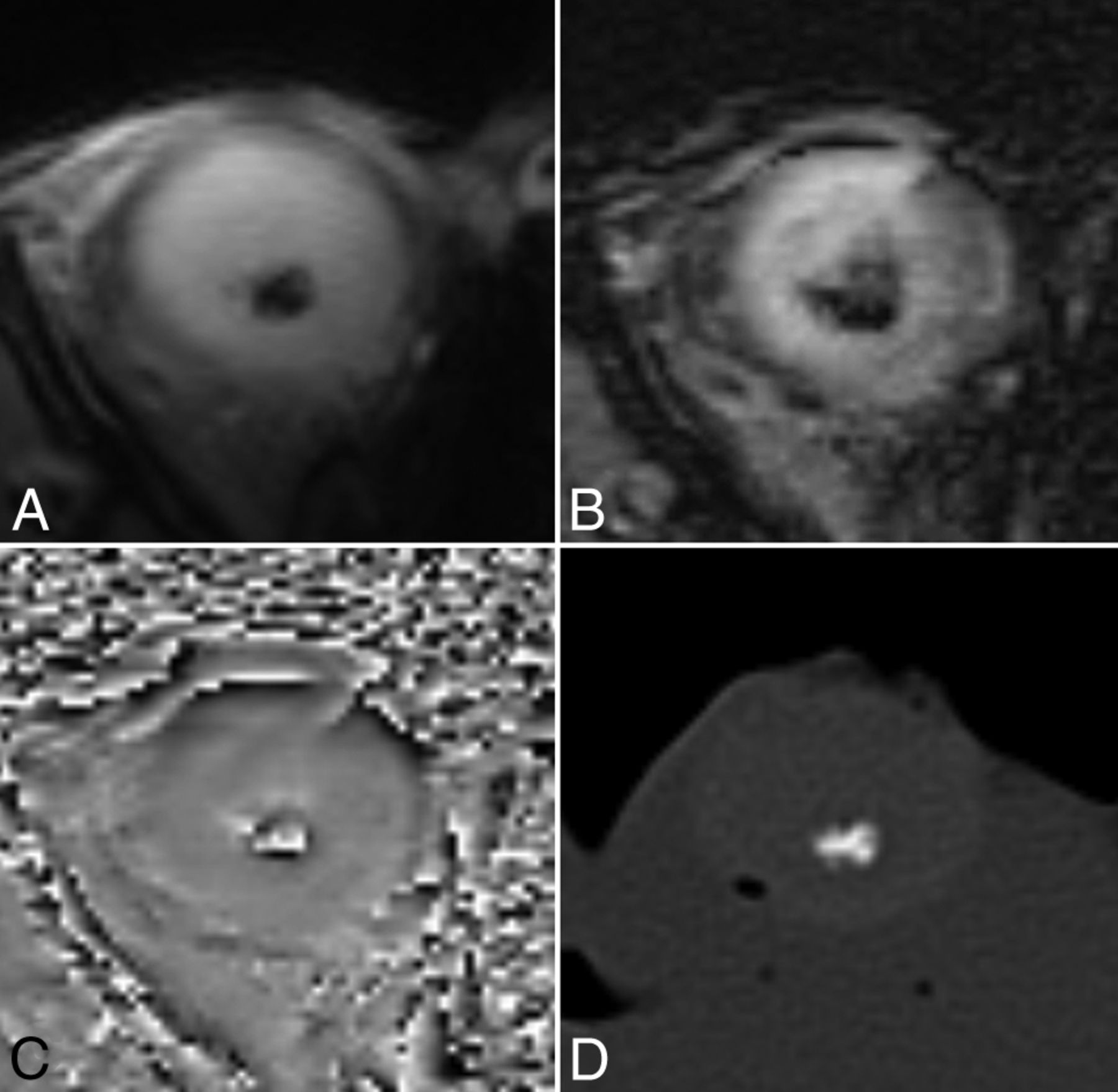
    The value of phase imaging in identifying calcification in retinoblastoma (patient 22). Signal-intensity void spots can be seen on the T2*-weighted (A) and SWI minimum intensity projection (B). A phase image (C) shows high signal intensity centrally identifying calcification, confirmed on the ex vivo high-resolution CT image (D).

Tables

  • Figures
  • Patient findings and correlation of CT with MRI in calcium detection

    Patient (Lat)Age (mo)aInt MRI-En (day)Corr T2*WI-CTSWICorr T2*WI-SWI
    1 (U)11ModerateNoNA
    2 (U)48ModerateNoNA
    3 (U)466ModerateNoNA
    4 (U)30ModerateNoNA
    5 (U)266ModerateNoNA
    6 (U)731GoodNoNA
    7 (B)84GoodNoNA
    8 (U)115GoodNoNA
    9 (U)81GoodNoNA
    10 (U)161GoodNoNA
    11 (U)38GoodNoNA
    12 (B)128GoodNoNA
    13 (B)358GoodNoNA
    14 (U)291GoodNoNA
    15 (B)298GoodNoNA
    16 (B)136GoodNoNA
    17 (U)458GoodNoNA
    18 (U)58GoodYesEqual
    19 (U)97GoodYesBetter
    20 (U)381GoodYesBetter
    21 (U)58GoodYesBetter
    22 (U)375GoodYesBetter
    • Note:—Lat indicates tumor laterality; Int MRI-En, interval MRI and enucleation; Corr, correlation; U, unilateral; B, bilateral; NA, not applicable.

    • ↵a Median age, 12.5 mo; mean age, 20.72 mo.

PreviousNext
Back to top

In this issue

American Journal of Neuroradiology: 36 (2)
American Journal of Neuroradiology
Vol. 36, Issue 2
1 Feb 2015
  • Table of Contents
  • Index by author
  • Complete Issue (PDF)
Advertisement
Print
Download PDF
Email Article

Thank you for your interest in spreading the word on American Journal of Neuroradiology.

NOTE: We only request your email address so that the person you are recommending the page to knows that you wanted them to see it, and that it is not junk mail. We do not capture any email address.

Enter multiple addresses on separate lines or separate them with commas.
Detection of Calcifications in Retinoblastoma Using Gradient-Echo MR Imaging Sequences: Comparative Study between In Vivo MR Imaging and Ex Vivo High-Resolution CT
(Your Name) has sent you a message from American Journal of Neuroradiology
(Your Name) thought you would like to see the American Journal of Neuroradiology web site.
CAPTCHA
This question is for testing whether or not you are a human visitor and to prevent automated spam submissions.
Cite this article
F. Rodjan, P. de Graaf, P. van der Valk, T. Hadjistilianou, A. Cerase, P. Toti, M.C. de Jong, A.C. Moll, J.A. Castelijns, P. Galluzzi
Detection of Calcifications in Retinoblastoma Using Gradient-Echo MR Imaging Sequences: Comparative Study between In Vivo MR Imaging and Ex Vivo High-Resolution CT
American Journal of Neuroradiology Feb 2015, 36 (2) 355-360; DOI: 10.3174/ajnr.A4163

Citation Manager Formats

  • BibTeX
  • Bookends
  • EasyBib
  • EndNote (tagged)
  • EndNote 8 (xml)
  • Medlars
  • Mendeley
  • Papers
  • RefWorks Tagged
  • Ref Manager
  • RIS
  • Zotero
0 Responses
Respond to this article
Share
Bookmark this article
Detection of Calcifications in Retinoblastoma Using Gradient-Echo MR Imaging Sequences: Comparative Study between In Vivo MR Imaging and Ex Vivo High-Resolution CT
F. Rodjan, P. de Graaf, P. van der Valk, T. Hadjistilianou, A. Cerase, P. Toti, M.C. de Jong, A.C. Moll, J.A. Castelijns, P. Galluzzi
American Journal of Neuroradiology Feb 2015, 36 (2) 355-360; DOI: 10.3174/ajnr.A4163
del.icio.us logo Twitter logo Facebook logo Mendeley logo
  • Tweet Widget
  • Facebook Like
  • Google Plus One
Purchase

Jump to section

  • Article
    • Abstract
    • ABBREVIATIONS:
    • Materials and Methods
    • Results
    • Discussion
    • Conclusions
    • Footnotes
    • REFERENCES
  • Figures & Data
  • Info & Metrics
  • Responses
  • References
  • PDF

Related Articles

  • No related articles found.
  • PubMed
  • Google Scholar

Cited By...

  • No citing articles found.
  • Crossref (17)
  • Google Scholar

This article has been cited by the following articles in journals that are participating in Crossref Cited-by Linking.

  • Conservative management of retinoblastoma: Challenging orthodoxy without compromising the state of metastatic grace. “Alive, with good vision and no comorbidity”
    Francis L. Munier, Maja Beck-Popovic, Guillermo L. Chantada, David Cobrinik, Tero T. Kivelä, Dietmar Lohmann, Philippe Maeder, Annette C. Moll, Angel Montero Carcaboso, Alexandre Moulin, Paula Schaiquevich, Ciara Bergin, Paul J. Dyson, Susan Houghton, Francesco Puccinelli, Yvan Vial, Marie-Claire Gaillard, Christina Stathopoulos
    Progress in Retinal and Eye Research 2019 73
  • Retinoblastoma, the visible CNS tumor: A review
    Helen Dimaras, Timothy W. Corson
    Journal of Neuroscience Research 2019 97 1
  • AJCC Cancer Staging Manual
    2017
  • MR Imaging Features to Differentiate Retinoblastoma from Coats’ Disease and Persistent Fetal Vasculature
    Robin W. Jansen, Christiaan M. de Bloeme, Hervé J. Brisse, Paolo Galluzzi, Liesbeth Cardoen, Sophia Göricke, Philippe Maeder, Nathalie Cassoux, Arnaud Gauthier, Sabrina Schlueter, Theodora Hadjistilianou, Francis L. Munier, Jonas A. Castelijns, Paul van der Valk, Annette C. Moll, Marcus C. de Jong, Pim de Graaf
    Cancers 2020 12 12
  • The potential of 3T high-resolution magnetic resonance imaging for diagnosis, staging, and follow-up of retinoblastoma
    Marcus C. de Jong, Pim de Graaf, Hervé J. Brisse, Paolo Galluzzi, Sophia L. Göricke, Annette C. Moll, Francis L. Munier, Maja Beck Popovic, Alexandre P. Moulin, Stefano Binaghi, Jonas A. Castelijns, Philippe Maeder, Jonas A. Castelijns, Pim de Graaf, Marcus C. de Jong, Hervé J. Brisse, Paolo Galluzzi, Sophia L. Göricke, Philippe Maeder
    Survey of Ophthalmology 2015 60 4
  • Guidelines for magnetic resonance imaging in pediatric head and neck pathologies: a multicentre international consensus paper
    Felice D’Arco, Livja Mertiri, Pim de Graaf, Bert De Foer, Katarina S. Popovič, Maria I. Argyropoulou, Kshitij Mankad, Hervé J. Brisse, Amy Juliano, Mariasavina Severino, Sofie Van Cauter, Mai-Lan Ho, Caroline D. Robson, Ata Siddiqui, Steve Connor, Sotirios Bisdas, Alessandro Bozzao, Jan Sedlacik, Camilla Rossi Espagnet, Daniela Longo, Alessia Carboni, Lorenzo Ugga, Stefania Picariello, Giacomo Talenti, Sniya V. Sudahakar, Martina Di Stasi, Ulrike Löbel, Robert Nash, Kaukab Rajput, Olivia Carney, Davide Farina, Richard Hewitt, Olga Slater, Jessica Cooper, Gennaro D’Anna, Gul Moonis, Andrea Rossi, Domenico Tortora, Cesar Augusto Alves, Asif Mazumder, Faraan Khan, Teresa Nunes, Owen Arthurs, Hisham Dahmoush, Renato Cuocolo, Pablo Caro-Dominguez, Arastoo Vossough, William T. O’Brien, Asthik Biswas, Catriona Duncan, Lennyn Alban
    Neuroradiology 2022 64 6
  • Neonatal Retinoblastoma
    Frank Y. Lin, Murali M. Chintagumpala
    Clinics in Perinatology 2021 48 1
  • 9.4T and 17.6T MRI of Retinoblastoma: Ex Vivo evaluation of microstructural anatomy and disease extent compared with histopathology
    Marcus C. de Jong, Pim de Graaf, Petra J.W. Pouwels, Jan‐Willem Beenakker, Robin W. Jansen, Jeroen J.G. Geurts, Annette C. Moll, Jonas A. Castelijns, Paul van der Valk, Louise van der Weerd
    Journal of Magnetic Resonance Imaging 2018 47 6
  • Diagnostic Imaging for Retinoblastoma Cancer Staging: Guide for Providing Essential Insights for Ophthalmologists and Oncologists
    Vivek Pai, Prakash Muthusami, Birgit Ertl-Wagner, Manohar M. Shroff, Carmen Parra-Fariñas, Kanchan Sainani, Stephanie Kletke, Marie-Anne Brundler, Ashwin Mallipatna
    RadioGraphics 2024 44 4
  • Retinoblastoma - Past, Present and Future
    Fahad Albader, Dalal Fatani
    2019

More in this TOC Section

  • NI-RADS for HEAD&NECK Cancer Recurrence
  • WHO Classification Update: Nasal&Skull Base Tumors
  • Peritumoral Signal in Vestibular Schwannomas
Show more HEAD & NECK

Similar Articles

Advertisement

Indexed Content

  • Current Issue
  • Accepted Manuscripts
  • Article Preview
  • Past Issues
  • Editorials
  • Editors Choice
  • Fellow Journal Club
  • Letters to the Editor

Cases

  • Case Collection
  • Archive - Case of the Week
  • Archive - Case of the Month
  • Archive - Classic Case

Special Collections

  • Special Collections

Resources

  • News and Updates
  • Turn around Times
  • Submit a Manuscript
  • Author Policies
  • Manuscript Submission Guidelines
  • Evidence-Based Medicine Level Guide
  • Publishing Checklists
  • Graphical Abstract Preparation
  • Imaging Protocol Submission
  • Submit a Case
  • Become a Reviewer/Academy of Reviewers
  • Get Peer Review Credit from Publons

Multimedia

  • AJNR Podcast
  • AJNR SCANtastic
  • Video Articles

About Us

  • About AJNR
  • Editorial Board
  • Not an AJNR Subscriber? Join Now
  • Alerts
  • Feedback
  • Advertise with us
  • Librarian Resources
  • Permissions
  • Terms and Conditions

American Society of Neuroradiology

  • Not an ASNR Member? Join Now

© 2025 by the American Society of Neuroradiology All rights, including for text and data mining, AI training, and similar technologies, are reserved.
Print ISSN: 0195-6108 Online ISSN: 1936-959X

Powered by HighWire