Advertisement

Main menu

  • Home
  • Content
    • Current Issue
    • Accepted Manuscripts
    • Article Preview
    • Past Issue Archive
    • AJNR Case Collection
    • Case of the Week Archive
    • Classic Case Archive
    • Case of the Month Archive
  • Special Collections
    • Spinal CSF Leak Articles (Jan 2020-June 2024)
    • 2024 AJNR Journal Awards
    • Most Impactful AJNR Articles
  • Multimedia
    • AJNR Podcast
    • AJNR Scantastics
    • Video Articles
  • For Authors
    • Submit a Manuscript
    • Author Policies
    • Fast publishing of Accepted Manuscripts
    • Graphical Abstract Preparation
    • Manuscript Submission Guidelines
    • Imaging Protocol Submission
    • Submit a Case for the Case Collection
  • About Us
    • About AJNR
    • Editorial Board
  • More
    • Become a Reviewer/Academy of Reviewers
    • Subscribers
    • Permissions
    • Alerts
    • Feedback
    • Advertisers
    • ASNR Home
  • Other Publications
    • ajnr

User menu

  • Alerts
  • Log in

Search

  • Advanced search
American Journal of Neuroradiology

American Journal of Neuroradiology

ASHNR American Society of Functional Neuroradiology ASHNR American Society of Pediatric Neuroradiology ASSR
  • Alerts
  • Log in

Advanced Search

  • Home
  • Content
    • Current Issue
    • Accepted Manuscripts
    • Article Preview
    • Past Issue Archive
    • AJNR Case Collection
    • Case of the Week Archive
    • Classic Case Archive
    • Case of the Month Archive
  • Special Collections
    • Spinal CSF Leak Articles (Jan 2020-June 2024)
    • 2024 AJNR Journal Awards
    • Most Impactful AJNR Articles
  • Multimedia
    • AJNR Podcast
    • AJNR Scantastics
    • Video Articles
  • For Authors
    • Submit a Manuscript
    • Author Policies
    • Fast publishing of Accepted Manuscripts
    • Graphical Abstract Preparation
    • Manuscript Submission Guidelines
    • Imaging Protocol Submission
    • Submit a Case for the Case Collection
  • About Us
    • About AJNR
    • Editorial Board
  • More
    • Become a Reviewer/Academy of Reviewers
    • Subscribers
    • Permissions
    • Alerts
    • Feedback
    • Advertisers
    • ASNR Home
  • Follow AJNR on Twitter
  • Visit AJNR on Facebook
  • Follow AJNR on Instagram
  • Join AJNR on LinkedIn
  • RSS Feeds

Welcome to the new AJNR, Updated Hall of Fame, and more. Read the full announcements.


AJNR is seeking candidates for the position of Associate Section Editor, AJNR Case Collection. Read the full announcement.


Getting new auth cookie, if you see this message a lot, tell someone!
Research ArticleSpine

Intramedullary Spinal Cord Metastases: Prognostic Value of MRI and Clinical Features from a 13-Year Institutional Case Series

F.E. Diehn, J.B. Rykken, J.T. Wald, C.P. Wood, L.J. Eckel, C.H. Hunt, K.M. Schwartz, R.K. Lingineni, R.E. Carter and T.J. Kaufmann
American Journal of Neuroradiology March 2015, 36 (3) 587-593; DOI: https://doi.org/10.3174/ajnr.A4160
F.E. Diehn
aFrom the Division of Neuroradiology (F.E.D., J.B.R., J.T.W., C.P.W., L.J.E., C.H.H., K.M.S., T.J.K.), Department of Radiology
  • Find this author on Google Scholar
  • Find this author on PubMed
  • Search for this author on this site
J.B. Rykken
aFrom the Division of Neuroradiology (F.E.D., J.B.R., J.T.W., C.P.W., L.J.E., C.H.H., K.M.S., T.J.K.), Department of Radiology
  • Find this author on Google Scholar
  • Find this author on PubMed
  • Search for this author on this site
J.T. Wald
aFrom the Division of Neuroradiology (F.E.D., J.B.R., J.T.W., C.P.W., L.J.E., C.H.H., K.M.S., T.J.K.), Department of Radiology
  • Find this author on Google Scholar
  • Find this author on PubMed
  • Search for this author on this site
C.P. Wood
aFrom the Division of Neuroradiology (F.E.D., J.B.R., J.T.W., C.P.W., L.J.E., C.H.H., K.M.S., T.J.K.), Department of Radiology
  • Find this author on Google Scholar
  • Find this author on PubMed
  • Search for this author on this site
L.J. Eckel
aFrom the Division of Neuroradiology (F.E.D., J.B.R., J.T.W., C.P.W., L.J.E., C.H.H., K.M.S., T.J.K.), Department of Radiology
  • Find this author on Google Scholar
  • Find this author on PubMed
  • Search for this author on this site
C.H. Hunt
aFrom the Division of Neuroradiology (F.E.D., J.B.R., J.T.W., C.P.W., L.J.E., C.H.H., K.M.S., T.J.K.), Department of Radiology
  • Find this author on Google Scholar
  • Find this author on PubMed
  • Search for this author on this site
K.M. Schwartz
aFrom the Division of Neuroradiology (F.E.D., J.B.R., J.T.W., C.P.W., L.J.E., C.H.H., K.M.S., T.J.K.), Department of Radiology
  • Find this author on Google Scholar
  • Find this author on PubMed
  • Search for this author on this site
R.K. Lingineni
bDepartment of Health Sciences Research (R.K.L., R.E.C.), Mayo Clinic, Rochester, Minnesota.
  • Find this author on Google Scholar
  • Find this author on PubMed
  • Search for this author on this site
R.E. Carter
bDepartment of Health Sciences Research (R.K.L., R.E.C.), Mayo Clinic, Rochester, Minnesota.
  • Find this author on Google Scholar
  • Find this author on PubMed
  • Search for this author on this site
T.J. Kaufmann
aFrom the Division of Neuroradiology (F.E.D., J.B.R., J.T.W., C.P.W., L.J.E., C.H.H., K.M.S., T.J.K.), Department of Radiology
  • Find this author on Google Scholar
  • Find this author on PubMed
  • Search for this author on this site
  • Article
  • Figures & Data
  • Info & Metrics
  • Responses
  • References
  • PDF
Loading

Article Figures & Data

Figures

  • Tables
  • Fig 1.
    • Download figure
    • Open in new tab
    • Download powerpoint
    Fig 1.

    Kaplan-Meier plot. Decreased survival for patients with lung or breast primary malignancy compared with all other types of primary malignancy. The median survival for patients with lung or breast primaries was 57 days and for patients with nonlung/nonbreast primaries was 308 days (P < .001, log-rank test; N = 49 patients).

  • Fig 2.
    • Download figure
    • Open in new tab
    • Download powerpoint
    Fig 2.

    The presence of multiple ISCMs was a poor prognostic indicator. A 76-year-old man with metastatic non-small cell lung cancer. Lung or breast carcinoma was a poor prognostic feature compared with all other primary malignancy types. MR images of the cervical and lumbar spine with postcontrast fat-saturated sagittal (A, D, and E) and axial (B) T1-weighted and axial T2-weighted (C) images are shown. ISCMs are present at C2 (arrows, A and B) and L1 (arrows, C and D). Compared with solitary ISCMs, multiple ISCMs were associated with decreased survival. This patient survived 59 days from the diagnosis of ISCMs. The median survival of patients with multiple ISCMs was 53 days, compared with 121 days for those with a solitary ISCM. The conus lesion demonstrates the flame sign1 inferiorly (arrowhead, D); the presence of this sign was associated with a non-statistically significant trend toward shorter survival. Note that multiple enhancing noncord CNS and spinal (osseous) metastases are present, including in the cerebellum (E); their presence did not correlate with a shorter survival.

  • Fig 3.
    • Download figure
    • Open in new tab
    • Download powerpoint
    Fig 3.

    The presence of extensive edema was a poor prognostic indicator. A 55-year-old man with metastatic cerebral glioblastoma. MR images of the thoracic spine with sagittal T2- (A) and postcontrast T1-weighted sagittal (B) and axial (C) images are shown. An ISCM is present at T4 (arrows, A–C). Extensive edema for lesion size spans 5 vertebral segments, from the T1–T2 to T6–T7 levels (arrowheads, A). Spinal cord edema spanning multiple segments was associated with decreased survival in this series. This patient survived 19 days from the diagnosis of ISCMs. The median survival of patients with cord edema spanning ≥3 segments was 111 days, compared with 184 days for patients in whom it spanned ≤2 (note that the T7 vertebral body lesion is an incidental hemangioma).

  • Fig 4.
    • Download figure
    • Open in new tab
    • Download powerpoint
    Fig 4.

    The ancillary presence of primary malignancy and/or non-CNS metastases on reference MR imaging was a poor prognostic indicator. A 39-year-old man with metastatic non-small cell lung cancer. MR images of the cervical, thoracic, and lumbar spine with sagittal T2- (A), postcontrast T1-weighted (B), fat-saturated T1-weighted (C), and axial T2-weighted (D) images, and an axial postcontrast chest CT image (E) are shown. An ISCM is present at C3 (arrows, A and B). The primary lung carcinoma is visualized in the right upper lobe (arrowheads, D and E), as are several presumed pulmonary metastases (eg, arrows, D and E). The presence of the primary malignancy and/or non-CNS metastases was associated with decreased survival in this series. This patient survived 24 days from the diagnosis of ISCM. The median survival of patients with visible primary malignancy/non-CNS metastases was 96 days, compared with 316 days for patients in whom these were not visible. Note that leptomeningeal metastases are present (block arrows, B and C); their presence did not correlate with a shorter survival.
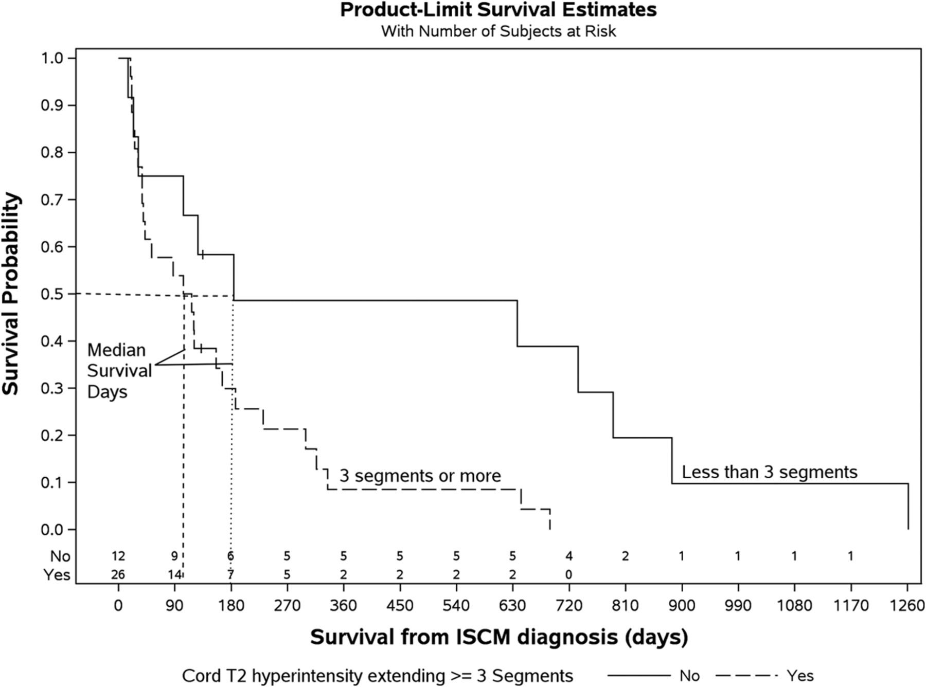
  • Fig 5.
    • Download figure
    • Open in new tab
    • Download powerpoint
    Fig 5.

    Kaplan-Meier plot. Decreased survival of patients with ISCM-related cord T2 hyperintensity extending ≥3 spinal segments. The median survival for T2 hyperintensity of ≥3 segments was 111 days and for <3 segments was 184 days (P = .018, log-rank test; n = 38 patients with solitary ISCMs with available sagittal T2WI).

Tables

  • Figures
    • View popup
    Table 1:

    Survival analysis of clinical features for all patients with ISCM (N = 49)

    VariableMedian Survival (daysa) (95% CI)Hazard Ratio (95% CI)P Valueb
    Age at diagnosis (yr) (N = 49)108 (48–156)1.02 (0.99–1.04).091
    Primary malignancy.005
        Lung (n = 24)58 (31–120)–
        Breast (n = 7)48 (21–456)–
        Melanoma (n = 5)87 (15–734)–
        CNS (n = 4)763 (19–1261)–
        Renal (n = 3)299 (231–643)–
        Other (n = 6)503 (24–884)–
    Lung/breast primary malignancy<.001
        Nonlung/nonbreast (n = 18)308 (87–689)–
        Lung/breast (n = 31)57 (33–117)–
    Melanoma.635
        Nonmelanoma (n = 44)115 (40–166)–
        Melanoma (n = 5)87 (15–734)–
    CNS primary malignancy.018
        Non-CNS (n = 45)104 (42–127)–
        CNS (n = 4)763 (19–1261)–
    ISCM diagnosis precedes primary malignancy diagnosis.361
        No (n = 44)104 (42–184)–
        Yes (n = 5)127 (21–187)–
    • Note:— – indicates not applicable.

    • ↵a From the date of ISCM diagnosis.

    • ↵b P values are for log-rank tests for categoric variables and Wald tests for continuous variables.

    • View popup
    Table 2:

    Survival analysis of MRI features for patients with solitary ISCMs (n = 39)

    VariableMedian Survival (daysa) (95% CI)Hazard Ratio (95% CI)P Valueb
    No. of ISCMs.022
        Solitary (n = 39)121 (42–187)–
        Multiple (n = 10)53 (12–88)–
    Noncord CNS/spinal metastases.879
        No (n = 19)166 (87–334)–
        Yes (n = 20)79 (26–184)–
    Leptomeningeal metastases.274
        No (n = 27)127 (38–231)–
        Yes (n = 12)113 (24–790)–
    Primary tumor/non-CNS metastases.012
        No (n = 15)316 (38–790)–
        Yes (n = 24)96 (32–166)–
    Cord T2 hyperintensity ≥2 segmentsc.037
        No (n = 10)184 (24–884)–
        Yes (n = 28)111 (38–166)–
    Cord T2 hyperintensity ≥3 segmentsc.018
        No (n = 12)184 (24–790)–
        Yes (n = 26)111 (38–166)–
    Cord T2 hyperintensity (No. of segments)c (n = 38)121 (42–187)1.02 (0.96–1.09).541
    ISCM enhancement, superoinferior size (mm)c (n = 35)120 (42–187)1.01 (0.99–1.02).138
    ISCM enhancement, extent (No. of vertebral segments)c (n = 35)120 (42–187)1.08 (0.85–1.38).524
    Ratio, longitudinal extent of cord T2 hyperintensity to enhancementc (n = 34)119 (40–187)1.03 (0.94–1.13).477
    Rim signc.621
        No (n = 18)79 (26–643)–
        Yes (n = 17)127 (40–299)–
    Flame signc.068
        No (n = 19)127 (42–734)–
        Yes (n = 16)96 (26–299)–
    Rim/flame signsc.143
        Neither (n = 13)121 (32–884)–
        Rim sign only (n = 6)147 (21–790)–
        Flame sign only (n = 5)26 (15–643)–
        Both signs (n = 11)120 (38–316)–
    • Note:— – indicates not applicable.

    • ↵a From date of ISCM diagnosis.

    • ↵b P values are for log-rank tests for categoric variables and Wald tests for continuous variables.

    • ↵c Missing patients (n < 39) due to MRI not having sagittal T2- or gadolinium-enhanced sagittal T1-weighted images.

PreviousNext
Back to top

In this issue

American Journal of Neuroradiology: 36 (3)
American Journal of Neuroradiology
Vol. 36, Issue 3
1 Mar 2015
  • Table of Contents
  • Index by author
  • Complete Issue (PDF)
Advertisement
Print
Download PDF
Email Article

Thank you for your interest in spreading the word on American Journal of Neuroradiology.

NOTE: We only request your email address so that the person you are recommending the page to knows that you wanted them to see it, and that it is not junk mail. We do not capture any email address.

Enter multiple addresses on separate lines or separate them with commas.
Intramedullary Spinal Cord Metastases: Prognostic Value of MRI and Clinical Features from a 13-Year Institutional Case Series
(Your Name) has sent you a message from American Journal of Neuroradiology
(Your Name) thought you would like to see the American Journal of Neuroradiology web site.
CAPTCHA
This question is for testing whether or not you are a human visitor and to prevent automated spam submissions.
Cite this article
F.E. Diehn, J.B. Rykken, J.T. Wald, C.P. Wood, L.J. Eckel, C.H. Hunt, K.M. Schwartz, R.K. Lingineni, R.E. Carter, T.J. Kaufmann
Intramedullary Spinal Cord Metastases: Prognostic Value of MRI and Clinical Features from a 13-Year Institutional Case Series
American Journal of Neuroradiology Mar 2015, 36 (3) 587-593; DOI: 10.3174/ajnr.A4160

Citation Manager Formats

  • BibTeX
  • Bookends
  • EasyBib
  • EndNote (tagged)
  • EndNote 8 (xml)
  • Medlars
  • Mendeley
  • Papers
  • RefWorks Tagged
  • Ref Manager
  • RIS
  • Zotero
0 Responses
Respond to this article
Share
Bookmark this article
Intramedullary Spinal Cord Metastases: Prognostic Value of MRI and Clinical Features from a 13-Year Institutional Case Series
F.E. Diehn, J.B. Rykken, J.T. Wald, C.P. Wood, L.J. Eckel, C.H. Hunt, K.M. Schwartz, R.K. Lingineni, R.E. Carter, T.J. Kaufmann
American Journal of Neuroradiology Mar 2015, 36 (3) 587-593; DOI: 10.3174/ajnr.A4160
del.icio.us logo Twitter logo Facebook logo Mendeley logo
  • Tweet Widget
  • Facebook Like
  • Google Plus One
Purchase

Jump to section

  • Article
    • Abstract
    • ABBREVIATION:
    • Materials and Methods
    • Results
    • Discussion
    • Conclusions
    • Footnotes
    • REFERENCES
  • Figures & Data
  • Info & Metrics
  • Responses
  • References
  • PDF

Related Articles

  • No related articles found.
  • PubMed
  • Google Scholar

Cited By...

  • Spinal intramedullary uterine carcinosarcoma metastasis
  • Crossref (15)
  • Google Scholar

This article has been cited by the following articles in journals that are participating in Crossref Cited-by Linking.

  • Spinal Hematomas: What a Radiologist Needs to Know
    Jennifer L. Pierce, Joseph H. Donahue, Nicholas C. Nacey, Cody R. Quirk, Michael T. Perry, Nicholas Faulconer, Gene A. Falkowski, Michael D. Maldonado, Catherine A. Shaeffer, Francis H. Shen
    RadioGraphics 2018 38 5
  • Experimental Study of Almonertinib Crossing the Blood-Brain Barrier in EGFR-Mutant NSCLC Brain Metastasis and Spinal Cord Metastasis Models
    Yuhan Zhang, Yaoshuai Zhang, Wenwen Niu, Xianming Ge, Fuhao Huang, Jinlong Pang, Xian Li, Yu Wang, Wei Gao, Fangtian Fan, Shanshan Li, Hao Liu
    Frontiers in Pharmacology 2021 12
  • Syphilitic meningomyelitis misdiagnosed as spinal cord tumor: Case and review
    Huiqing Dong, Zheng Liu, Yunyun Duan, Dawei Li, Zhandong Qiu, Yaou Liu, Jing Huang, Chaodong Wang
    The Journal of Spinal Cord Medicine 2021 44 5
  • Clinical Features, Treatments, and Prognosis of Intramedullary Spinal Cord Metastases From Lung Cancer: A Case Series and Systematic Review
    Liang Wu, Li'ao Wang, Jun Yang, Wenqing Jia, Yulun Xu
    Neurospine 2022 19 1
  • Metastatic Complications of Cancer Involving the Central and Peripheral Nervous Systems
    Joe S. Mendez, Lisa M. DeAngelis
    Neurologic Clinics 2018 36 3
  • Image-Guided Robotic Radiosurgery for the Management of Intramedullary Spinal Cord Metastases—A Multicenter Experience
    Felix Ehret, Carolin Senger, Markus Kufeld, Christoph Fürweger, Melina Kord, Alfred Haidenberger, Paul Windisch, Daniel Rueß, David Kaul, Maximilian Ruge, Christian Schichor, Jörg-Christian Tonn, Alexander Muacevic
    Cancers 2021 13 2
  • Neuroimaging of Spinal Cord and Cauda Equina Disorders
    Felix E. Diehn, Karl N. Krecke
    CONTINUUM: Lifelong Learning in Neurology 2021 27 1
  • Intramedullary spinal cord metastasis from Ewing sarcoma
    Cagri Yurtsever, Cahit Kafadar, Guner Sonmez, Hakan Mutlu
    The Spine Journal 2016 16 11
  • Intramedullary Spinal Cord Metastases from Differentiated Thyroid Cancer, a Case Report
    Fabio Volpe, Leandra Piscopo, Mariarosaria Manganelli, Maria Falzarano, Federica Volpicelli, Carmela Nappi, Massimo Imbriaco, Alberto Cuocolo, Michele Klain
    Life 2022 12 6
  • Spinal biopsies: a clinicopathologic review of 53 cases diagnosed between 2011 and 2018 at a tertiary hospital in Kampala, Uganda
    James J. Yahaya
    Egyptian Journal of Neurosurgery 2021 36 1

More in this TOC Section

  • Bern Score Validity for SIH
  • MP2RAGE 7T in MS Lesions of the Cervical Spine
  • Deep Learning for STIR Spine MRI Quality
Show more Spine

Similar Articles

Advertisement

Indexed Content

  • Current Issue
  • Accepted Manuscripts
  • Article Preview
  • Past Issues
  • Editorials
  • Editors Choice
  • Fellow Journal Club
  • Letters to the Editor

Cases

  • Case Collection
  • Archive - Case of the Week
  • Archive - Case of the Month
  • Archive - Classic Case

Special Collections

  • Special Collections

Resources

  • News and Updates
  • Turn around Times
  • Submit a Manuscript
  • Author Policies
  • Manuscript Submission Guidelines
  • Evidence-Based Medicine Level Guide
  • Publishing Checklists
  • Graphical Abstract Preparation
  • Imaging Protocol Submission
  • Submit a Case
  • Become a Reviewer/Academy of Reviewers
  • Get Peer Review Credit from Publons

Multimedia

  • AJNR Podcast
  • AJNR SCANtastic
  • Video Articles

About Us

  • About AJNR
  • Editorial Board
  • Not an AJNR Subscriber? Join Now
  • Alerts
  • Feedback
  • Advertise with us
  • Librarian Resources
  • Permissions
  • Terms and Conditions

American Society of Neuroradiology

  • Not an ASNR Member? Join Now

© 2025 by the American Society of Neuroradiology All rights, including for text and data mining, AI training, and similar technologies, are reserved.
Print ISSN: 0195-6108 Online ISSN: 1936-959X

Powered by HighWire