
Article Figures & Data
Figures
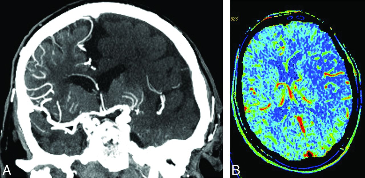
- Fig 1.
Coronal CTA MIP image and CBV map in a patient with SPAN-100-positivity at presentation. Coronal CTA MIP image (A) in this SPAN-100-positive patient with acute right-sided hemiparesis and subsequent unfavorable outcome demonstrates abrupt occlusion of the main stem left MCA with a collateral score of zero. The corresponding CBV map (B) demonstrates a large CBV deficit.
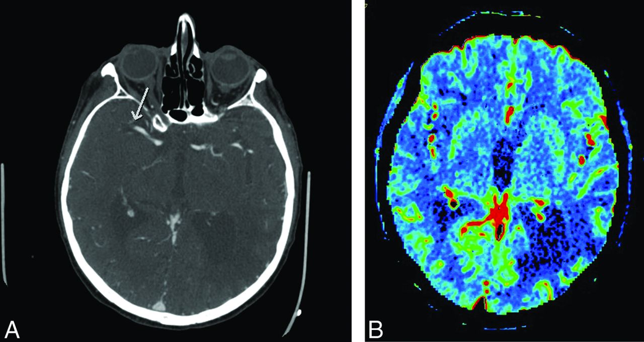
- Fig 2.
Axial CTA MIP image and CBV map in a patient with SPAN-100-positivity with favorable outcome. Axial CTA MIP (A) in this SPAN-100-positive patient with left-sided acute stroke and favorable outcome shows abrupt occlusion of the distal main stem right MCA. The extent of clot burden is low and underscores the utility of imaging in prognostication. The CBV map (B) shows a cortical-subcortical insular/subinsular defect in the right MCA distribution, with relative sparing of the basal ganglia. Chronic infarction is incidentally seen in the left parietal lobe.


Tables
- Table 1:
Comparing demographics/clinical factors between patients with positive SPAN-100 and patients with negative SPAN-100
SPAN-100-Negative (n = 218) SPAN-100-Positive (n = 55) P Value Age (yr) 68.2 ± 12.5 85.5 ± 5.07 <.001 NIHSS (median, IQR) 13 (7–18) 21 (17–24) <.001 Male sex 121 (55.5%) 22 (40) .04 SBP 157.7 (139–172) 156.04 (138–177) .77 DBP 84.9 (71–95) 76.1 (64–87) .01 Glucose (admission) 8.1 (5.8–8.1) 7.6 (6.0–9.0) .18 Risk factors Hypertension 127 (58.26) 45 (81.8) .001 Diabetes mellitus 43 (19.72) 10 (18.18) .79 Hypercholesterolemia 72 (33.03) 25 (45.4) .08 Coronary artery disease 53 (24.3) 12 (21.8) .69 Atrial fibrillation 64 (29.36) 19 (34.5) .45 Smoker 44 (20.1) 6 (10.9) .11 Stroke 1 (0.46) 1 (1.82) .29 Hyperdense sign 114 (52.53) 35 (63.64) .13 ASPECTS (median) (IQR) 7 (6–9) 7 (5–8) .39 Clot burden score 6 (4–9) 6 (5–9) .29 Collateral score 2 (2–3) 2 (1–2) <.001 CBV (median) (IQR) 14.7 (4.7–34.7) 34.7 (13.8–60.5) <.001 CBF 101.6 (55.3–133.1) 98.2 (74.6–129.5) .75 MTT 104.5 (58.5–133.4) 98.4 (74.7–130.5) .69 Time and thrombolysis rtPA dose 63.0 (54–73) 63.0 (54–72) .75 Onset to CT 104.0 (80–148) 108.0 (75–127) .62 Onset to rtPA 161.7 (147) 143.0 (131) .03 Outcome Recanalization 119 (55.35) 33 (61.1) .44 mRS (at follow-up) 3 (1–4) 5 (4–6) <.001 mRS ≤ 2 92 (42.2) 6 (10.9) <.001 NIHSS improves >3 in 24 hours 101 (46.3) 27 (49) .71 Hemorrhagic transformation 78 (38.6) 28 (56.0) .02 Hemorrhage infarct 64 (29.4) 23 (41.8) .07 Parenchymal hemorrhage 26 (11.9) 9 (16.4) .37 Note:—IQR indicates interquartile range; SBP, systolic blood pressure; DBP, diastolic blood pressure.
- Table 2:
Univariate logistic regression analysis of good clinical outcome (mRS ≤ 2) on demographic and clinical factors and imaging parameters
mRS ≤ 2 (n = 98), Favorable Outcome mRS > 2 (n = 175), Poor Outcome P Value OR (95% CI) Age (yr) 67 (57–78) 76 (66–83) <.001 0.96 (0.94–0.98) NIHSS pre-rtPA (median, IQR) 17 (12–21) 9.0 (4–15) <.001 0.86 (0.82–0.90) Female sex 42 (42.86) 88 (50.29) .23 1.35 (0.82–2.23) SBP 151 (138.5–173.5) 155 (137–171.5) .93 1.00 (0.99–1.01) DBP 81 (72.5–92) 80 (70–90) .26 1.01 (0.99–1.02) Glucose (admission)a 6.4 (5.4–7.3) 6.4 (5.4–7.3) .04 0.42 (0.17–0.92) Risk factors Hypertension 54 (55.10) 118 (67.43%) .04 0.59 (0.36–0.99) Diabetes mellitus 15 (15.31) 38 (21.71) .20 0.65 (0.33–1.24) Hypercholesterolemia 29 (29.59) 68 (38.86) .12 0.66 (0.39–1.12) Coronary artery disease 23 (24.3) 42 (24) .92 0.97 (0.54–1.73) Atrial fibrillation 29 (29.59) 54 (30.86) .82 0.94 (0.55–1.61) Smoker 22 (22.45) 28 (16) .18 1.52 (0.81–2.83) Stroke 1 (1.02) 1 (0.57) .68 1.79 (0.07–45.66) Hyperdense sign 48 (48.98%) 101 (58.05) .14 0.69 (0.42–1.14) EIC 84 (85.91) 158 (90.29) .25 0.65 (0.30–1.39) ASPECTS (median, IQR) 8 (6–9) 7 (5–8) .001 1.24 (1.09–1.42) Clot burden score 7 (6–9) 6 (4–9) .002 1.14 (1.05–1.25) Collateral score 2 (2–3) 2 (1–3) .02 1.46 (1.06–2.04) CBV (median, IQR)a 8.77 (2.5–24.09) 25.14 (9.1–49.60) <.001 0.58 (0.46–0.72) CBF 88.12 (44.7–122.1) 104.17 (74.3–143.6) <.001 0.99 (0.99–1.00) MTT 87.96 (43.2–125.9) 105.4 (77.5–138.8) <.001 0.99 (0.99–1.00) Time and thrombolysis rtPA given (No. of the patients) 80 (81.63%) 139 (79.43%) .66 1.15 (0.62–2.20) rtPA dose (mg) 64.0 (50–73) 62.0 (54–72) .94 1.00 (0.98–1.02) Onset to CT (min) (median, IQR)a 102.0 (74–151) 105.0 (80.5–141.5) .73 1.08 (0.68–1.72) Onset to rtPA (min) (median IQR) 145 (120–179) 146 (126–175) .70 1.00 (1.00–1.01) Outcome Recanalization 119 (55.35%) 33 (61.1%) <.001 3.58 (2.09–6.30) Hemorrhagic transformation 28 (30.43%) 78 (48.75%) .004 0.46 (0.26–0.78) NIHSS improves >3 in 24 hours 101 (46.3%) 27 (49%) .20 1.38 (0.84–2.27) SPAN-100-positive 6 (6.12%) 49 (28%) <.001 0.17 (0.06–0.38) Note:—EIC indicates early ischemic changes.
↵a Natural log-transformation was applied for normalizing the distribution.
AIC −2 (Log-Likelihood) R2a AUC OR 95% CI P Value Favorable clinical outcome Model of SPAN-100 only 316.64 312.64 0.054 0.599 0.56–0.64 SPAN-100 0.2 0.1–0.4 Model of SPAN-100, CBS 307.42 301.42 0.088 0.676 0.61–0.74 .004 SPAN-100 0.2 0.1–0.3 CBS 1.2 1.1–1.3 Model of SPAN-100, CBS, CBV 294.09 286.09 0.135 0.742 0.68–0.80 <.001 SPAN-100 0.2 0.1–0.5 CBS 1.1 1.0–1.2 CBV 0.6 0.5–0.8 Hemorrhagic transformation Model of SPAN-100 only 342.06 338.06 0.019 0.55 0.49–0.60 SPAN-100 2.0 1.1–3.8 Model of SPAN-100, CBS 339.39 333.392 0.037 0.62 0.54.5–0.69 .02 SPAN-100 2.2 1.2–4.1 CBS 0.9 0.8–0.9 Note:—AIC indicates Akaike information criterion.
↵a R2 is the proportion of variability in a dataset that is accounted for by the statistical model.


