
Article Figures & Data
Figures
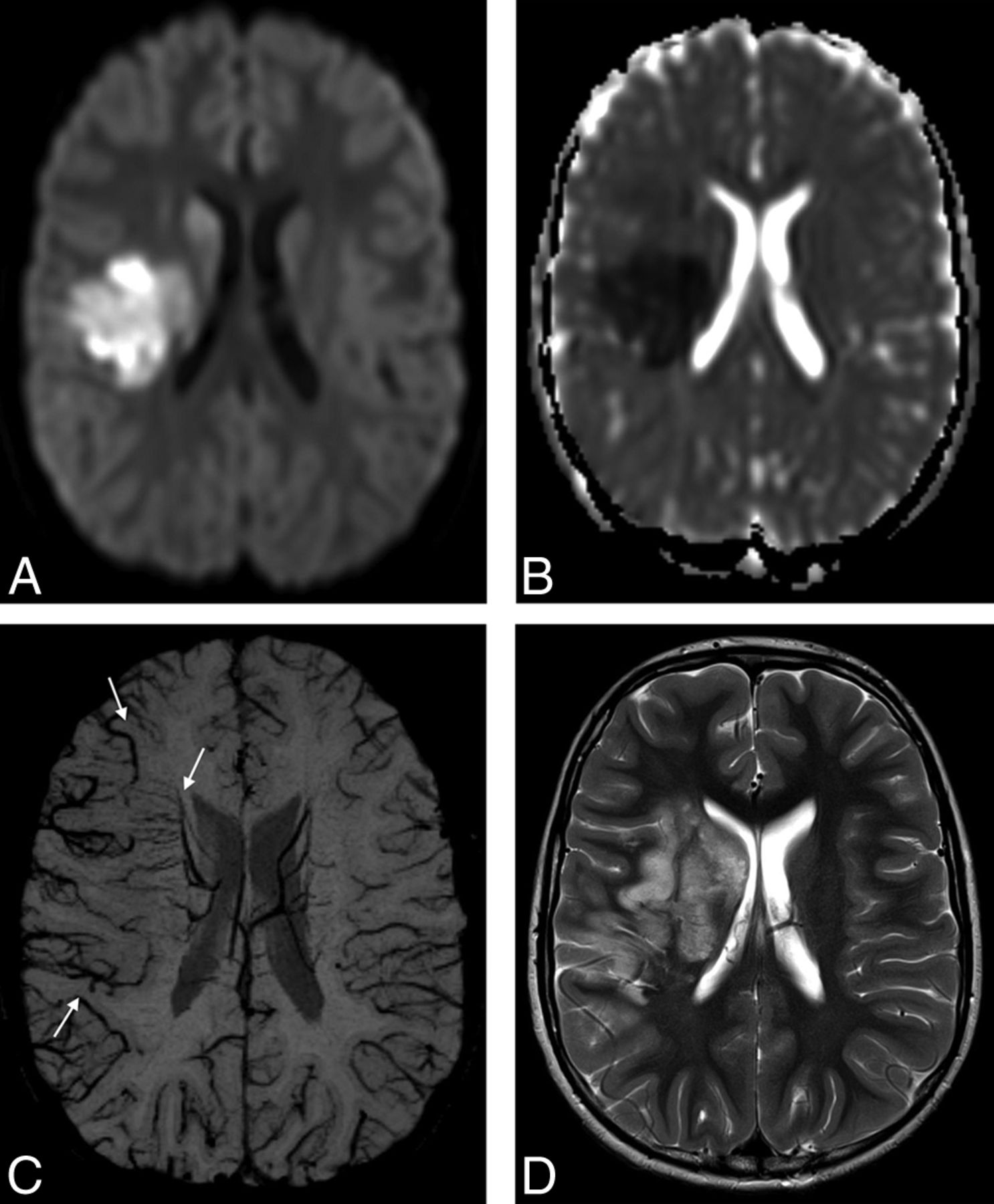
- Fig 1.
An 8-year-old boy with elevated lipoprotein A and AIS involving the right MCA territory. Trace of diffusion (A) and ADC (B) maps show areas of restricted diffusion in the right basal ganglia and part of the subcortical white matter and cortical gray matter in the right MCA territory, representing acute ischemia. C, mIP-SWI map shows markedly hypointense sulcal and intramedullary veins within the larger right MCA territory (arrows). D, Follow-up axial T2-weighted image 6 days after AIS shows hyperintense signal in the infarcted brain tissue that extends beyond the vascular territories with restricted diffusion and matches the area with SWI-hypointense veins on acute neuroimaging.
- Fig 2.
Fused axial ADC and mIP-SWI map images for the same child as in Fig 1 show that markedly hypointense sulcal and intramedullary veins on SWI are draining an area that extends beyond the region of restricted diffusion on the ADC map in the right MCA territory.
- Fig 3.
A 7-year-old boy with AIS involving the right ACA and partial bilateral MCA territories. A, ADC map shows areas of restricted diffusion in the right ACA, M1, M2, M4, and M5 territories as well as the left M2, M4, and M5 territories. B, mIP-SWI shows markedly hyperintense sulcal veins in the right ACA, M1, M2, M4, and M5 territories and hypointense sulcal veins in the left M1, M2, M4, and M5 territories. C and D, Follow-up axial CT image 2 days after AIS shows stroke evolution in the right ACA, M1, M2, M4, and M5 as well as in the left M2, M4, and M5 territories. In addition, there is increasing mass effect with effacement of both frontal horns of the lateral ventricles, the third and fourth ventricles, and prepontine cistern, compatible with malignant edema.


Tables
SWI Findings (n = 24) DWI/DTI Findings (n = 24) ASL Findings (n = 7) Iso Hyper Mildly Hypo Markedly Hypo Normal Decreased Normal Perfusion ↓ Perfusion ↑ ACA R 18 4 1 1 20 4 6 1 0 L 16 5 2 1 20 4 7 0 0 MCA M1 R 16 2 4 2 17 7 7 0 0 L 15 4 4 1 17 7 6 1 0 M2 R 14 2 6 2 17 7 6 1 0 L 14 2 6 2 15 9 6 1 0 M3 R 18 2 2 2 20 4 6 1 0 L 20 2 1 1 16 8 7 0 0 M4 R 17 2 3 2 16 8 6 1 0 L 15 4 3 2 16 8 6 0 1 M5 R 13 3 6 2 13 11 6 1 0 L 13 2 5 4 13 11 5 0 2 M6 R 18 3 1 2 16 8 6 1 0 L 19 2 2 1 18 6 7 0 0 IMV R 17 0 6 1 L 20 0 2 2 PCA R 15 4 3 2 20 4 5 2 0 L 12 6 5 1 18 6 4 1 2 Note:—IMV indicates intramedullary veins; Iso, isointense; Hyper, hyperintense; Hypo, hypointense; ↑, increased; ↓, decreased; R, right; L, left; PCA, posterior cerebral artery.


Jump to section
Related Articles
Cited By...
- No citing articles found.